
Diagnosis and Management of Hyperglycemic Crises Diabetic Ketoacidosis
Diagnosis and Management of Hyperglycemic Crises Diabetic Ketoacidosis Hyperglycemic Hyperosmolar State 1

OVERVIEW 2
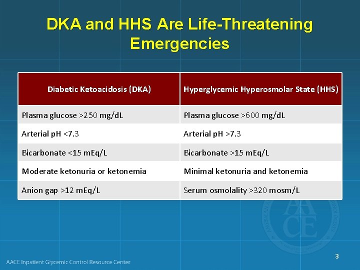
DKA and HHS Are Life-Threatening Emergencies Diabetic Ketoacidosis (DKA) Hyperglycemic Hyperosmolar State (HHS) Plasma glucose >250 mg/d. L Plasma glucose >600 mg/d. L Arterial p. H <7. 3 Arterial p. H >7. 3 Bicarbonate <15 m. Eq/L Bicarbonate >15 m. Eq/L Moderate ketonuria or ketonemia Minimal ketonuria and ketonemia Anion gap >12 m. Eq/L Serum osmolality >320 mosm/L 3
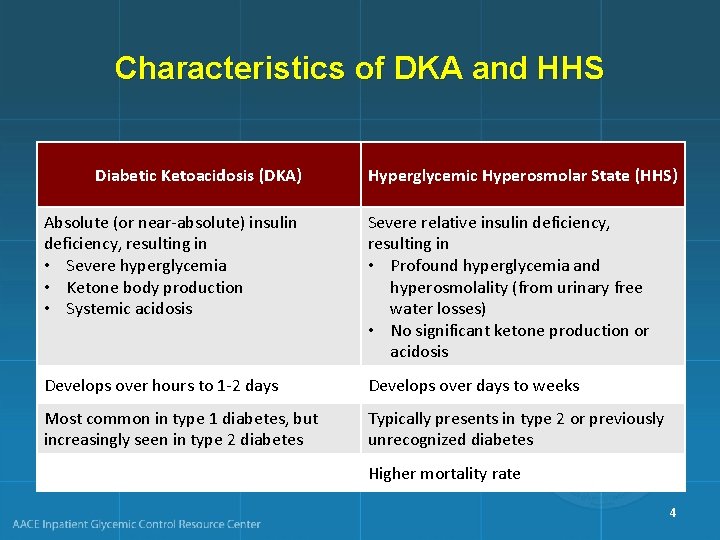
Characteristics of DKA and HHS Diabetic Ketoacidosis (DKA) Hyperglycemic Hyperosmolar State (HHS) Absolute (or near-absolute) insulin deficiency, resulting in • Severe hyperglycemia • Ketone body production • Systemic acidosis Severe relative insulin deficiency, resulting in • Profound hyperglycemia and hyperosmolality (from urinary free water losses) • No significant ketone production or acidosis Develops over hours to 1 -2 days Develops over days to weeks Most common in type 1 diabetes, but increasingly seen in type 2 diabetes Typically presents in type 2 or previously unrecognized diabetes Higher mortality rate 4
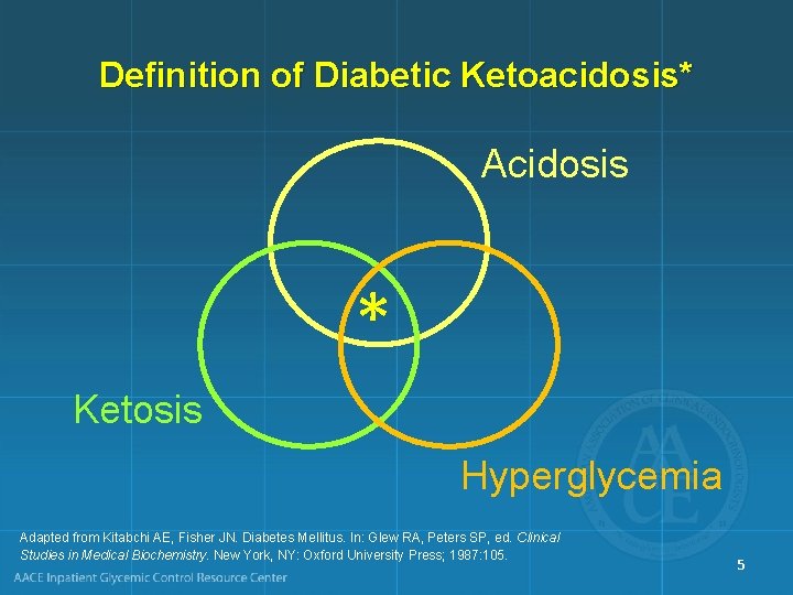
Definition of Diabetic Ketoacidosis* Acidosis * Ketosis Hyperglycemia Adapted from Kitabchi AE, Fisher JN. Diabetes Mellitus. In: Glew RA, Peters SP, ed. Clinical Studies in Medical Biochemistry. New York, NY: Oxford University Press; 1987: 105. 5
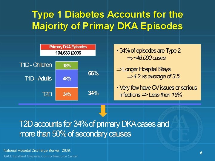
Type 1 Diabetes Accounts for the Majority of Primay DKA Episodes National Hospital Discharge Survey. 2006. 6
Hospital Discharges for Diabetic Ketoacidosis (DKA) in the US • In 2005, diagnosis of DKA was present on – 120, 000 discharges – 7. 4 discharges per 1000 DM patient population • There was a higher rate of DKA for persons <age 45 – 55. 4 discharges/1000 DM patient population (1987) – 31. 6 discharges/1000 DM patient population (2005) CDC. Diabetes Data and Trends. Hospitalization. Available from: http: //www. cdc. gov/diabetes/statistics/hospitalization_national. htm#5 7
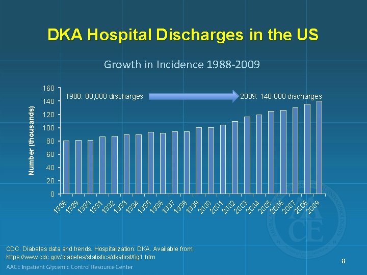
DKA Hospital Discharges in the US Growth in Incidence 1988 -2009 160 1988: 80, 000 discharges Number (thousands) 140 2009: 140, 000 discharges 120 100 80 60 40 20 89 19 90 19 91 19 92 19 93 19 94 19 95 19 96 19 97 19 98 19 99 20 00 20 01 20 02 20 03 20 04 20 05 20 06 20 07 20 08 20 09 19 19 88 0 CDC. Diabetes data and trends. Hospitalization: DKA. Available from: https: //www. cdc. gov/diabetes/statistics/dkafirst/fig 1. htm 8
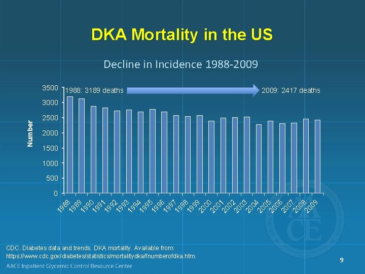
DKA Mortality in the US Decline in Incidence 1988 -2009 3500 1988: 3189 deaths 2009: 2417 deaths Number 3000 2500 2000 1500 1000 500 19 8 89 19 0 9 19 1 9 19 2 9 19 3 9 19 4 9 19 5 9 19 6 9 19 7 9 19 8 9 20 9 00 20 1 0 20 2 0 20 3 0 20 4 0 20 5 0 20 6 0 20 7 0 20 8 09 0 CDC. Diabetes data and trends. DKA mortality. Available from: https: //www. cdc. gov/diabetes/statistics/mortalitydka/fnumberofdka. htm. 9
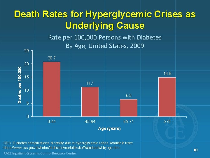
Death Rates for Hyperglycemic Crises as Underlying Cause 25 Rate per 100, 000 Persons with Diabetes By Age, United States, 2009 20. 7 Deaths per 100, 000 20 14. 8 15 11. 1 10 6. 5 5 0 0 -44 45 -64 65 -71 ≥ 75 Age (years) CDC. Diabetes complications. Mortality due to hyperglycemic crises. Available from: https: //www. cdc. gov/diabetes/statistics/mortalitydka/fratedkadiabbyage. htm. 10
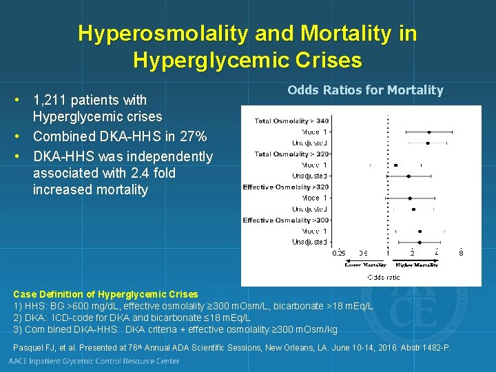
Hyperosmolality and Mortality in Hyperglycemic Crises • 1, 211 patients with Hyperglycemic crises • Combined DKA-HHS in 27% • DKA-HHS was independently associated with 2. 4 fold increased mortality Odds Ratios for Mortality Case Definition of Hyperglycemic Crises 1) HHS: BG >600 mg/d. L, effective osmolality ≥ 300 m. Osm/L, bicarbonate >18 m. Eq/L 2) DKA: ICD-code for DKA and bicarbonate ≤ 18 m. Eq/L 3) Com bined DKA-HHS: DKA criteria + effective osmolality ≥ 300 m. Osm/kg Pasquel FJ, et al. Presented at 76 th Annual ADA Scientific Sessions, New Orleans, LA. June 10 -14, 2016. Abstr 1482 -P.
Causes of Morbidity and Mortality in DKA • Shock • Hypokalemia during treatment • Hypoglycemia during treatment • Cerebral edema during treatment • Hypophosphatemia • Acute renal failure • Adult respiratory distress syndrome • Vascular thrombosis • Precipitating illness, including MI, stroke, sepsis, pancreatitis, pneumonia 12

PATHOGENESIS AND PATHOPHYSIOLOGY 13

Diabetic Ketoacidosis: Pathophysiology Unchecked gluconeogenesis Hyperglycemia Osmotic diuresis Dehydration Unchecked ketogenesis Ketosis Dissociation of ketone bodies into hydrogen ion and anions Anion-gap metabolic acidosis Often a precipitating event is identified (infection, lack of insulin administration) 14
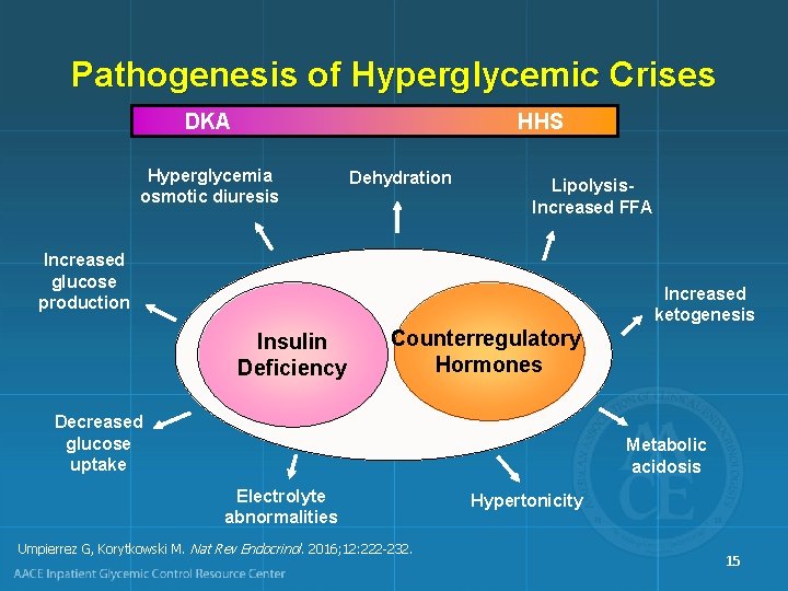
Pathogenesis of Hyperglycemic Crises DKA HHS Hyperglycemia osmotic diuresis Dehydration Lipolysis. Increased FFA Increased glucose production Increased ketogenesis Insulin Deficiency Counterregulatory Hormones Decreased glucose uptake Metabolic acidosis Electrolyte abnormalities Umpierrez G, Korytkowski M. Nat Rev Endocrinol. 2016; 12: 222 -232. Hypertonicity 15
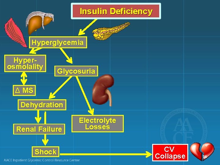
Insulin Deficiency Hyperglycemia Hyperosmolality Glycosuria Δ MS Dehydration Renal Failure Shock Electrolyte Losses CV Collapse 16
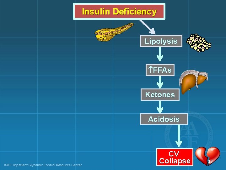
Insulin Deficiency Lipolysis FFAs Ketones Acidosis CV Collapse 17
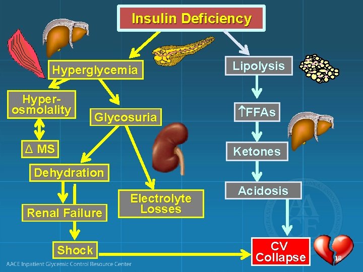
Insulin Deficiency Hyperglycemia Hyperosmolality Glycosuria Δ MS Lipolysis FFAs Ketones Dehydration Renal Failure Shock Electrolyte Losses Acidosis CV Collapse 18
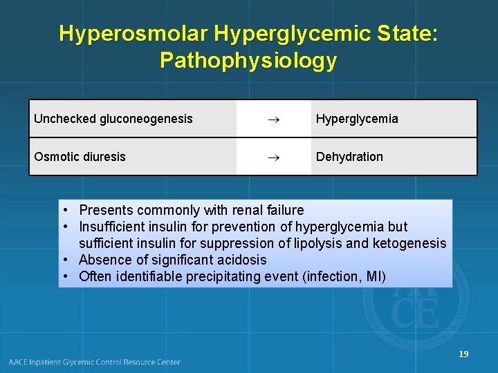
Hyperosmolar Hyperglycemic State: Pathophysiology Unchecked gluconeogenesis Hyperglycemia Osmotic diuresis Dehydration • Presents commonly with renal failure • Insufficient insulin for prevention of hyperglycemia but sufficient insulin for suppression of lipolysis and ketogenesis • Absence of significant acidosis • Often identifiable precipitating event (infection, MI) 19

Diabetic Hyperglycemic Crises Diabetic Ketoacidosis (DKA) Hyperglycemic Hyperosmolar State (HHS) Younger, type 1 diabetes Older, type 2 diabetes No hyperosmolality Hyperosmolality Volume depletion Electrolyte disturbances Acidosis No acidosis 20
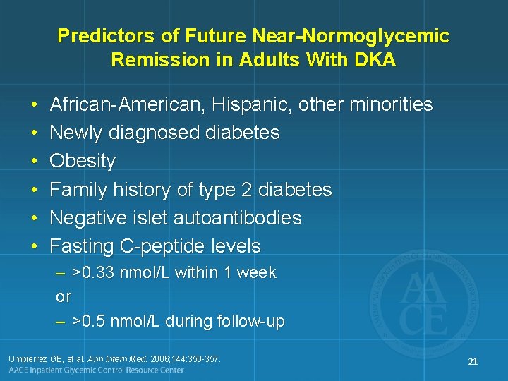
Predictors of Future Near-Normoglycemic Remission in Adults With DKA • • • African-American, Hispanic, other minorities Newly diagnosed diabetes Obesity Family history of type 2 diabetes Negative islet autoantibodies Fasting C-peptide levels – >0. 33 nmol/L within 1 week or – >0. 5 nmol/L during follow-up Umpierrez GE, et al. Ann Intern Med. 2006; 144: 350 -357. 21

FOCUS ON ACIDOSIS 22
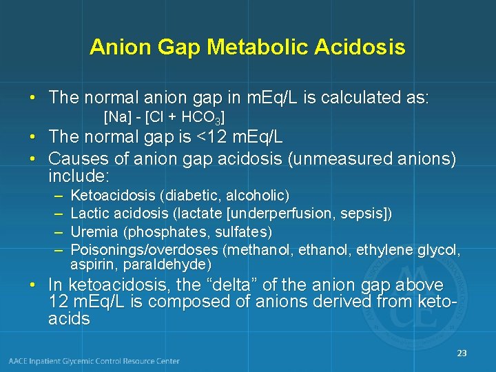
Anion Gap Metabolic Acidosis • The normal anion gap in m. Eq/L is calculated as: [Na] - [Cl + HCO 3] • The normal gap is <12 m. Eq/L • Causes of anion gap acidosis (unmeasured anions) include: – – Ketoacidosis (diabetic, alcoholic) Lactic acidosis (lactate [underperfusion, sepsis]) Uremia (phosphates, sulfates) Poisonings/overdoses (methanol, ethylene glycol, aspirin, paraldehyde) • In ketoacidosis, the “delta” of the anion gap above 12 m. Eq/L is composed of anions derived from ketoacids 23
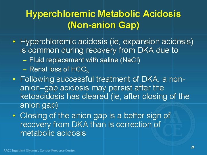
Hyperchloremic Metabolic Acidosis (Non-anion Gap) • Hyperchloremic acidosis (ie, expansion acidosis) is common during recovery from DKA due to – Fluid replacement with saline (Na. Cl) – Renal loss of HCO 3 • Following successful treatment of DKA, a nonanion–gap acidosis may persist after the ketoacidosis has cleared (ie, after closing of the anion gap) • Closing of the anion gap is a better sign of recovery from DKA than is correction of metabolic acidosis 24
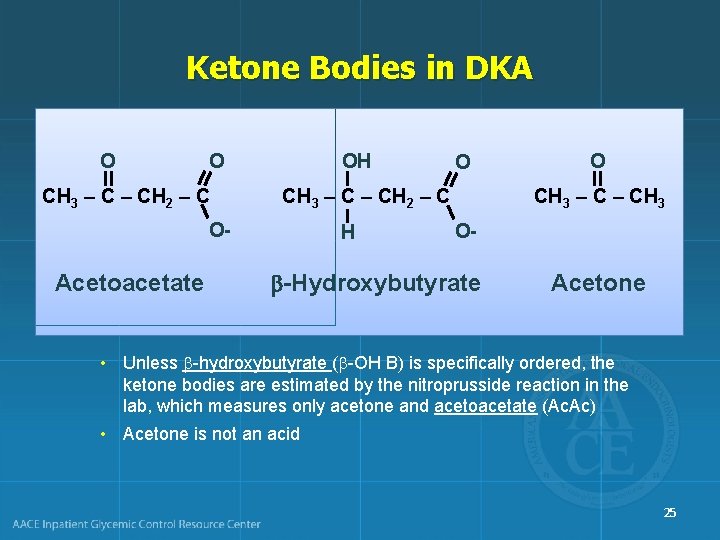
Ketone Bodies in DKA O O CH 3 – CH 2 – C O- Acetoacetate OH O CH 3 – CH 2 – C H O CH 3 – CH 3 O- -Hydroxybutyrate Acetone • Unless -hydroxybutyrate ( -OH B) is specifically ordered, the ketone bodies are estimated by the nitroprusside reaction in the lab, which measures only acetone and acetoacetate (Ac. Ac) • Acetone is not an acid 25
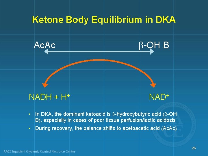
Ketone Body Equilibrium in DKA Ac. Ac NADH + H+ -OH B NAD+ • In DKA, the dominant ketoacid is -hydroxybutyric acid ( -OH B), especially in cases of poor tissue perfusion/lactic acidosis • During recovery, the balance shifts to acetoacetic acid (Ac. Ac) 26
Significance of Ketone Measurements • -hydroxybutyrate can only be measured using specialized equipment not available in most in-house laboratories • During recovery, results from the nitroprusside test might wrongly indicate that the ketone concentration is not improving or is even getting worse • The best biochemical indicator of resolution of ketoacid excess is simply the anion gap • There is no rationale for follow-up ketone measurements after the initial measurement has returned high 27
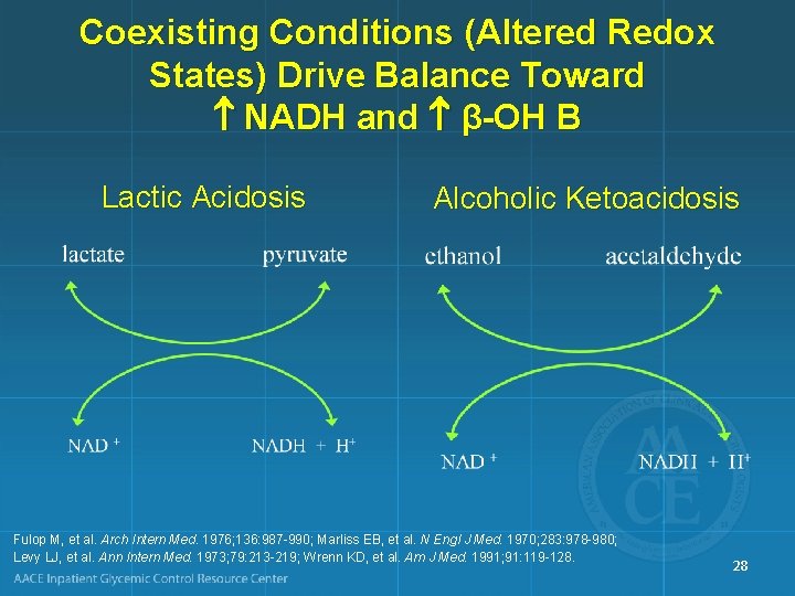
Coexisting Conditions (Altered Redox States) Drive Balance Toward NADH and β-OH B Lactic Acidosis Alcoholic Ketoacidosis Fulop M, et al. Arch Intern Med. 1976; 136: 987 -990; Marliss EB, et al. N Engl J Med. 1970; 283: 978 -980; Levy LJ, et al. Ann Intern Med. 1973; 79: 213 -219; Wrenn KD, et al. Am J Med. 1991; 91: 119 -128. 28
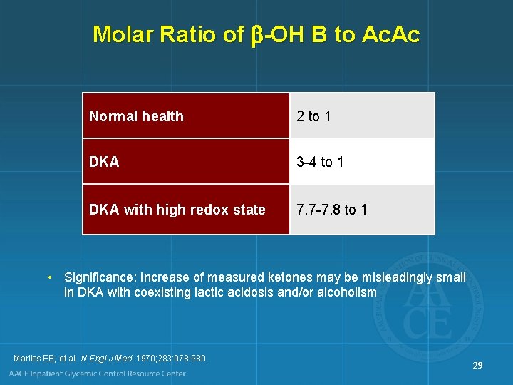
Molar Ratio of -OH B to Ac. Ac Normal health 2 to 1 DKA 3 -4 to 1 DKA with high redox state 7. 7 -7. 8 to 1 • Significance: Increase of measured ketones may be misleadingly small in DKA with coexisting lactic acidosis and/or alcoholism Marliss EB, et al. N Engl J Med. 1970; 283: 978 -980. 29

PATIENT PRESENTATION 30
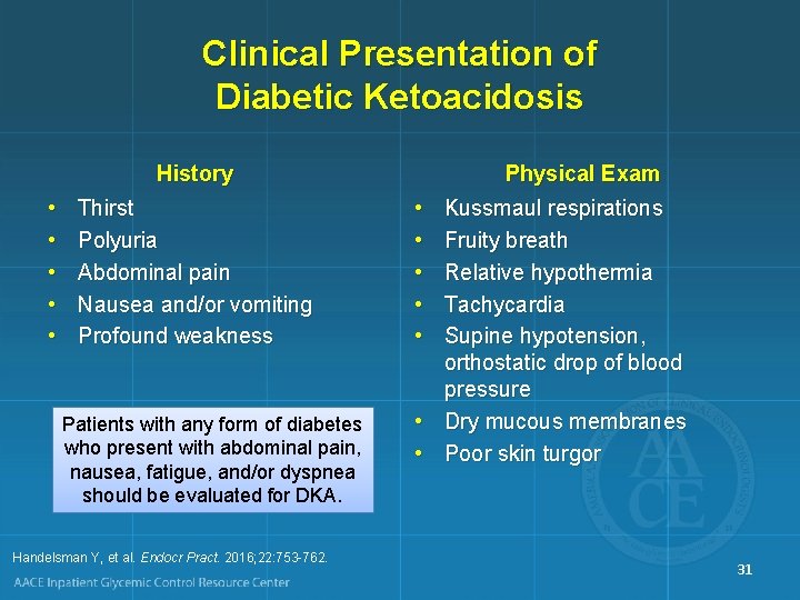
Clinical Presentation of Diabetic Ketoacidosis History • • • Thirst Polyuria Abdominal pain Nausea and/or vomiting Profound weakness Patients with any form of diabetes who present with abdominal pain, nausea, fatigue, and/or dyspnea should be evaluated for DKA. Handelsman Y, et al. Endocr Pract. 2016; 22: 753 -762. Physical Exam • • • Kussmaul respirations Fruity breath Relative hypothermia Tachycardia Supine hypotension, orthostatic drop of blood pressure • Dry mucous membranes • Poor skin turgor 31
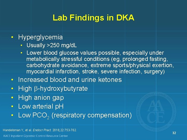
Lab Findings in DKA • Hyperglycemia • Usually >250 mg/d. L • Lower blood glucose values possible, especially under metabolically stressful conditions (eg, prolonged fasting, carbohydrate avoidance, extreme sports/physical exertion, myocardial infarction, stroke, severe infection, surgery) • • • Increased blood and urine ketones High -hydroxybutyrate High anion gap Low arterial p. H Low PCO 2 (respiratory compensation) Handelsman Y, et al. Endocr Pract. 2016; 22: 753 -762. 32
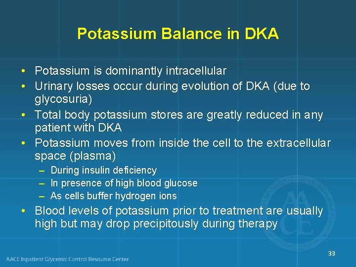
Potassium Balance in DKA • Potassium is dominantly intracellular • Urinary losses occur during evolution of DKA (due to glycosuria) • Total body potassium stores are greatly reduced in any patient with DKA • Potassium moves from inside the cell to the extracellular space (plasma) – – – During insulin deficiency In presence of high blood glucose As cells buffer hydrogen ions • Blood levels of potassium prior to treatment are usually high but may drop precipitously during therapy 33
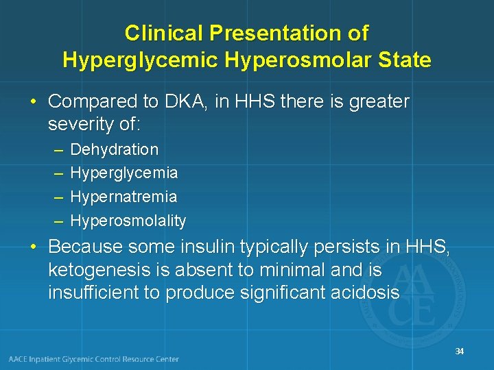
Clinical Presentation of Hyperglycemic Hyperosmolar State • Compared to DKA, in HHS there is greater severity of: – – Dehydration Hyperglycemia Hypernatremia Hyperosmolality • Because some insulin typically persists in HHS, ketogenesis is absent to minimal and is insufficient to produce significant acidosis 34
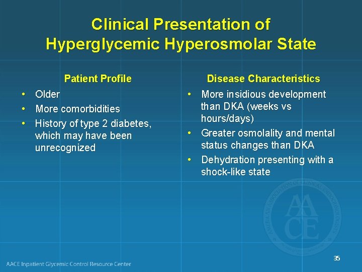
Clinical Presentation of Hyperglycemic Hyperosmolar State Patient Profile • • • Older More comorbidities History of type 2 diabetes, which may have been unrecognized Disease Characteristics • More insidious development than DKA (weeks vs hours/days) • Greater osmolality and mental status changes than DKA • Dehydration presenting with a shock-like state 35
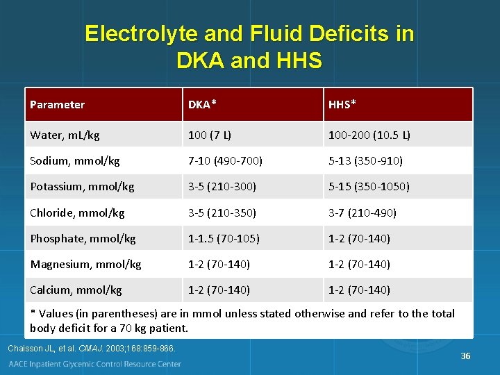
Electrolyte and Fluid Deficits in DKA and HHS Parameter DKA* HHS* Water, m. L/kg 100 (7 L) 100 -200 (10. 5 L) Sodium, mmol/kg 7 -10 (490 -700) 5 -13 (350 -910) Potassium, mmol/kg 3 -5 (210 -300) 5 -15 (350 -1050) Chloride, mmol/kg 3 -5 (210 -350) 3 -7 (210 -490) Phosphate, mmol/kg 1 -1. 5 (70 -105) 1 -2 (70 -140) Magnesium, mmol/kg 1 -2 (70 -140) Calcium, mmol/kg 1 -2 (70 -140) * Values (in parentheses) are in mmol unless stated otherwise and refer to the total body deficit for a 70 kg patient. Chaisson JL, et al. CMAJ. 2003; 168: 859 -866. 36
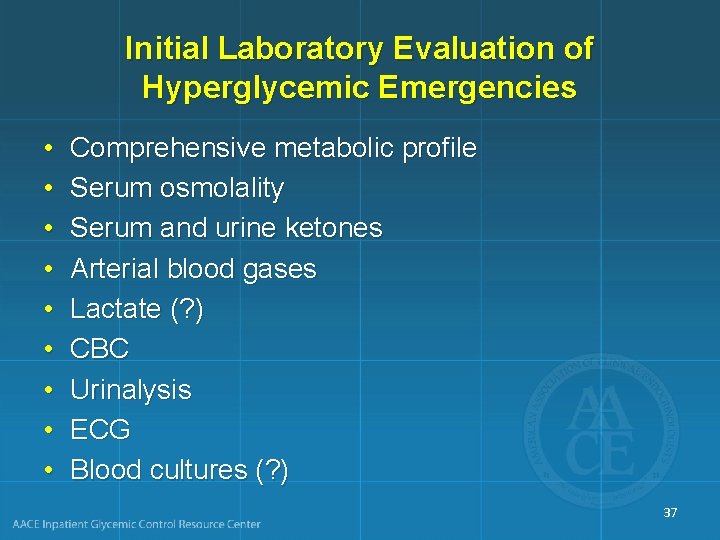
Initial Laboratory Evaluation of Hyperglycemic Emergencies • • • Comprehensive metabolic profile Serum osmolality Serum and urine ketones Arterial blood gases Lactate (? ) CBC Urinalysis ECG Blood cultures (? ) 37
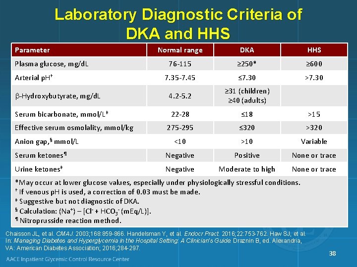
Laboratory Diagnostic Criteria of DKA and HHS Parameter Normal range DKA HHS 76 -115 ≥ 250* ≥ 600 7. 35 -7. 45 ≤ 7. 30 >7. 30 -Hydroxybutyrate, mg/d. L 4. 2 -5. 2 ≥ 31 (children) ≥ 40 (adults) Serum bicarbonate, mmol/L‡ 22 -28 ≤ 18 >15 275 -295 ≤ 320 >320 <10 >10 Variable Serum ketones¶ Negative Positive None or trace Urine ketones‡ Negative Moderate to high None or trace Plasma glucose, mg/d. L Arterial p. H† Effective serum osmolality, mmol/kg Anion gap, § mmol/L *May occur at lower glucose values, especially under physiologically stressful conditions. † If venous p. H is used, a correction of 0. 03 must be made. ‡ Suggestive but not diagnostic of DKA. § Calculation: (Na+) – [Cl- + HCO - (m. Eq/L)]. 3 ¶ Nitroprusside reaction method. Chaisson JL, et al. CMAJ. 2003; 168: 859 -866. Handelsman Y, et al. Endocr Pract. 2016; 22: 753 -762. Haw SJ, et al. In: Managing Diabetes and Hyperglycemia in the Hospital Setting: A Clinician’s Guide. Draznin B, ed. Alexandria, VA: American Diabetes Association; 2016; 284 -297. 38
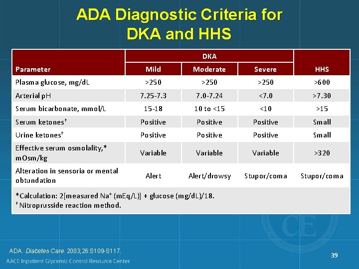
ADA Diagnostic Criteria for DKA and HHS DKA Parameter Mild Moderate Severe HHS Plasma glucose, mg/d. L >250 >600 7. 25 -7. 3 7. 0 -7. 24 <7. 0 >7. 30 15 -18 10 to <15 <10 >15 Serum ketones† Positive Small Urine ketones† Positive Small Effective serum osmolality, * m. Osm/kg Variable >320 Alert/drowsy Stupor/coma Arterial p. H Serum bicarbonate, mmol/L Alteration in sensoria or mental obtundation *Calculation: 2[measured Na+ (m. Eq/L)] + glucose (mg/d. L)/18. † Nitroprusside reaction method. ADA. Diabetes Care. 2003; 26: S 109 -S 117. 39
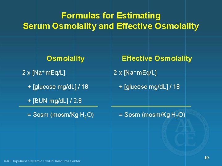
Formulas for Estimating Serum Osmolality and Effective Osmolality 2 x [Na+ m. Eq/L] + [glucose mg/d. L] / 18 + [BUN mg/d. L] / 2. 8 = Sosm (mosm/Kg H 2 O) 40
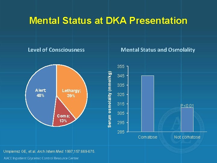
Mental Status at DKA Presentation Level of Consciousness Mental Status and Osmolality Alert; 48% Lethargy; 39% Coma; 13% Serum osmolality (mmol/kg) 355 345 335 325 315 P<0. 01 305 295 285 Comatose Umpierrez GE, et al. Arch Intern Med. 1997; 157: 669 -675. Not comatose
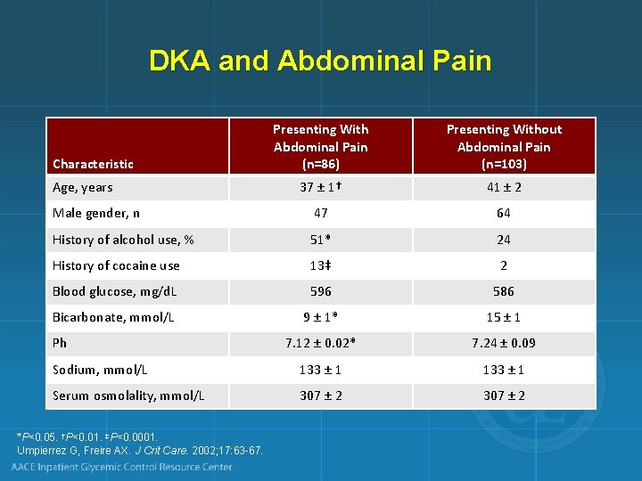
DKA and Abdominal Pain Presenting With Abdominal Pain (n=86) Presenting Without Abdominal Pain (n=103) 37 ± 1† 41 ± 2 Male gender, n 47 64 History of alcohol use, % 51* 24 History of cocaine use 13‡ 2 Blood glucose, mg/d. L 596 586 Bicarbonate, mmol/L 9 ± 1* 15 ± 1 7. 12 ± 0. 02* 7. 24 ± 0. 09 Sodium, mmol/L 133 ± 1 Serum osmolality, mmol/L 307 ± 2 Characteristic Age, years Ph *P<0. 05. †P<0. 01. ‡P<0. 0001. Umpierrez G, Freire AX. J Crit Care. 2002; 17: 63 -67.
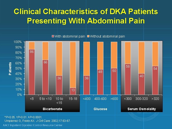
Clinical Characteristics of DKA Patients Presenting With Abdominal Pain With abdominal pain Without abdominal pain 100% 90% 86 Patients 70% 66 60% 58 50% 48 40% 36 30% 54 50 40 36 20% 13 0% <5 5 to <10 10 to <15 15 -18 Bicarbonate *P<0. 05. †P<0. 01. ‡P<0. 0001. Umpierrez G, Freire AX. J Crit Care. 2002; 17: 63 -67. <400 400 -600 Glucose >600 <300 300 -320 >320 Serum Osmolality

TREATMENT RECOMMENDATIONS 44
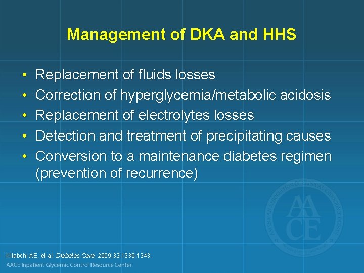
Management of DKA and HHS • • • Replacement of fluids losses Correction of hyperglycemia/metabolic acidosis Replacement of electrolytes losses Detection and treatment of precipitating causes Conversion to a maintenance diabetes regimen (prevention of recurrence) Kitabchi AE, et al. Diabetes Care. 2009; 32: 1335 -1343.
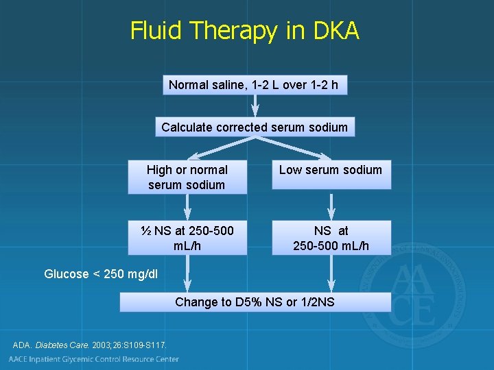
Fluid Therapy in DKA Normal saline, 1 -2 L over 1 -2 h Calculate corrected serum sodium High or normal serum sodium Low serum sodium ½ NS at 250 -500 m. L/h Glucose < 250 mg/dl Change to D 5% NS or 1/2 NS ADA. Diabetes Care. 2003; 26: S 109 -S 117.
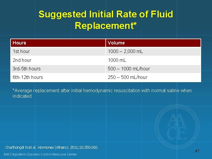
Suggested Initial Rate of Fluid Replacement* Hours Volume 1 st hour 1000 – 2, 000 m. L 2 nd hour 1000 m. L 3 rd-5 th hours 500 – 1000 m. L/hour 6 th-12 th hours 250 – 500 m. L/hour *Average replacement after initial hemodynamic resuscitation with normal saline when indicated Chaithongdi N et al. Hormones (Athens). 2011; 10: 250 -260. 47
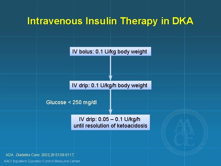
Intravenous Insulin Therapy in DKA IV bolus: 0. 1 U/kg body weight IV drip: 0. 1 U/kg/h body weight Glucose < 250 mg/dl IV drip: 0. 05 – 0. 1 U/kg/h until resolution of ketoacidosis ADA. Diabetes Care. 2003; 26: S 109 -S 117.
Potassium Repletion in DKA • Life-threatening hypokalemia can develop during insulin treatment • Potassium reenters cells with insulinization and correction of acidosis • The small extracellular compartment experiences a precipitous drop of potassium concentration • Anticipatory potassium replacement during treatment of DKA is almost always required 49
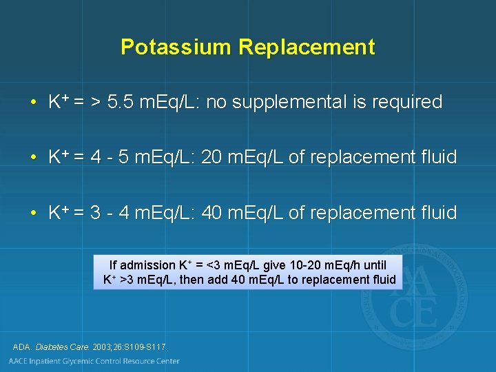
Potassium Replacement • K+ = > 5. 5 m. Eq/L: no supplemental is required • K+ = 4 - 5 m. Eq/L: 20 m. Eq/L of replacement fluid • K+ = 3 - 4 m. Eq/L: 40 m. Eq/L of replacement fluid If admission K+ = <3 m. Eq/L give 10 -20 m. Eq/h until K+ >3 m. Eq/L, then add 40 m. Eq/L to replacement fluid ADA. Diabetes Care. 2003; 26: S 109 -S 117.
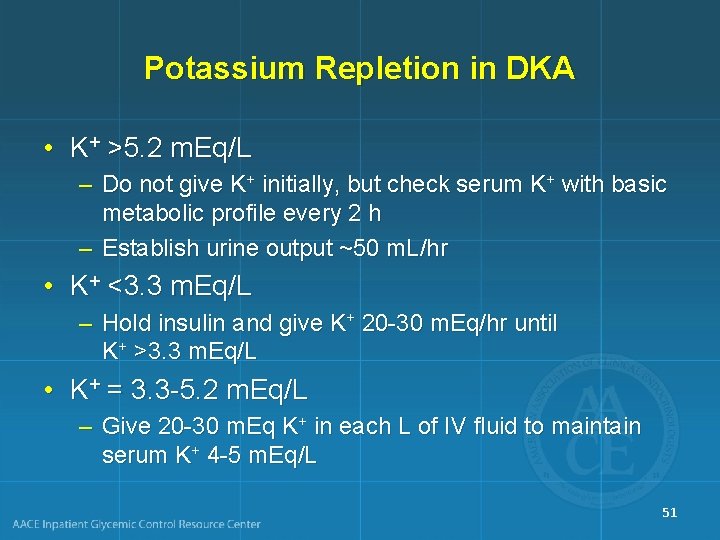
Potassium Repletion in DKA • K+ >5. 2 m. Eq/L – Do not give K+ initially, but check serum K+ with basic metabolic profile every 2 h – Establish urine output ~50 m. L/hr • K+ <3. 3 m. Eq/L – Hold insulin and give K+ 20 -30 m. Eq/hr until K+ >3. 3 m. Eq/L • K+ = 3. 3 -5. 2 m. Eq/L – Give 20 -30 m. Eq K+ in each L of IV fluid to maintain serum K+ 4 -5 m. Eq/L 51
Phosphorus Repletion in DKA • A sharp drop of serum phosphorus can also occur during insulin treatment • Treatment is usually not required – Caregiver can give some K+ as K- phos 52
Bicarbonate Administration • p. H > 7. 0: no bicarbonate • p. H < 7. 0 and bicarbonate < 5 m. Eq/L: 44. 6 m. Eq in 500 m. L 0. 45% saline over 1 h until p. H > 7. 0 ADA. Diabetes Care. 2003; 26: S 109 -S 117.
Phosphorus Administration • Not routinely recommended • If serum phosphorus < 1 mg/d. L: 30 -40 mmol K-Phos over 24 h • Monitor serum calcium level ADA. Diabetes Care. 2003; 26: S 109 -S 117.
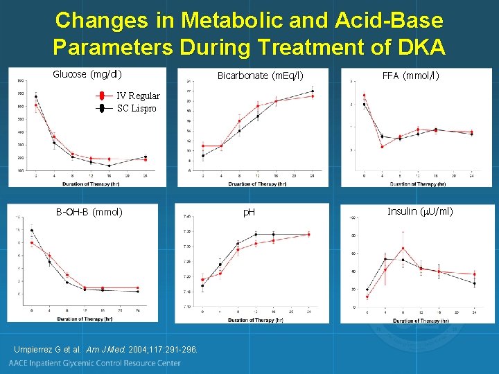
Changes in Metabolic and Acid-Base Parameters During Treatment of DKA Glucose (mg/dl) Bicarbonate (m. Eq/l) FFA (mmol/l) IV Regular SC Lispro B-OH-B (mmol) Umpierrez G et al. Am J Med. 2004; 117: 291 -296. p. H Insulin ( U/ml)
Conventional Insulin Guidelines • Initiate the correction of hypovolemic shock with fluids, and correct hypokalemia if present, before starting insulin • When starting insulin, initially infuse 0. 1 to 0. 14 units/kg/h • If plasma glucose does not decrease by 50 -75 mg in the first hour, increase the infusion rate of insulin • Continue insulin infusion until anion gap closes • Initiate subcutaneous insulin at least 2 h before interruption of insulin infusion Kitabchi AE, et al. Diabetes Care. 2009; 32: 1335 -1343. 56
Subcutaneous Rapid Acting Insulin or Intravenous Regular Insulin for DKA Treatment Systematic Review (N=5 RCTs) • No substantial difference in time to resolution of DKA between SC lispro or aspart vs IV regular insulin in adults – In single study including children and adolescents, DKA resolution slower with SC rapid acting analogs than with IV regular insulin • Rates of hypoglycemia and duration of hospital stay comparable between rapid acting insulin analogs and regular insulin in adults and children Andrade-Castellanos CA, et al. Cochrane Database Syst Rev. 2016 Jan 21; (1): CD 011281. 57
Subcutaneous Insulin Protocols Rapid Acting Insulin Every 1 Hour • Initial dose Rapid Acting Insulin Every 2 Hours • Initial dose – 0. 2 U/kg of body weight, followed by 0. 1 U/kg/h – 0. 3 U/kg of body weight, followed by 0. 2 U/kg 1 h later, then – Rapid acting insulin at 0. 2 U/kg every 2 h • When BG <250 mg/d. L – Change IVF to D 5%-0. 45% saline – Reduce rapid acting insulin to 0. 05 unit/kg/h – Keep glucose ≈ 200 mg/d. L until resolution of DKA • When BG <250 mg/d. L Haw SJ, et al. In: Managing Diabetes and Hyperglycemia in the Hospital Setting: A Clinician’s Guide. Draznin B, ed. Alexandria, VA: American Diabetes Association; 2016; 284 -297. – Change IVF to D 5%-0. 45% saline – Reduce rapid acting insulin to 0. 1 U/kg every 2 h – Keep glucose ≈ 200 mg/d. L until resolution of DKA 58
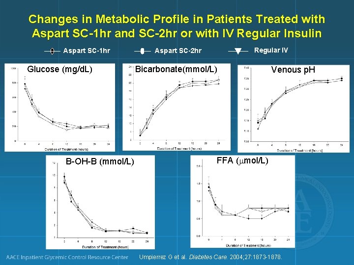
Changes in Metabolic Profile in Patients Treated with Aspart SC-1 hr and SC-2 hr or with IV Regular Insulin Aspart SC-1 hr Glucose (mg/d. L) B-OH-B (mmol/L) Regular IV Aspart SC-2 hr Bicarbonate(mmol/L) Venous p. H FFA ( mol/L) Umpierrez G et al. Diabetes Care. 2004; 27: 1873 -1878.
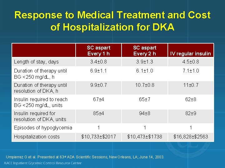
Response to Medical Treatment and Cost of Hospitalization for DKA SC aspart Every 1 h SC aspart Every 2 h IV regular insulin Length of stay, days 3. 4± 0. 8 3. 9± 1. 3 4. 5± 0. 8 Duration of therapy until BG <250 mg/d. L, h 6. 9± 1. 1 6. 1± 1. 0 7. 1± 1. 0 Duration of therapy until resolution of DKA, h 9. 9± 0. 7 10. 7± 0. 8 11± 0. 7 Insulin required to reach BG <250 mg/d. L, units 67± 4 65± 7 62± 8 Insulin required for resolution of DKA, units 85± 4 94± 8 82± 9 1 1 1 $10, 733±$2017 $10, 473±$1738 $16, 828±$2563 Episodes of hypoglycemia Hospitalization costs Umpierrez G et al. Presented at 63 rd ADA Scientific Sessions, New Orleans, LA; June 14, 2003.
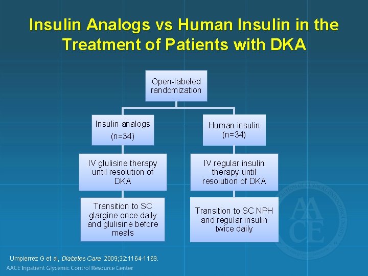
Insulin Analogs vs Human Insulin in the Treatment of Patients with DKA Open-labeled randomization Insulin analogs (n=34) Human insulin (n=34) IV glulisine therapy until resolution of DKA IV regular insulin therapy until resolution of DKA Transition to SC glargine once daily and glulisine before meals Transition to SC NPH and regular insulin twice daily Umpierrez G et al, Diabetes Care. 2009; 32: 1164 -1169.
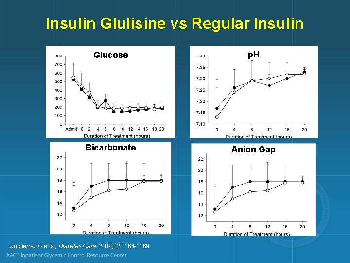
Insulin Glulisine vs Regular Insulin Glucose p. H Bicarbonate Anion Gap Umpierrez G et al, Diabetes Care. 2009; 32: 1164 -1169.
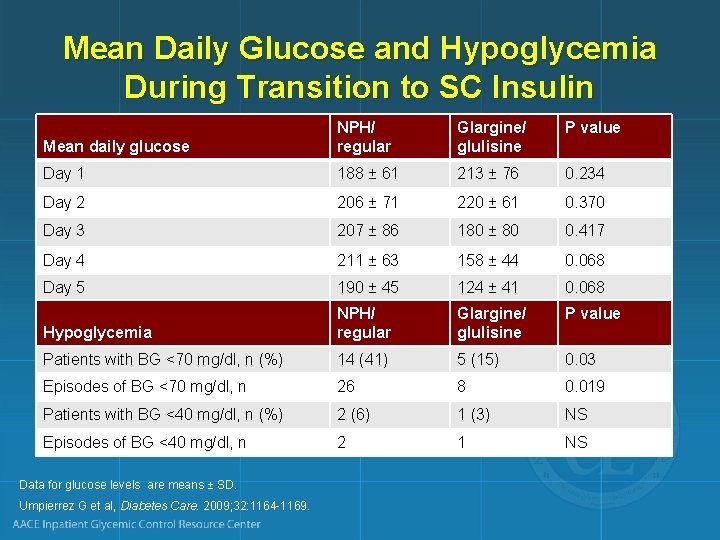
Mean Daily Glucose and Hypoglycemia During Transition to SC Insulin Glargine/ glulisine P value Mean daily glucose NPH/ regular Day 1 188 ± 61 213 ± 76 0. 234 Day 2 206 ± 71 220 ± 61 0. 370 Day 3 207 ± 86 180 ± 80 0. 417 Day 4 211 ± 63 158 ± 44 0. 068 Day 5 190 ± 45 124 ± 41 0. 068 Glargine/ glulisine P value Hypoglycemia NPH/ regular Patients with BG <70 mg/dl, n (%) 14 (41) 5 (15) 0. 03 Episodes of BG <70 mg/dl, n 26 8 0. 019 Patients with BG <40 mg/dl, n (%) 2 (6) 1 (3) NS Episodes of BG <40 mg/dl, n 2 1 NS Data for glucose levels are means ± SD. Umpierrez G et al, Diabetes Care. 2009; 32: 1164 -1169.
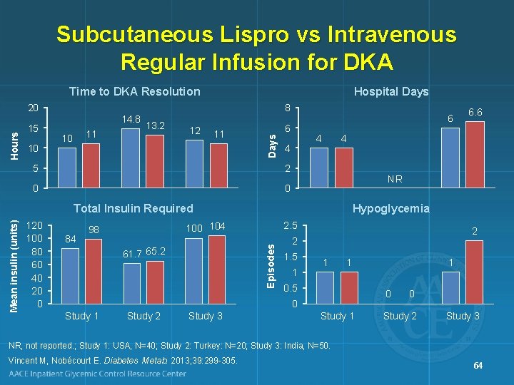
Subcutaneous Lispro vs Intravenous Regular Infusion for DKA Time to DKA Resolution 15 10 14. 8 11 10 8 13. 2 12 11 6 6 Days Hours 20 Hospital Days 4 4 5 2 0 0 4 NR 84 Hypoglycemia 2. 5 100 104 98 Episodes Mean insulin (units) Total Insulin Required 120 100 80 60 40 20 0 61. 7 65. 2 2 2 1. 5 1 1 1 0. 5 Study 2 Study 3 1 0 0 Study 1 6. 6 Study 1 0 Study 2 Study 3 NR, not reported. ; Study 1: USA, N=40; Study 2: Turkey: N=20; Study 3: India, N=50. Vincent M, Nobécourt E. Diabetes Metab. 2013; 39: 299 -305. 64
Rationale for a Dynamic Insulin Protocol for DKA and HHS • Even with low-dose insulin therapy 1, 2 – Hypokalemia and hypoglycemia may continue to occur – Failure to reduce insulin infusion rate as the blood glucose approaches target may lead to hypoglycemia • There is a lag between the change in intravenous insulin infusion rate and the resulting effects 3 1. Umpierrez GE, et al. Arch Intern Med. 1997; 157: 669 -675. 2. Burghen GA, et al. Diabetes Care. 1980; 3: 15 -20. 3. Mudaliar S, et al. Diabetes Care. 2002; 25: 1597 -1602. 65
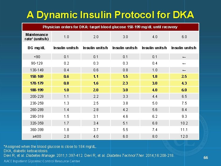
A Dynamic Insulin Protocol for DKA Physician orders for DKA: target blood glucose 150 -199 mg/d. L until recovery Maintenance rate* (units/h) 1. 0 2. 0 3. 0 4. 0 6. 0 BG mg/d. L Insulin units/h Insulin units/h <90 0. 1 ← 90 -129 0. 2 0. 3 0. 4 ← 130 -149 0. 4 0. 6 0. 8 1. 0 ← 150 -169 0. 6 1. 1 1. 5 1. 8 2. 5 170 -179 0. 8 1. 6 2. 3 3. 0 4. 3 180 -199 1. 0 2. 0 3. 0 4. 0 6. 0 200 -229 1. 1 2. 2 3. 3 4. 4 6. 5 230 -259 1. 3 2. 5 3. 8 5. 0 7. 5 260 -289 1. 4 2. 8 4. 2 5. 6 8. 4 290 -319 1. 5 3. 1 4. 6 6. 2 9. 3 320 -359 1. 7 3. 4 5. 1 6. 8 10. 2 360 -399 1. 8 3. 7 5. 5 7. 4 11. 1 ≥ 400 2. 0 4. 0 6. 0 8. 0 12. 0 *Assigned when the blood glucose is close to 184 mg/d. L. DKA, diabetic ketoacidosis. Devi R, et al. Diabetes Manage. 2011; 1: 397 -412. Devi R, et al. Diabetes Technol Ther. 2014; 16: 208 -218. 66
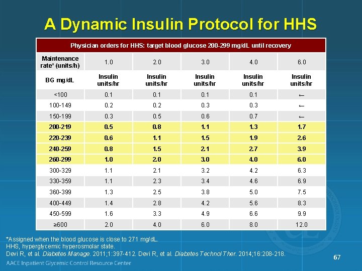
A Dynamic Insulin Protocol for HHS Physician orders for HHS: target blood glucose 200 -299 mg/d. L until recovery Maintenance rate* (units/h) 1. 0 2. 0 3. 0 4. 0 6. 0 BG mg/d. L Insulin units/hr Insulin units/hr <100 0. 1 ← 100 -149 0. 2 0. 3 ← 150 -199 0. 3 0. 5 0. 6 0. 7 ← 200 -219 0. 5 0. 8 1. 1 1. 3 1. 7 220 -239 0. 6 1. 1 1. 5 1. 9 2. 6 240 -259 0. 8 1. 5 2. 1 2. 7 3. 9 260 -299 1. 0 2. 0 3. 0 4. 0 6. 0 300 -329 1. 1 2. 1 3. 2 4. 2 6. 3 330 -359 1. 1 2. 3 3. 4 4. 6 6. 9 360 -399 1. 3 2. 5 3. 8 5. 0 7. 5 400 -449 1. 4 2. 8 4. 2 5. 6 8. 3 450 -599 1. 6 3. 3 4. 9 6. 6 9. 9 ≥ 600 2. 0 4. 0 6. 0 8. 0 12. 0 *Assigned when the blood glucose is close to 271 mg/d. L. HHS, hyperglycemic hyperosmolar state. Devi R, et al. Diabetes Manage. 2011; 1: 397 -412. Devi R, et al. Diabetes Technol Ther. 2014; 16: 208 -218. 67
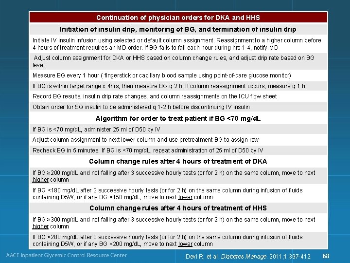
Continuation of physician orders for DKA and HHS Initiation of insulin drip, monitoring of BG, and termination of insulin drip Initiate IV insulin infusion using selected or default column assignment. Reassignment to a higher column before 4 hours of treatment requires an MD order. If BG fails to fall each hour during hrs 1 -4, notify MD Adjust column assignment for DKA or HHS based on column change rules, and adjust drip rate based on BG level Measure BG every 1 hour ( fingerstick or capillary blood sample using point-of-care glucose monitor) If BG is within target range x 4 hrs, then measure BG q 2 h. If column reassignment occurs, measure q 1 h Record BG results, insulin drip rate changes, and column reassignments on the ICU flow sheet Obtain order for SQ insulin to be administered q 1 -2 h before discontinuing IV insulin Algorithm for order to treat patient if BG <70 mg/d. L If BG is <70 mg/d. L, administer 25 ml of D 50 by IV Adjust column assignment to next lower column and use pretreatment BG to assign row Recheck BG in 5 minutes. If BG is <70 mg/d. L, repeat administration of 25 ml of D 50 by IV Column change rules after 4 hours of treatment of DKA If BG ≥ 200 mg/d. L and not falling after 3 successive hourly tests (or for 2 h) on the same column, move to next higher column If BG <180 mg/d. L after 3 successive hourly tests (or for 2 h) on the same column during infusion of fluids containing D 5 W, or if any BG <150 mg/d. L, move to next lower column Column change rules after 4 hours of treatment of HHS If BG ≥ 300 mg/d. L and not falling after 3 successive hourly tests (or for 2 h) on the same column, move to next higher column If BG <280 mg/d. L after 3 successive hourly tests (or for 2 h) on the same column during infusion of fluids containing D 5 W, or if any BG <200 mg/d. L, move to next lower column Devi R, et al. Diabetes Manage. 2011; 1: 397 -412. 68
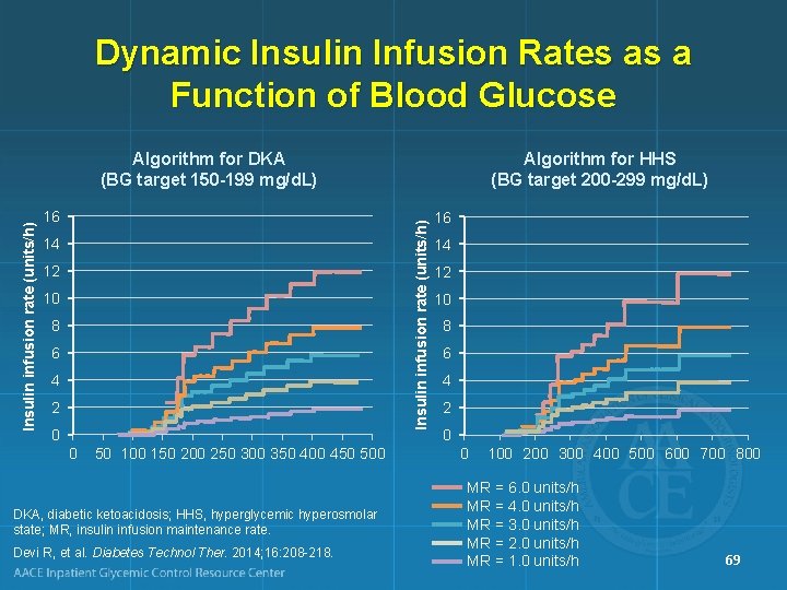
Dynamic Insulin Infusion Rates as a Function of Blood Glucose 16 Algorithm for HHS (BG target 200 -299 mg/d. L) Insulin infusion rate (units/h) Algorithm for DKA (BG target 150 -199 mg/d. L) 14 12 10 8 6 4 2 0 0 50 100 150 200 250 300 350 400 450 500 DKA, diabetic ketoacidosis; HHS, hyperglycemic hyperosmolar state; MR, insulin infusion maintenance rate. Devi R, et al. Diabetes Technol Ther. 2014; 16: 208 -218. 16 14 12 10 8 6 4 2 0 0 100 200 300 400 500 600 700 800 MR = 6. 0 units/h MR = 4. 0 units/h MR = 3. 0 units/h MR = 2. 0 units/h MR = 1. 0 units/h 69
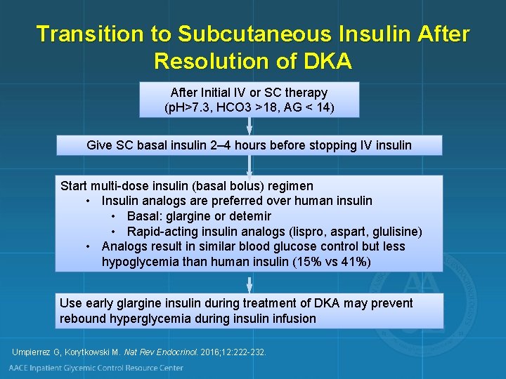
Transition to Subcutaneous Insulin After Resolution of DKA After Initial IV or SC therapy (p. H>7. 3, HCO 3 >18, AG < 14) Give SC basal insulin 2– 4 hours before stopping IV insulin Start multi-dose insulin (basal bolus) regimen • Insulin analogs are preferred over human insulin • Basal: glargine or detemir • Rapid-acting insulin analogs (lispro, aspart, glulisine) • Analogs result in similar blood glucose control but less hypoglycemia than human insulin (15% vs 41%) Use early glargine insulin during treatment of DKA may prevent rebound hyperglycemia during insulin infusion Umpierrez G, Korytkowski M. Nat Rev Endocrinol. 2016; 12: 222 -232.
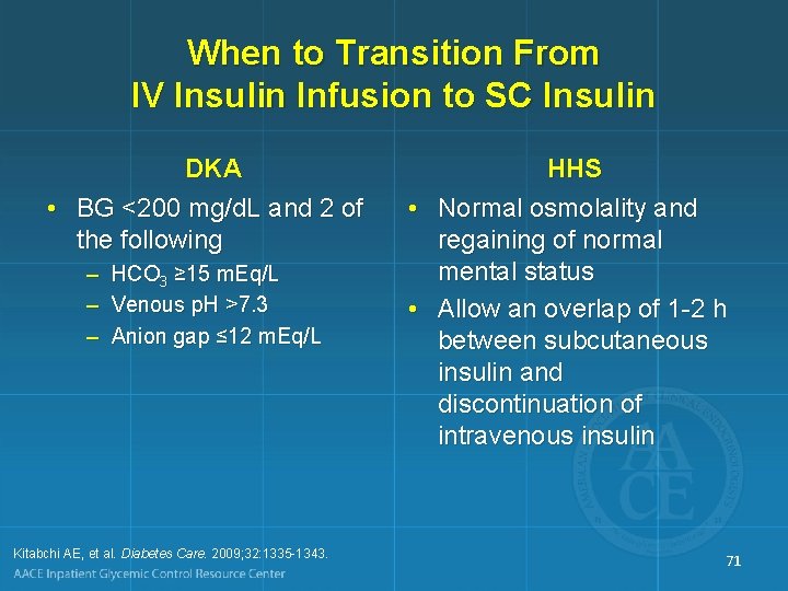
When to Transition From IV Insulin Infusion to SC Insulin DKA • BG <200 mg/d. L and 2 of the following – HCO 3 ≥ 15 m. Eq/L – Venous p. H >7. 3 – Anion gap ≤ 12 m. Eq/L Kitabchi AE, et al. Diabetes Care. 2009; 32: 1335 -1343. HHS • Normal osmolality and regaining of normal mental status • Allow an overlap of 1 -2 h between subcutaneous insulin and discontinuation of intravenous insulin 71
Cerebral Edema • Cerebral edema is a dreaded complication of DKA in childhood 1 • Mortality may be 24%, with significant morbidity among survivors 2 • One pediatric study found that rates of fluid administration and insulin administration were not associated with cerebral edema 3 • In another case control pediatric study, insulin dose in first 2 h was significantly associated with the risk of cerebral edema 4 1. Muir AB, et al. Diabetes Care. 2004; 27: 1541 -1546. 2. Edge JA, et al. Arch Dis Child. 2001; 85: 16 -22. 3. Glaser N, et al. N Engl J Med. 2001; 344: 264 -269. 4. Edge J, et al. Diabetologia. 2006; 49: 2002 -2009. 72
Fluid and Electrolyte Management in HHS • Treatment of HHS requires more free water and greater volume replacement than needed for patients with DKA • To avoid heart failure, caution is required in the elderly with preexisting heart disease • Potassium – Usually not significantly elevated on admission (unless in renal failure) – Replacement required during treatment 73
DKA Management Pitfalls • Not assessing for and/or treating underlying cause of the DKA • Not watching K+ closely enough and/or not replacing K+ aggressively enough • Following serial serum ketone concentrations • Following serum bicarbonate instead of the anion gap, with misinterpretation of expansion acidosis as “persistent ketoacidosis” • Interrupting IV insulin too soon (eg, patient not yet eating, anion gap not yet closed) 74
DKA Management Pitfalls • Occurrence of rebound ketosis consequent to inadequate insulin dosing at transition (eg, failure to give SC insulin when glucose is “low” or injudicious use of sliding scale insulin) • Inappropriate extension of hospitalization to “fine -tune” an outpatient regimen • Inadequate patient education and training • Inadequate follow-up care 75

FINDING THE CAUSE AND PREVENTING RECURRENCE 76
Possible Precipitating Causes or Factors in DKA: Type 1 Diabetes • Nonadherence to insulin regimen or psychiatric issues • Insulin error or insulin pump malfunction • Poor “sick-day” management • Infection (intra-abdominal, pyelonephritis, flu) • Myocardial infarction • Pancreatitis • Other endocrinopathy (rare) • Steroid therapy, other drugs or substances 77
Possible Precipitating Causes or Factors in DKA: Type 2 Diabetes • • Nonadherence to medication regimen Poor “sick-day’ management Dehydration Renal insufficiency Infection (intra-abdominal, pyelonephritis, flu) Myocardial infarction, stroke Other endocrinopathy (rare) Steroid therapy, other drugs or substances 78
DKA and SGLT 2 Inhibitor Therapy AACE Recommendations Findings • In T 1 D and T 2 D, metabolic changes shift substrate metabolism from carbohydrate to fat metabolism, predisposing patients to development of ketonemia and DKA during SGLT 2 inhibitor use • Normal or modestly elevated BG does not exclude the diagnosis of DKA during SGLT 2 inhibitor use Handelsman Y, et al. Endocr Pract. 2016; 22: 753 -762. Recommendations • Stop SGLT 2 inhibitor immediately – Symptoms of DKA – Emergency surgery • Stop SGLT 2 inhibitor ≥ 24 hours before – Planned invasive procedures – Anticipated stressful physical activity (eg, marathon) • Measure blood rather than urine ketones for DKA diagnosis • Advise patients taking SGLT 2 inhibitors to avoid excess alcohol and low-carbohydrate/ ketogenic diets 79
Predischarge Checklist • Diet information • Glucose monitor and strips (and associated prescription) • Medications, insulin, needles (and associated prescription) • Treatment goals • Contact phone numbers • “Medic-Alert” bracelet • “Survival Skills” training 80
Education in Type 1 Diabetes to Prevent DKA • Recognize symptoms and findings that require contact with a healthcare provider • Prevent ketoacidosis through self-management skills: – – Glucose testing Appropriate use of urine acetone testing Appropriate maintenance of insulin on sick days Use of supplemental insulin during illness • Address social factors 81
Summary • DKA and HHS are life-threatening emergencies • Management involves – – – Attention to precipitating cause Fluid and electrolyte management Insulin therapy Patient monitoring Prevention of metabolic complications during recovery Transition to long-term therapy • Patient education and discharge planning should aim at prevention of recurrence 82
- Slides: 82