Diabetic retinopathy diagnostics therapy classification Diabetes mellitus definition
")
Diabetic retinopathy (diagnostics, therapy, classification)
 due")
Diabetes mellitus- definition • Diabetes mellitus is disease with high glucose level (hyperglycaemia) due to absolute or relative lack of insulin produced in beta cells of Langerhans pancreatic islets

Diabetes mellitus- classification Ø Diabetes type 1 Ø Diabetes type 2 Ø Gestational diabetes
 • • - 800 000 8% of population")
Epidemiology of diabetes (CZ in 2009) • • - 800 000 8% of population DM type 1 DM type 2 8% 92%

Pathogenesis of diabetes type 1 • The destruction of insulin-producing betacells of pancreatic islets of Langerhans (autoimmune process, a genetic predisposition, external environmental factor, in the second of identical twins diabetes arises only in 50% of cases)

Pathogenesis of diabetes type 2 • Failure of insulin secretion in pancreatic beta-cells Reduction of insulin action in target tissues (insulin resistance)
")
Insulin discovered 1921 (Banting, Best, Macleod, Collip)
 § hyperglycaemic ketoacidotic coma hyperglycaemic hyperosmolar coma lactacidotic coma hypoglycaemic")
Diabetes mellitus (acute complications) § hyperglycaemic ketoacidotic coma hyperglycaemic hyperosmolar coma lactacidotic coma hypoglycaemic coma
 1. 2. 3. 4. retinopathy nephropathy diabetic foot neuropathy")
Diabetes mellitus (late complications) 1. 2. 3. 4. retinopathy nephropathy diabetic foot neuropathy
 • • Education Diet Oral antidiabetics Insulin")
Diabetes mellitus (therapy) • • Education Diet Oral antidiabetics Insulin
 • Diabetic retinopathy is microangiopathy, ie. retinal vascular impairment in diabetic")
Diabetic retinopathy (definition) • Diabetic retinopathy is microangiopathy, ie. retinal vascular impairment in diabetic patients
 • DM first description– Ebers papyrus (1550 before Ch. ), Aretaios")
Diabetic retinopathy (history) • DM first description– Ebers papyrus (1550 before Ch. ), Aretaios from Kappadokia (2 th century) • DR was first described after Helmholtz ophthalmoscope discovery (1851) • First description of DR – Jäger (1851), Desmarres (1855), von Gräfe (1858) • First classification of DR – Ballantyn a Löwensteine (1943), nonproliferative and proliferative DR
 • First retinal fotocoagulation– Meyer-Schwickerath (1945), solar photocoagulator (heliostat) • Regression")
Diabetic retinopathy (history) • First retinal fotocoagulation– Meyer-Schwickerath (1945), solar photocoagulator (heliostat) • Regression of proliferative DR after postpartual hypophysal necrosis (1953) – Simmonds- Sheehan syndrome • Discovery of fluorescence angiography – Novotny, Alvis (1959)
 • First use of laser– Meyer-Schwickerath (1955 -1958), xenon lamp •")
Diabetic retinopathy (history) • First use of laser– Meyer-Schwickerath (1955 -1958), xenon lamp • • Rubine laser (1960) Argon laser (1968) Pars plana vitrectomy – Machemer, Parel (1970) Fluorophotometry – Cunha-Vaz (1975), preretinopathy
 • 1976 - Diabetic Retinopathy Study (DRS)- laser reduces risk of")
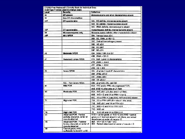
Diabetická retinopatie (historie) • 1976 - Diabetic Retinopathy Study (DRS)- laser reduces risk of blidness in proliferative DR • 1985 - Early Treatment Diabetic Retinopathy Study (ETDRS)- focal laser photocoagulation reduces risc of visual acuity lost in diabetic macular edema
 • • • Diabetes mellitus- 8% population 25% diabetics - DR")
Diabetic retinopathy (epidemiology) • • • Diabetes mellitus- 8% population 25% diabetics - DR 5% diabetics - proliferative DR DR rare until first 3 -5 years of duration of DM DR in 60 - 90% after 15 - 20 years of duration of DM • DR in 97% after 30 years of duration of DM

Patophysiology of diabetic retinopathy

Patophysiology of diabetic retinopathy • Microangiopathy • Lost of endothelial cells and pericites • Thickening of basal membrane of retinal capillaries (glycoproteins) • Failure of outer and inner blood retinal barrier

Patophysiology of diabetic retinopathy

Patophysiology of diabetic retinopathy

Patophysiology of diabetic retinopathy
 2. Proliferative DR (PDR) 3. Diabetic")
Classification of diabetic retinopathy 1. Nonproliferative DR (NPDR) 2. Proliferative DR (PDR) 3. Diabetic maculopathy (M) (each level of diabetic retinopathy may or may not beaccompanied by diabetic maculopathy)
 • • • Beginning Intermediate Advanced")
Nonproliferative DR (NPDR) • • • Beginning Intermediate Advanced

Beginning NPDR

Intermediate NPDR

Intermediate NPDR

Advanced NPDR
 • • • Light Intermediate Fully advanced VH- vitreous hemorrhage, PRHpreretinal")
Proliferative DR (PDR) • • • Light Intermediate Fully advanced VH- vitreous hemorrhage, PRHpreretinal hemorhage, TRD- retinal detachment at center of macula

Light PDR

Intermediate PDR

Fully advanced PDR

Fully advanced PDR

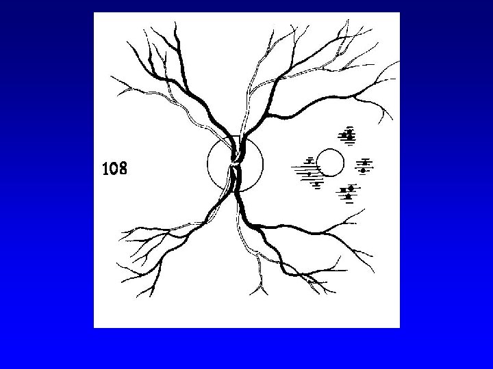
Vitreous hemorrhage in fully advanced PDR
 • affects 33% of diabetic patients after 8 -10 years of")
Diabetic maculopathy (M) • affects 33% of diabetic patients after 8 -10 years of duration of disease • the most common cause of vision loss in diabetic retinopathy
")
Diabetic makulopathy (M)
 • Macular area is a predilection site for edema formation Microangiopathy")
Diabetic makulopathy (M) • Macular area is a predilection site for edema formation Microangiopathy leads to ischemia, fluid accumulation, formation of microcysts and cysts Hard exudates (lipid accumulation) occur on the boundary of ischemic and normal retina
")
Diabetic makulopathy (M)
 • Focal edema • Difuse edema • Ischemic edema (rare)- avascular")
Diabetic makulopathy (classification) • Focal edema • Difuse edema • Ischemic edema (rare)- avascular zone in macula

Focal edema

Difuse edema

Ischemic edema

Therapy of diabetic retinopathy and maculopathy • Gold standard is laser photocoagulation of ischemic retinal parts • Laser can‘t be performed in central macular zone
 • Laser therapy of DR 1. focal 2. panretinal (scatter)")
Laser therapy (technique) • Laser therapy of DR 1. focal 2. panretinal (scatter)

Laser spots in retina suffered from NPDR
 • Laser therapy of diabetic maculopathy 1. focal 2. grid")
Laser therapy (technique) • Laser therapy of diabetic maculopathy 1. focal 2. grid

Focal laser


Grid laser
 • Reducing of risc of visual loss • Reducing of risc")
Laser therapy (positives) • Reducing of risc of visual loss • Reducing of risc of vitreous hemorrhage, neovascular glaucoma and tractional retinal detachment
 • Paliative treatment • Dark adaptation problems")
Laser therapy (negatives) • Paliative treatment • Dark adaptation problems
")
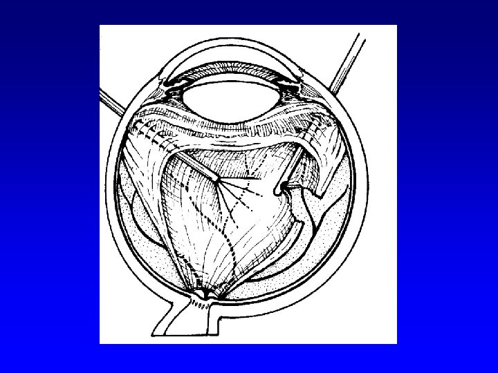
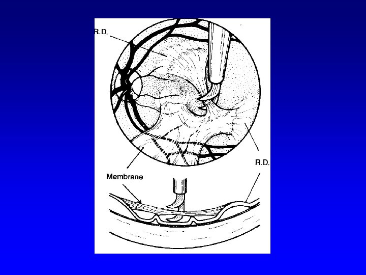
Surgical therapy of DR • Pars plana vitrectomy- (Machemer, Parel – 1970)
- Slides: 55