Diabetic macular oedema Dr Shahriar Nabili DMO Macular

Diabetic macular oedema Dr Shahriar Nabili

DMO • Macular edema in diabetes, defined as retinal thickening within 2 disc diameters of the center of the macula, results from retinal microvascular changes that compromise the blood-retinal barrier, causing leakage of plasma constituents into the surrounding retina and, consequently, retinal edema

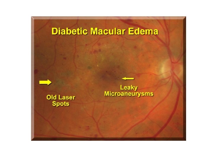
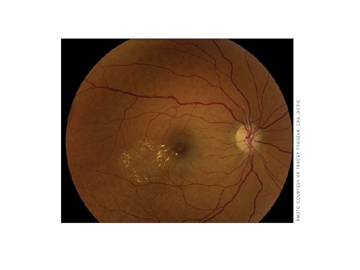
DMO • Focal edema is associated with hard exudate rings caused by leakage from microaneurysms. Diffuse edema is caused by leakage from microaneurysms, retinal capillaries, and arterioles • Diabetes is the leading cause of new blindness

DMO • Distinct entity from diabetic retinopathy • Leading cause of blindness in working age group • Prevention is important but treatment reduces the risk of visual loss by 50%

DMO Sources of Referrals • Screening service • Optometrists
 • Tight control of blood")
DMO Prevention • Diabetes Control and Complications Trial (DCCT) • Tight control of blood sugar • Treatment of hypertension • Treatment of Lipids • Proteinuria • Pregnancy
, as defined by the Early Treatment Diabetic")
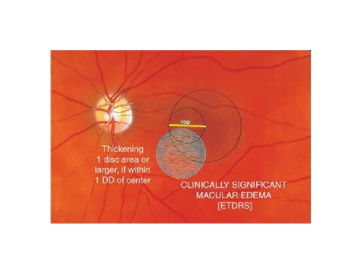
CSME • clinically significant macular edema (CSME), as defined by the Early Treatment Diabetic Retinopathy Study (ETDRS)

CSME • Retinal thickening within 500 µm of the center of the fovea • Hard, yellow exudates within 500 µm of the center of the fovea with adjacent retinal thickening • At least 1 disc area of retinal thickening, any part of which is within 1 disc diameter of the center of the fovea
 • Funduscopy under stereopsis and high magnification")
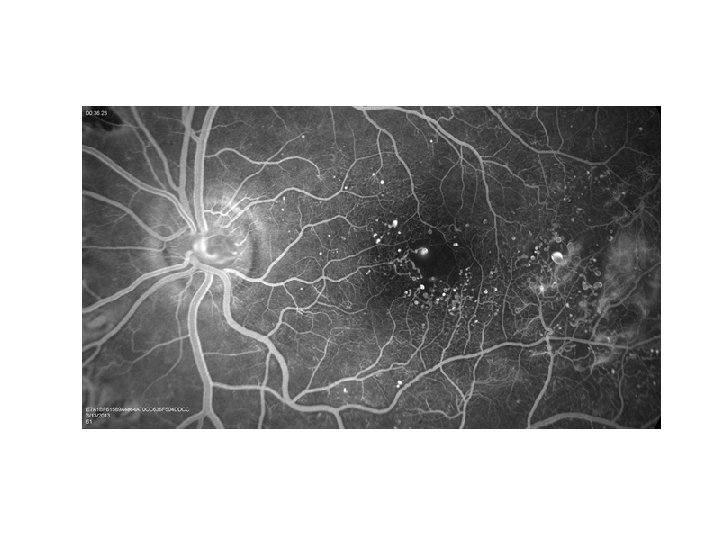
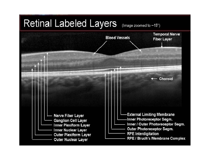
DMO Assessment • Visual acuity (EDTRS chart) • Funduscopy under stereopsis and high magnification • Optical coherence tomography (OCT) captures reflected light from retinal structures to create a cross-sectional image of the retina, which is comparable to histological sections as seen with a light microscope. • ? Fluorescein angiography


DMO Treatment • Argon Laser • Intra-vitreal injection of steriods • Intra-vitreal injections of anti Vascular endothelial growth factor (VEGF)

DMO Treatment • Laser either focal or grid • Reduces progression of diabetic macular edema; significant visual improvement is uncommon • Limited to the location of the leakage

DMO Treatment IVT Triamcinolone Acetonide • Reduces macular oedema and improves vision • Action is maximal at 1 week, lasting 3 -6 months • High risk of increase in intra-ocular pressure and cataract formation
 increases retinal vascular permeability, causes")
DMO Anti VEGF • Vascular endothelial growth factor (VEGF) increases retinal vascular permeability, causes breakdown of the blood-retina barrier, and results in retina edema. VEGF is up-regulated in diabetic retinopathy.

DMO Lucentis • Improves vision significantly • The gain is maintained • Initial loading dose of 3 injections. Once a month • Follow up treatment as needed • Possibly need 6 -8 injections in the first year • There is no end point
- Slides: 22