
Diabetic Eye Disease Evan Jake Waxman MD Ph
Diabetic Eye Disease Evan (Jake) Waxman MD Ph. D
Diabetic Eye Disease Key Points • Diabetes is a major cause of visual loss
Diabetic Eye Disease Key Points • Risk factor control can prevent and slow visual loss
Diabetic Eye Disease Key Points • Treatments exist but work best before vision is lost
Diabetic Eye Disease Key Points • Diabetes is a major cause of visual loss • Risk factor control can prevent and slow visual loss • Treatments exist but work best before vision is lost So … to prevent visual loss • Control patient risk factors • Insist your patients get yearly dilated eye exams with an ophthalmologist
Diabetic Eye Disease Case Presentation - History • • • 27 year old woman DM I for 16 years poor blood sugar ctrl Hgb. A 1 C = 10 c/o spot in L vision for one day • Sees “Eye Doctor” every year -- no previous eye disease diagnosed
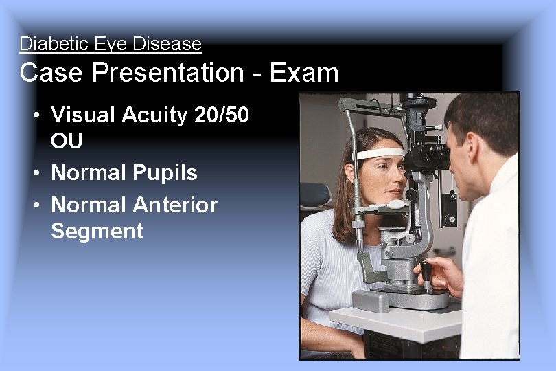
Diabetic Eye Disease Case Presentation - Exam • Visual Acuity 20/50 OU • Normal Pupils • Normal Anterior Segment
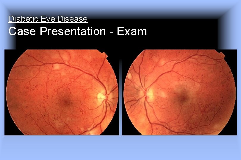
Diabetic Eye Disease Case Presentation - Exam
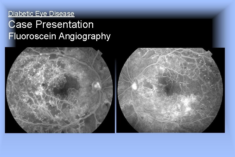
Diabetic Eye Disease Case Presentation Fluoroscein Angiography
Diabetic Eye Disease Case Presentation - Course • • Pan retinal photocoagulation OU Focal photocoagulation OS Vision dropped to 20/200 OD 1 month later Vit heme OS 2 months later Additional PRP Glaucoma surgery x 2 Current Acuity 20/400 OD 20/200 OS Prognosis Poor
Diabetic Eye Disease Background • Treatments work best before vision is lost • Many patients are diagnosed only after vision is lost • Vision loss is a late symptom of diabetic eye disease • Risk factor control is essential
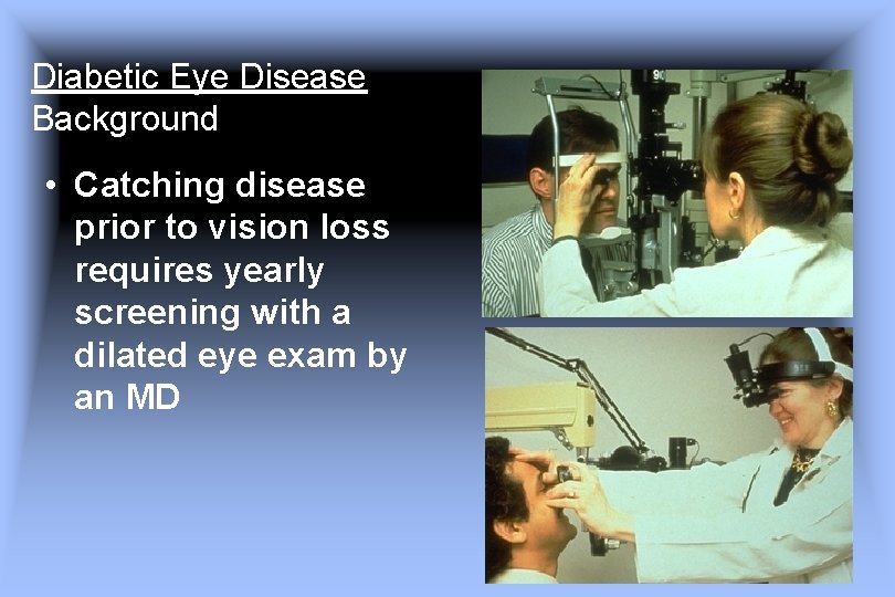
Diabetic Eye Disease Background • Catching disease prior to vision loss requires yearly screening with a dilated eye exam by an MD
Diabetic Eye Disease Key Points • Diabetes is a major cause of visual loss • Risk factor control can prevent and slow visual loss • Treatments exist but work best before vision is lost So … to prevent visual loss • Control patient risk factors • Insist your patients get yearly dilated eye exams with an ophthalmologist
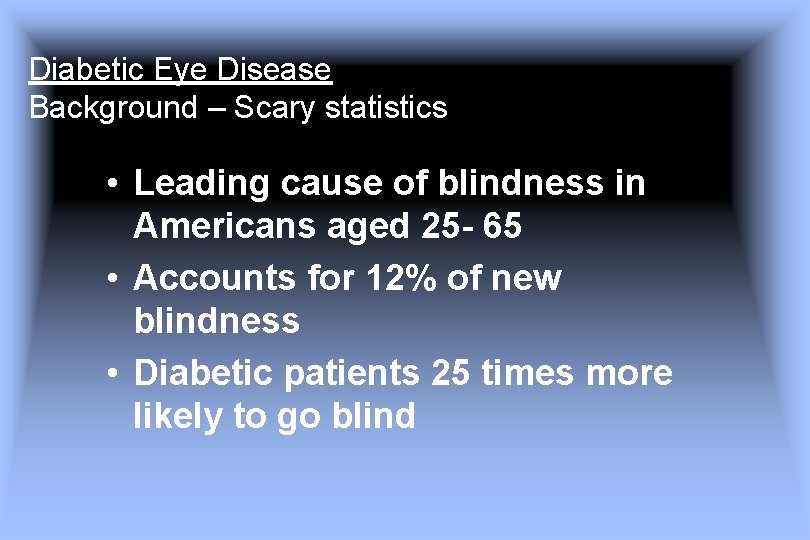
Diabetic Eye Disease Background – Scary statistics • Leading cause of blindness in Americans aged 25 - 65 • Accounts for 12% of new blindness • Diabetic patients 25 times more likely to go blind
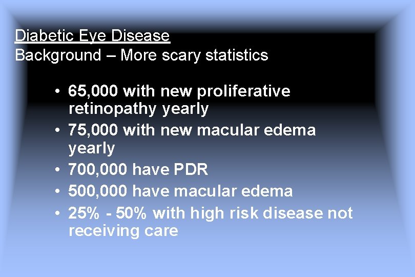
Diabetic Eye Disease Background – More scary statistics • 65, 000 with new proliferative retinopathy yearly • 75, 000 with new macular edema yearly • 700, 000 have PDR • 500, 000 have macular edema • 25% - 50% with high risk disease not receiving care
Diabetic Eye Disease Background Risk Factors • Duration • Poor Blood Sugar control • HTN • Hyperlipidemia • Barriers to care
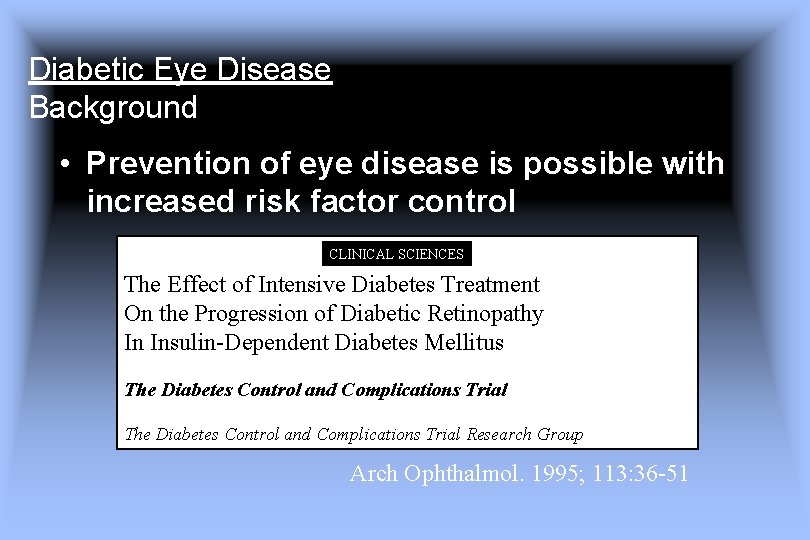
Diabetic Eye Disease Background • Prevention of eye disease is possible with increased risk factor control CLINICAL SCIENCES The Effect of Intensive Diabetes Treatment On the Progression of Diabetic Retinopathy In Insulin-Dependent Diabetes Mellitus The Diabetes Control and Complications Trial Research Group Arch Ophthalmol. 1995; 113: 36 -51
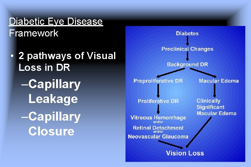
Diabetic Eye Disease Framework • 2 pathways of Visual Loss in DR –Capillary Leakage –Capillary Closure
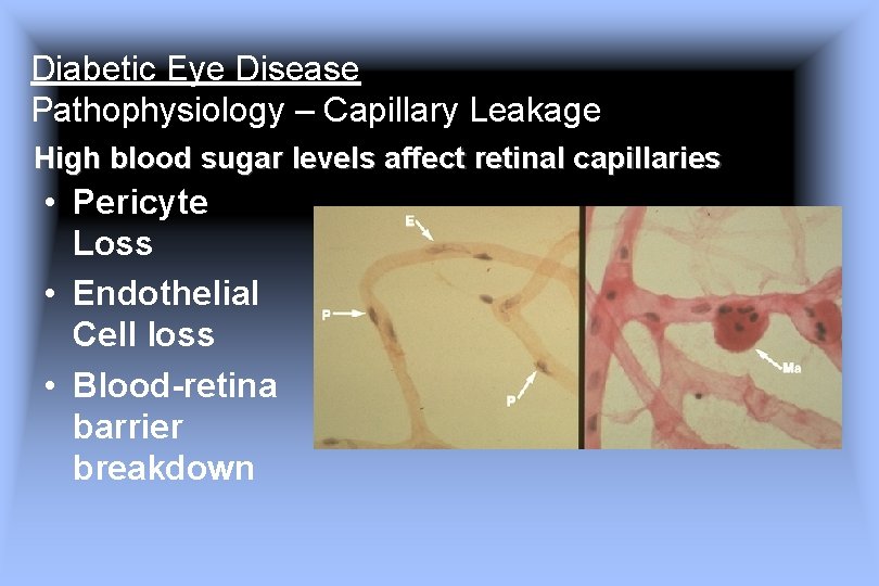
Diabetic Eye Disease Pathophysiology – Capillary Leakage High blood sugar levels affect retinal capillaries • Pericyte Loss • Endothelial Cell loss • Blood-retina barrier breakdown
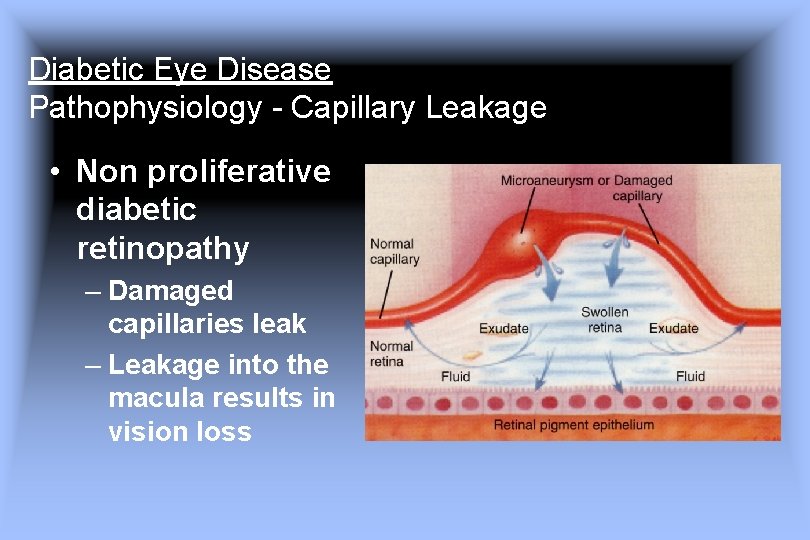
Diabetic Eye Disease Pathophysiology - Capillary Leakage • Non proliferative diabetic retinopathy – Damaged capillaries leak – Leakage into the macula results in vision loss
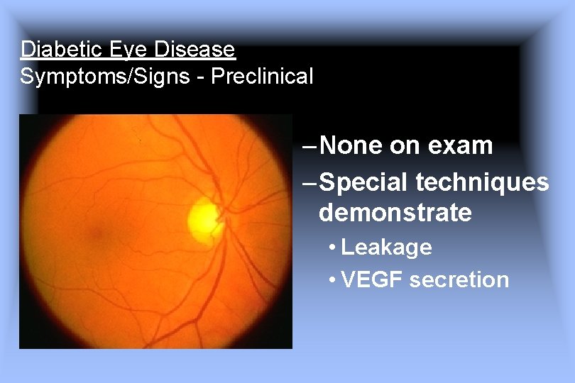
Diabetic Eye Disease Symptoms/Signs - Preclinical – None on exam – Special techniques demonstrate • Leakage • VEGF secretion
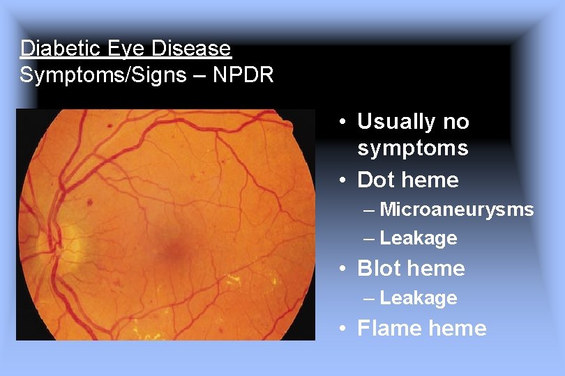
Diabetic Eye Disease Symptoms/Signs – NPDR • Usually no symptoms • Dot heme – Microaneurysms – Leakage • Blot heme – Leakage • Flame heme
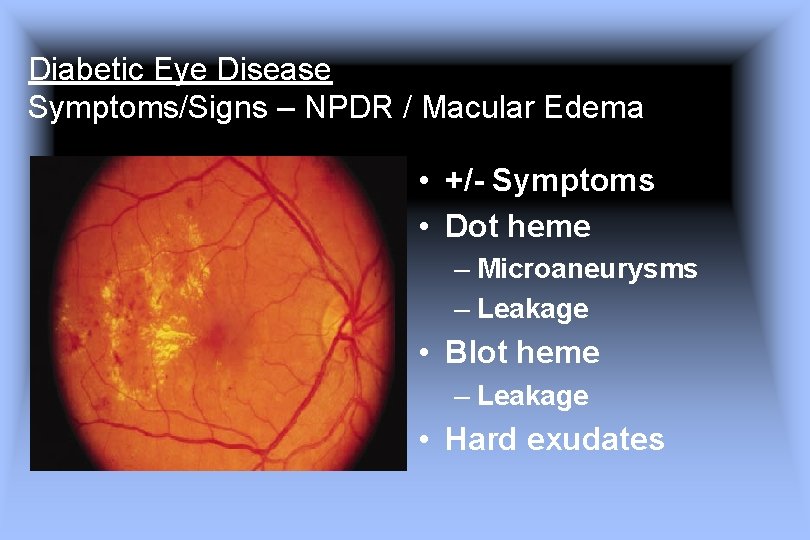
Diabetic Eye Disease Symptoms/Signs – NPDR / Macular Edema • +/- Symptoms • Dot heme – Microaneurysms – Leakage • Blot heme – Leakage • Hard exudates
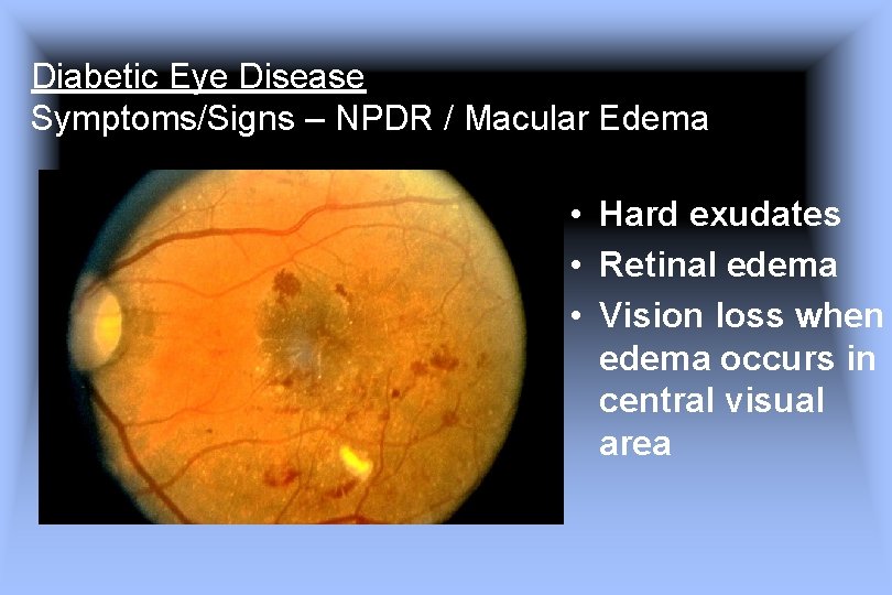
Diabetic Eye Disease Symptoms/Signs – NPDR / Macular Edema • Hard exudates • Retinal edema • Vision loss when edema occurs in central visual area
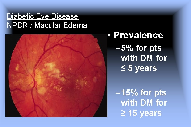
Diabetic Eye Disease NPDR / Macular Edema • Prevalence – 5% for pts with DM for ≤ 5 years – 15% for pts with DM for ≥ 15 years
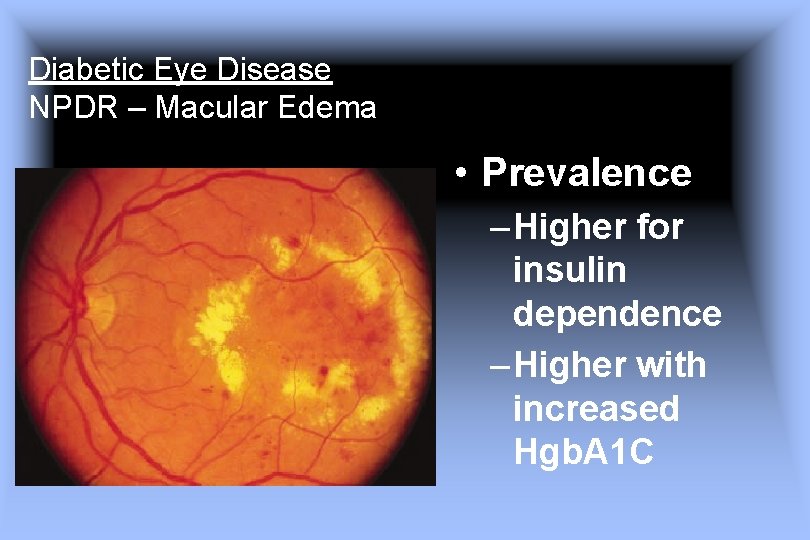
Diabetic Eye Disease NPDR – Macular Edema • Prevalence – Higher for insulin dependence – Higher with increased Hgb. A 1 C
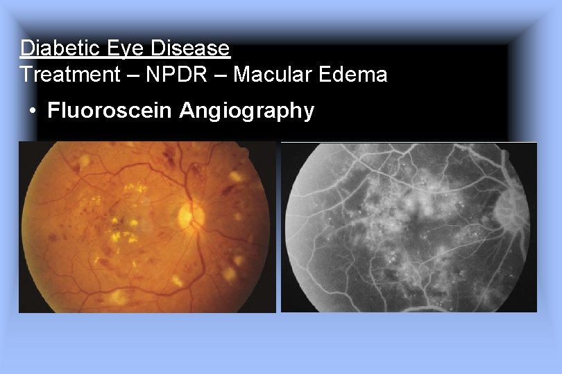
Diabetic Eye Disease Treatment – NPDR – Macular Edema • Fluoroscein Angiography
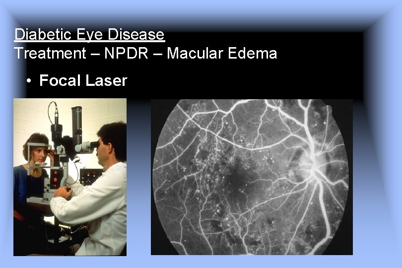
Diabetic Eye Disease Treatment – NPDR – Macular Edema • Focal Laser
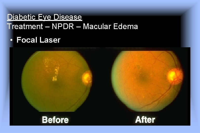
Diabetic Eye Disease Treatment – NPDR – Macular Edema • Focal Laser
Diabetic Eye Disease Treatment – NPDR – Macular Edema Focal Laser reduces risk of visual loss by 50% Early Photocoagulation for Diabetic Retinopathy ETDRS Report Number 9 EARLY TREATMENT DIABETIC RETINOPATHY STUDY RESEARCH GROUP Ophthalmology 1991; 98; 766 -785
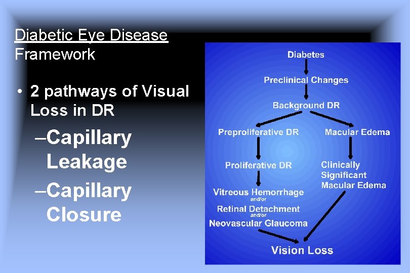
Diabetic Eye Disease Framework • 2 pathways of Visual Loss in DR –Capillary Leakage –Capillary Closure
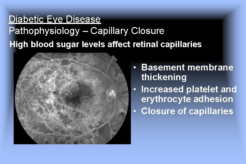
Diabetic Eye Disease Pathophysiology – Capillary Closure High blood sugar levels affect retinal capillaries • Basement membrane thickening • Increased platelet and erythrocyte adhesion • Closure of capillaries
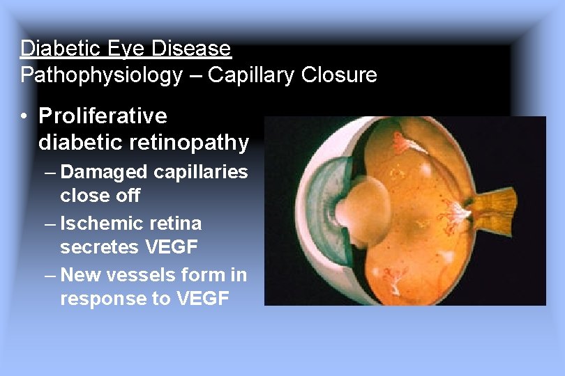
Diabetic Eye Disease Pathophysiology – Capillary Closure • Proliferative diabetic retinopathy – Damaged capillaries close off – Ischemic retina secretes VEGF – New vessels form in response to VEGF
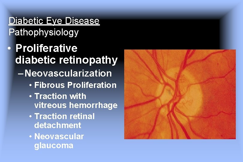
Diabetic Eye Disease Pathophysiology • Proliferative diabetic retinopathy – Neovascularization • Fibrous Proliferation • Traction with vitreous hemorrhage • Traction retinal detachment • Neovascular glaucoma
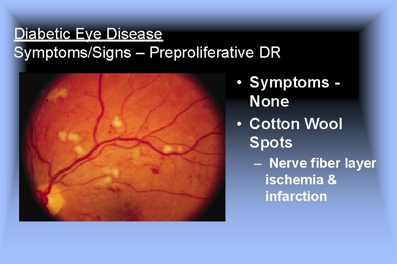
Diabetic Eye Disease Symptoms/Signs – Preproliferative DR • Symptoms None • Cotton Wool Spots – Nerve fiber layer ischemia & infarction
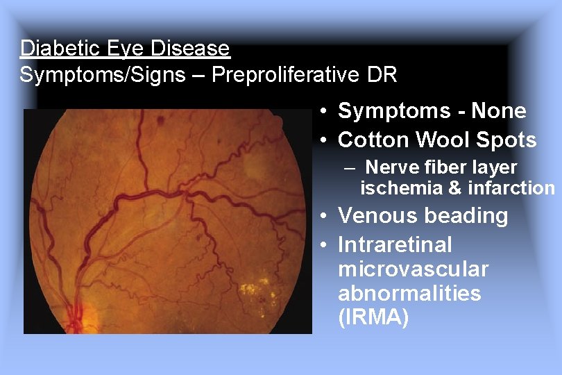
Diabetic Eye Disease Symptoms/Signs – Preproliferative DR • Symptoms - None • Cotton Wool Spots – Nerve fiber layer ischemia & infarction • Venous beading • Intraretinal microvascular abnormalities (IRMA)
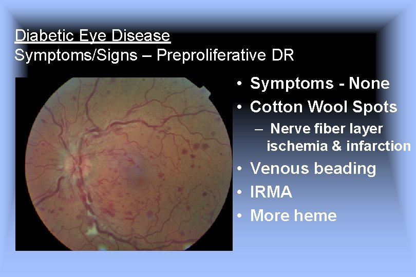
Diabetic Eye Disease Symptoms/Signs – Preproliferative DR • Symptoms - None • Cotton Wool Spots – Nerve fiber layer ischemia & infarction • Venous beading • IRMA • More heme
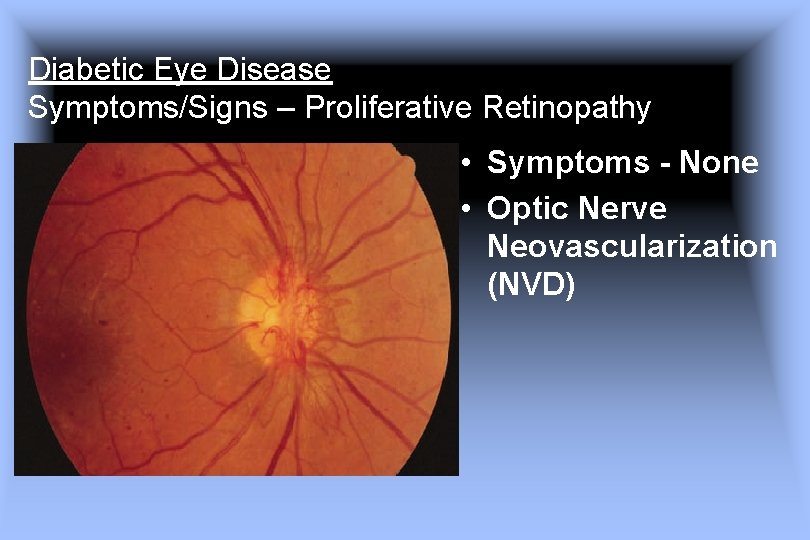
Diabetic Eye Disease Symptoms/Signs – Proliferative Retinopathy • Symptoms - None • Optic Nerve Neovascularization (NVD)
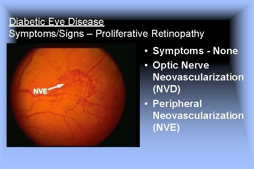
Diabetic Eye Disease Symptoms/Signs – Proliferative Retinopathy • Symptoms - None • Optic Nerve Neovascularization (NVD) • Peripheral Neovascularization (NVE)
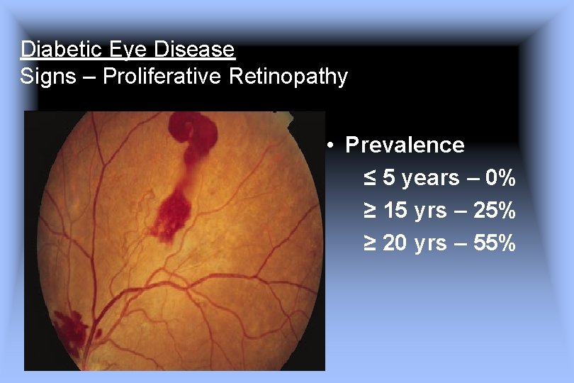
Diabetic Eye Disease Signs – Proliferative Retinopathy • Prevalence ≤ 5 years – 0% ≥ 15 yrs – 25% ≥ 20 yrs – 55%
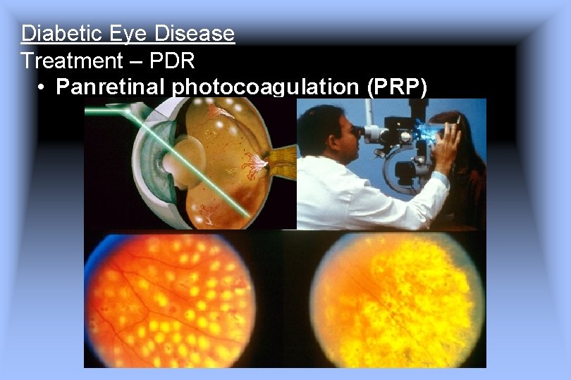
Diabetic Eye Disease Treatment – PDR • Panretinal photocoagulation (PRP)
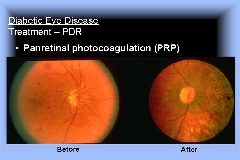
Diabetic Eye Disease Treatment – PDR • Panretinal photocoagulation (PRP) Before After
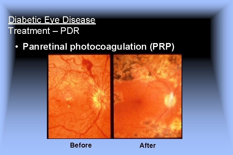
Diabetic Eye Disease Treatment – PDR • Panretinal photocoagulation (PRP) Before After
Diabetic Eye Disease Treatment – PDR PRP reduces the risk of severe vision loss by more than 50% Photocoagulation Treatment of Proliferative Diabetic Retinopathy Clinical Application of Diabetic Retinopathy Study (DRS) Findings, DRS Report Number 8 THE DIABETIC RETINOPATHY STUDY RESEARCH GROUP Ophthalmology 1991; 88; 583 -600
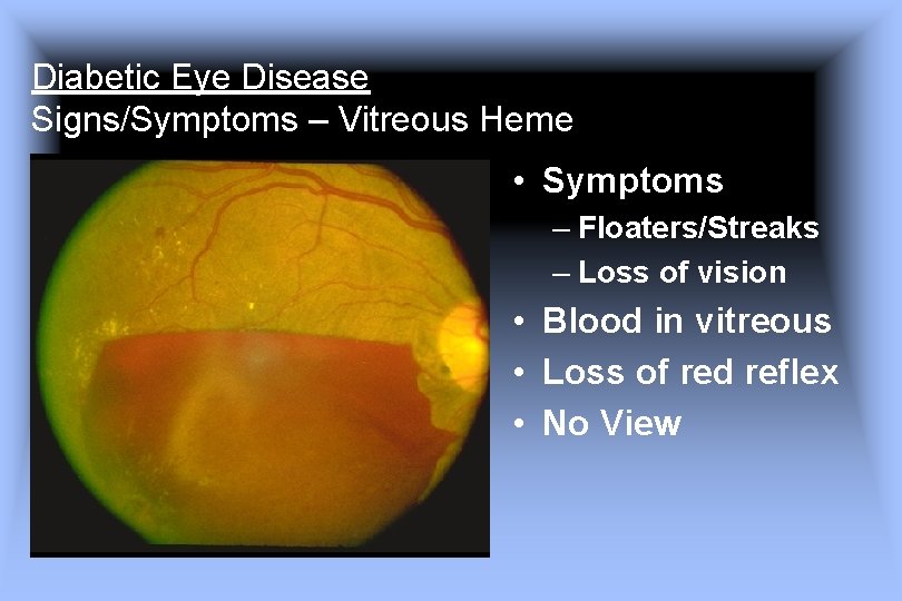
Diabetic Eye Disease Signs/Symptoms – Vitreous Heme • Symptoms – Floaters/Streaks – Loss of vision • Blood in vitreous • Loss of red reflex • No View
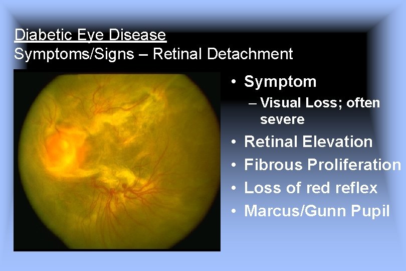
Diabetic Eye Disease Symptoms/Signs – Retinal Detachment • Symptom – Visual Loss; often severe • • Retinal Elevation Fibrous Proliferation Loss of red reflex Marcus/Gunn Pupil
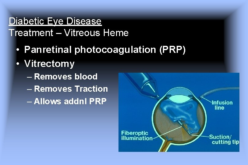
Diabetic Eye Disease Treatment – Vitreous Heme • Panretinal photocoagulation (PRP) • Vitrectomy – Removes blood – Removes Traction – Allows addnl PRP
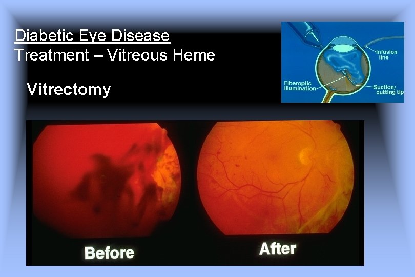
Diabetic Eye Disease Treatment – Vitreous Heme Vitrectomy
Diabetic Eye Disease Treatment – PDR Vitrectomy results in improved vision in patients with persistent vitreous hemorrhage Early Vitrectomy fo Severe Vitreous Hemorrhage in Diabetic Retinopathy Two-Year Results of a Randomized Trial Diabetic Retinopathy Virectomy Report 2 THE DIABETIC RETINOPATHY VITRECTOMY STUDY RESEARCH GROUP Arch Ophthalmol. 1985; 103 1644 -1652
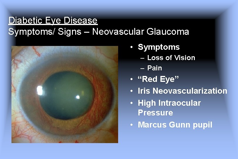
Diabetic Eye Disease Symptoms/ Signs – Neovascular Glaucoma • Symptoms – Loss of Vision – Pain • “Red Eye” • Iris Neovascularization • High Intraocular Pressure • Marcus Gunn pupil
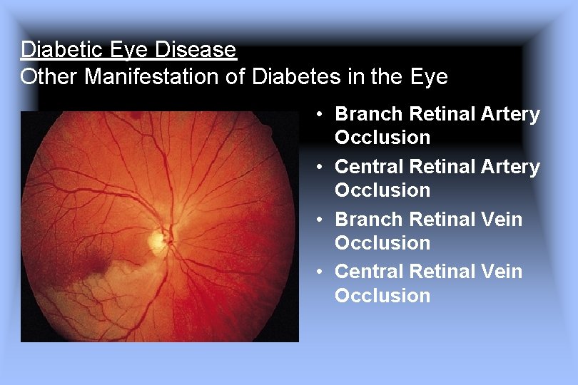
Diabetic Eye Disease Other Manifestation of Diabetes in the Eye • Branch Retinal Artery Occlusion • Central Retinal Artery Occlusion • Branch Retinal Vein Occlusion • Central Retinal Vein Occlusion
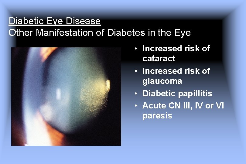
Diabetic Eye Disease Other Manifestation of Diabetes in the Eye • Increased risk of cataract • Increased risk of glaucoma • Diabetic papillitis • Acute CN III, IV or VI paresis
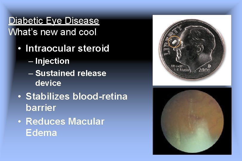
Diabetic Eye Disease What’s new and cool • Intraocular steroid – Injection – Sustained release device • Stabilizes blood-retina barrier • Reduces Macular Edema
Diabetic Eye Disease What’s new and cool • Anti VEGF drugs • Protein Kinase C beta inhibitors • Intravitreal hyaluronidase
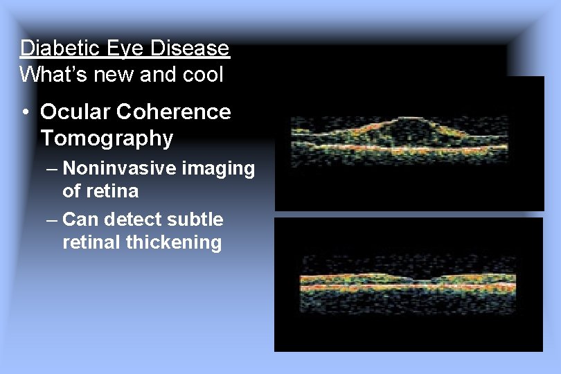
Diabetic Eye Disease What’s new and cool • Ocular Coherence Tomography – Noninvasive imaging of retina – Can detect subtle retinal thickening
Diabetic Eye Disease Key Points • Diabetes is a major cause of visual loss
Diabetic Eye Disease Key Points • Risk factor control can prevent and slow visual loss
Diabetic Eye Disease Key Points • Treatments exist but work best before vision is lost
Diabetic Eye Disease Key Points • Diabetes is a major cause of visual loss • Risk factor control can prevent and slow visual loss • Treatments exist but work best before vision is lost So … to prevent visual loss • Control patient risk factors • Insist your patients get yearly dilated eye exams with an ophthalmologist
- Slides: 59