Diabetes Mellitus WHO Diabetes mellitus is a chronic

Diabetes Mellitus

WHO • Diabetes mellitus is a chronic disease caused by inherited and/or acquired deficiency in production of insulin by the pancreas, or by the ineffectiveness of the insulin produced. • results in increased concentrations of glucose in the blood, which in turn damage the blood vessels and nerves.

• Diabetes mellitus is a group of metabolic disorders sharing the common feature of hyperglycemia

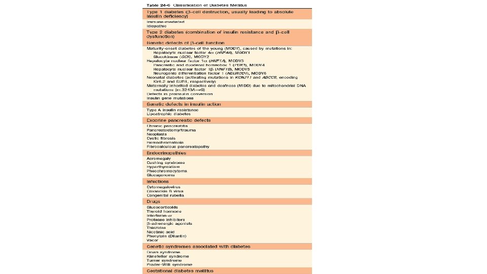
Classification • two principle forms of diabetes: 1. Type 1 diabetes (formerly known as insulin-dependent) 2. Type 2 diabetes (formerly named non-insulin-dependent)

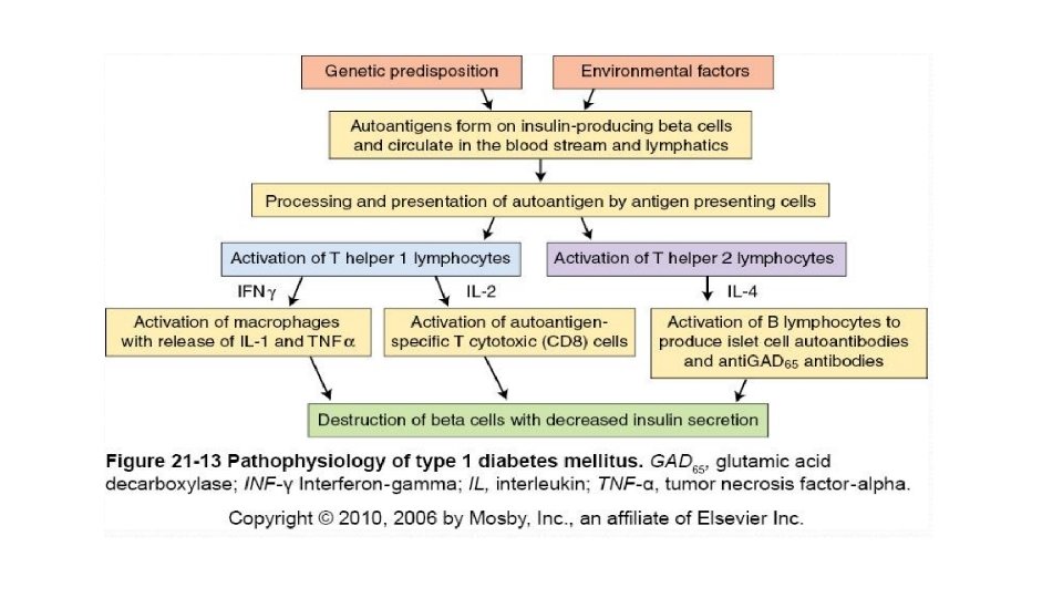
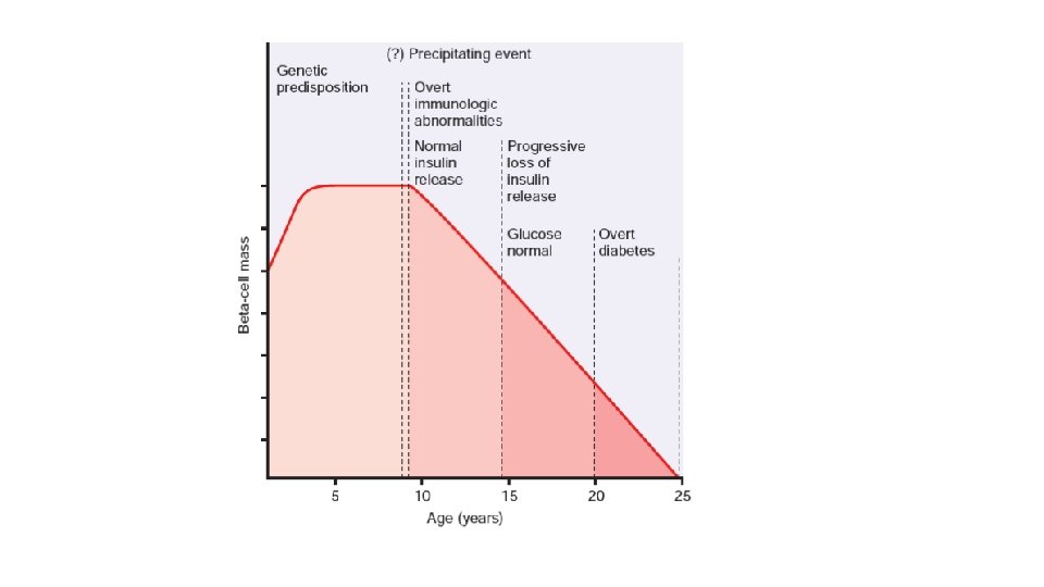
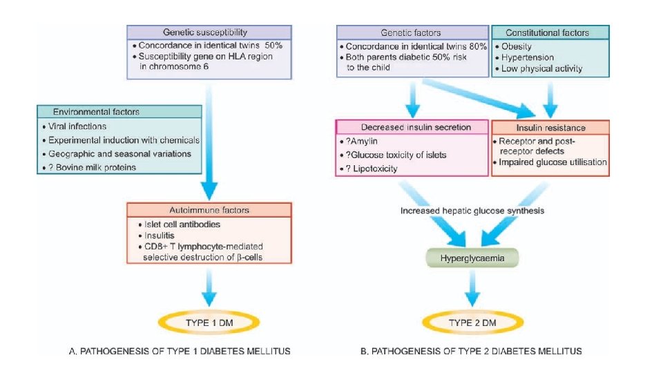
ETIO-PATHOGENESIS • PATHOGENESIS OF TYPE 1 DM. destruction of β-cell mass, usually leading to absolute insulin deficiency. 1. Genetic susceptibility- HLA gene cluster on chromosome 6 p 21, which according to some estimates contributes as much as 50% of the genetic susceptibility to type 1 diabetes. • Allele-HLA-DR 3 -DR 4 2. Autoimmune factors: fundamental immune abnormality in type 1 diabetes is a failure of self-tolerance in T cells specific for islet antigens • islet cell antibodies • insulitis • Selective destruction of β-cells

• TH 1 cells secrete-IFN-γ and TNF • Islet autoantigens-β cell enzyme glutamic acid decarboxylase (GAD), and islet cell autoantigen 512(ICA 512) • cell-mediated autoimmunity • Associated with other autoimmune diseases

3. Environmental factors • viral infections • Chemicals-alloxan, streptozotocin and pentamidine.

Etiopathogenesis in DM type 2
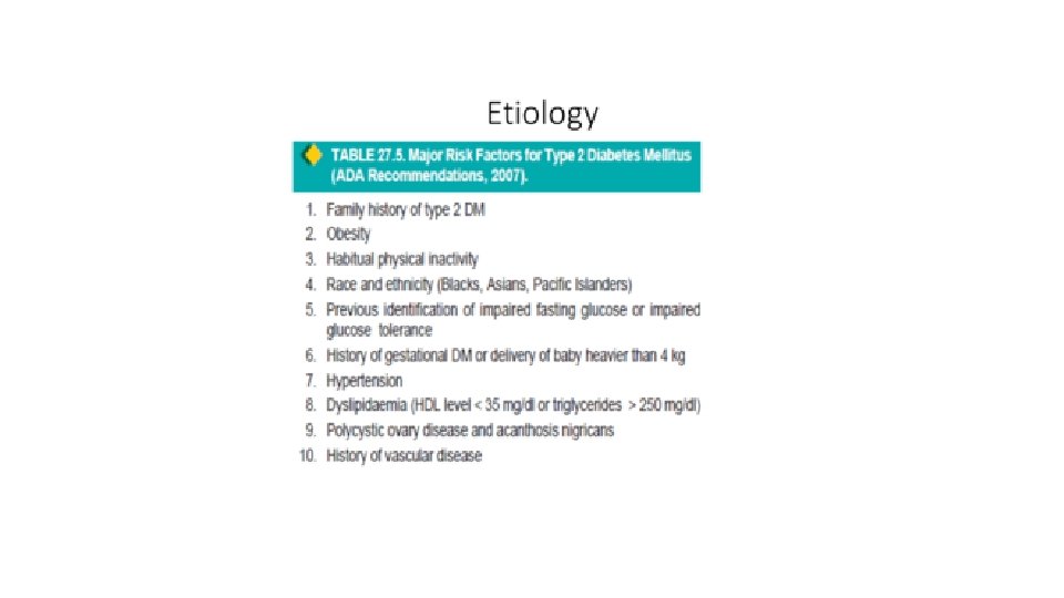

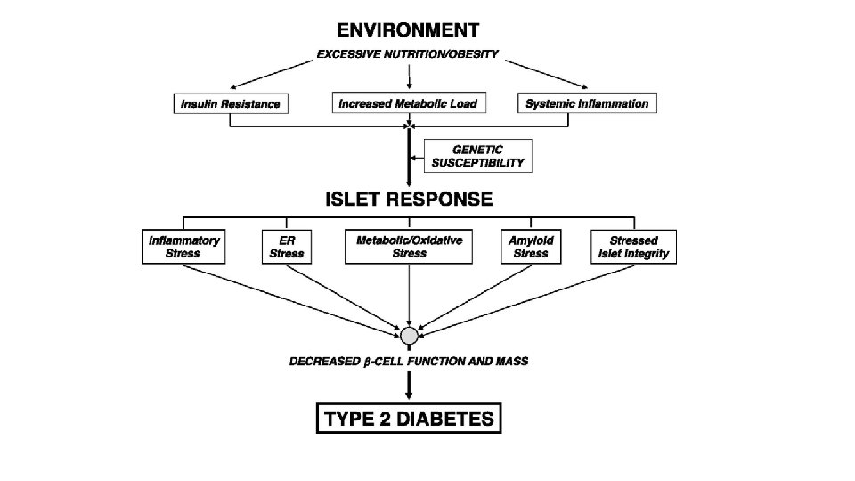
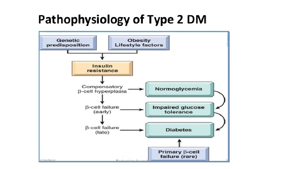
PATHOGENESIS OF TYPE 2 DM. complex disease that involves an interplay of genetic and environmental factors and a proinflammatory state. 1 -Genetic Factors-first-degree relatives have 5 - to 10 -fold higher risk 2 -Environmental Factors-Obesity, sedentary lifestyle
")
3 -Insulin resistance. Mechanism of hyperglycaemia in these cases is explained as under: i) impairs glucose utilisation and hence hyperglycaemia. ii) There is increased hepatic synthesis of glucose. iii) Hyperglycaemia in obesity is related to high levels of free fatty acids and cytokines

4 -Current consideration • Polymorphism in various post-receptor intracellular signal pathway molecules. • Elevated free fatty acids

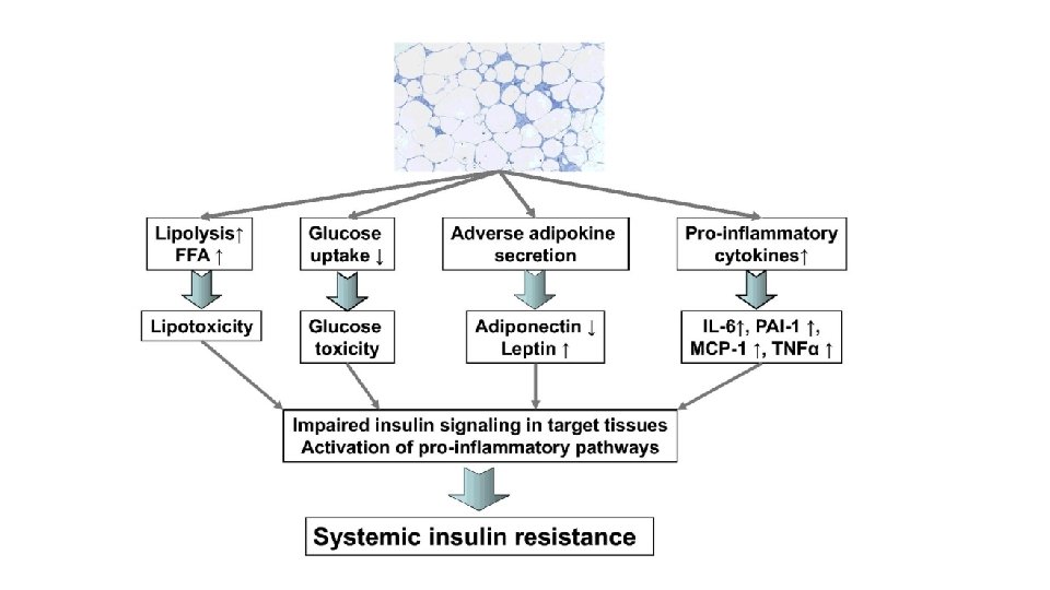
β-Cell Dysfunction Several mechanisms have been implicated in promoting β-cell dysfunction in type 2 diabetes, including: • Excess free fatty acids that compromise β cell function and attenuate insulin release (“lipotoxicity”) • impact of chronic hyperglycemia (“glucotoxicity”) • An abnormal “incretin effect, ” leading to reduced secretion of GIP and GLP-1, hormones that promote insulin release

• Amyloid deposition within islets 90% of diabetic islets cell in long standing • genetic predisposition
 • accumulation of cytoplasmic intermediates")
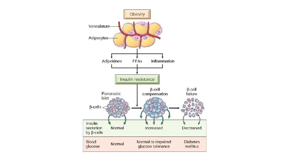
Obesity and Insulin Resistance v. Free fatty acids (FFAs) • accumulation of cytoplasmic intermediates like diacylglycerol (DAG) • DAG compete with glucose for substrate oxidation v. Adipokines- Adiponectin levels are reduced in obesity, thus contributing to insulin resistance v. Inflammation

Inflammation: • FFA & Beta cell • Inflammasome • Cytokines IL-1β, IL-1 • promote insulin resistance

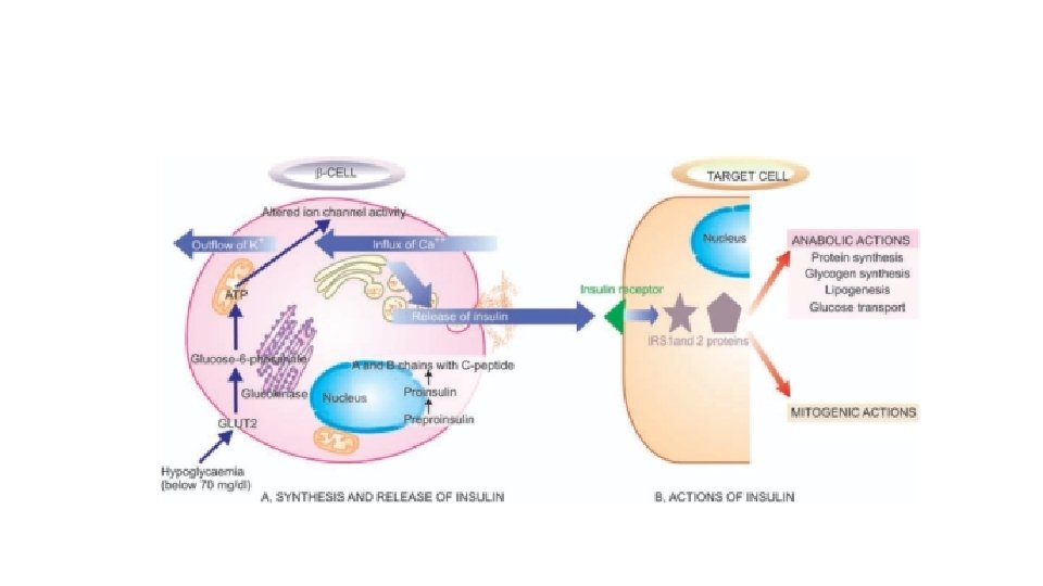
Metabolic actions of insulin in striated muscle, adipose tissue, and liver.

Pathophysiological basis of common signs and symptoms due to uncontrolled hyperglycaemia in diabetes mellitus

• Morphologic Features – 1. Pancreatic Islets 2. Diabetic Macrovascular Disease 3. Diabetic Microangiopathy 4. Diabetic Nephropathy 5. Diabetic Ocular Complications 6. Diabetic neuropathy

Morphologic Features Pancreatic Islets 1 -Insulitis: • In type 1 DMØlymphocytic infiltrate, macrophage and few polymorphs • In type 2 DMØvariable degree of fibrous tissue in the islets

2 -Islet cell mass: • Type-1 - loss of pancreatic β−cells and its hyalinisation • In type 2 DM-hyperplasia and hypertrophy of islets 3 -Amyloidosis: • type 1 DM- absent • Type-2 DM-around the capillaries of the islets causing compression and atrophy of islet tissue

• Diabetic Macrovascular DiseaseØhallmark of diabetic macrovascular disease is accelerated atherosclerosis involving the aorta and large- and medium-sized arteries ØMyocardial infarction ØGangrene of the lower extremities ØHyaline arteriolosclerosis

renal hyaline arteriolosclerosis

• Diabetic Microangiopathy- diffuse thickening of basement membranes. Øcapillaries of the skin, skeletal muscle, retina, renal glomeruli, and renal medulla Ø leaky
 glomerular lesions (2) renal vascular")
• Diabetic Nephropathy. Three lesions are encountered: (1) glomerular lesions (2) renal vascular lesions (3) pyelonephritis, including necrotizing papillitis

• Glomerular lesionØCapillary Basement Membrane

ØDiffuse Mesangial Sclerosis- consists of diffuse increase in mesangial matrix. ØNodular Glomerulosclerosis- also known as intercapillary glomerulosclerosis or Kimmelstiel-Wilson disease.
.")
Diffuse and nodular diabetic glomerulosclerosis (PAS stain).

• nodular lesions are frequently accompanied by prominent accumulations of hyaline material in capillary loops (“fibrin caps”) or adherent to Bowman capsules (“capsular drops”).

Nephrosclerosis

ØRenal atherosclerosis and arteriolosclerosis. Hyaline arteriolosclerosis affects not only the afferent but also the efferent arteriole ØPyelonephritis is an acute or chronic inflammation of the kidneys that usually begins in the interstitial tissue and then spreads to affect the tubules • necrotizing papillitis
 • proliferative retinopathy Background (non-proliferative) retinopathy.")
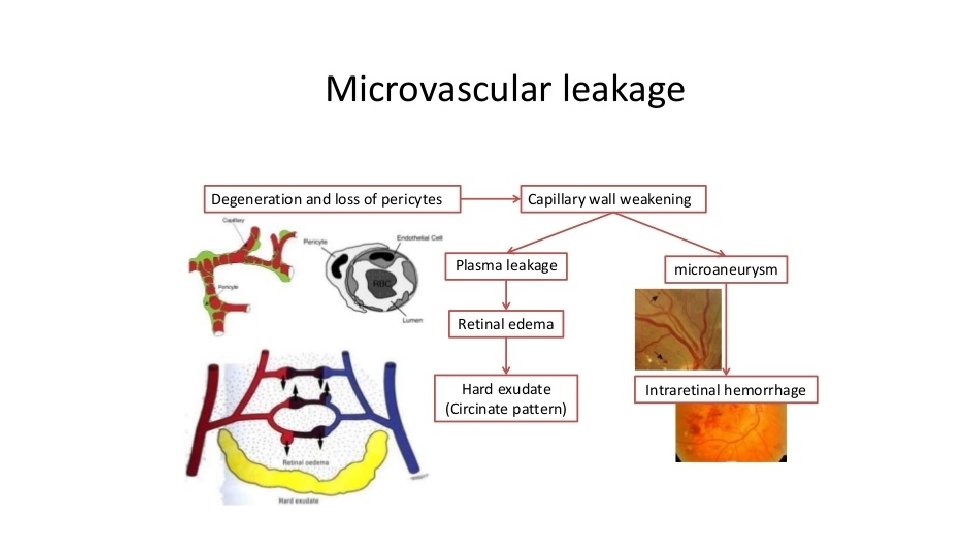
Diabetic Ocular Complications. Histologically, • Non proliferative (non-proliferative) • proliferative retinopathy Background (non-proliferative) retinopathy. initial retinal capillary microangiopathy
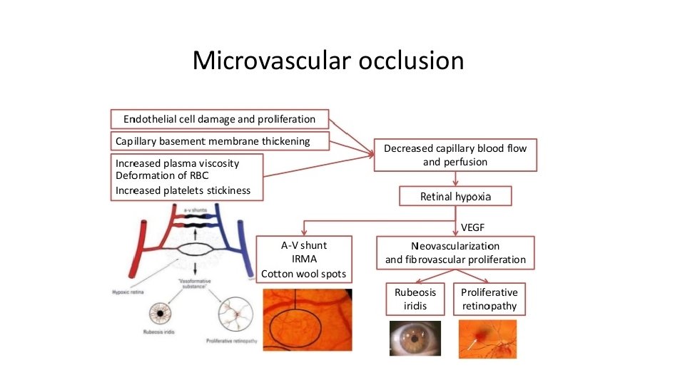
 Friability of neo vascularization results in vitreous haemorrhages. • iii) Proliferation")
• ii) Friability of neo vascularization results in vitreous haemorrhages. • iii) Proliferation of astrocytes and fibrous tissue around the new blood vessels. • iv) Fibrovascular and gliotic tissue contracts to cause retinal detachment and blindness.

• Diabetic Neuropathy duration of the disease; up to 50% of diabetics overall have peripheral neuropathy Activation of PKC and polyol pathway Accumulation of fructose and sorbitol in nerve Nonenzymatic glycosylation of structural nerve protein

Four distinct mechanisms
 are formed")
1 -Formation of Advanced Glycation End Products. Advanced glycation end products (AGEs) are formed as intracellular glucose derived dicarbonyl precursors+ amino groups advanced glycation end product(AGEs) (glyoxal, methylglyoxal, and 3 -deoxyglucosone)
 that is expressed on inflammatory")
• AGEs bind to a specific receptor (RAGE) that is expressed on inflammatory cells (macrophages and T cells), endothelium, and vascular smooth muscle.
-")
AGE-RAGE signalling axis • TGFβ-excess basement membrane material • vascular endothelial growth factor (VEGF)- neovasculerization • reactive oxygen species (ROS) in endothelial cells • procoagulant activity • Enhanced proliferation of vascular smooth muscle cells and synthesis of extracellular matrix
 is an important")
2 -Activation of Protein Kinase C. second messenger diacyl glycerol (DAG) is an important signal transduction pathway. Intracellular hyperglycemia--- de novo synthesis of DAG-- excessive PKC activation- vascular permeability and angiogenesis

3 -Oxidative Stress and Disturbances in Polyol Pathways • Sustained hyperglycemia---- aldol reductase-- progressive depletion of intracellular NADPH -- decreased rgeneration of reduced glutathione(GSH) - increasing cellular susceptibility to oxidative stress • Responsible for diabetic neuropathy

4 -Hexosamine Pathways and Generation of Fructose-6 - Phosphate Hyperglycemia --- increases intracellular levels of fructose-6 phosphate via HM- excess proteoglycans - abnormal expression of TGFβ or PAI-1 --- exacerbate the end-organ damage


Complications of Diabetes. I. Acute metabolic complications: • diabetic ketoacidosis • hyperosmolar nonketotic coma • hypoglycaemia

II. Late systemic complications: • atherosclerosis • diabetic microangiopathy • diabetic nephropathy • diabetic neuropathy • diabetic retinopathy and infections
, complication of type 1 DM. Lack of insulin Lypolysis Free")
1. Diabetic ketoacidosis (DKA), complication of type 1 DM. Lack of insulin Lypolysis Free fatty acid in plasma FFA+acetyl co enzyme A-- liver Ketone body
High Blood sugar High plasma osmolality Hyperglycemic diuresis Dehydrartion")
2. Hyperosmolar hyperglycaemic nonketotic coma (HHS)High Blood sugar High plasma osmolality Hyperglycemic diuresis Dehydrartion CNS complication

3. Hypoglycaemia • patients of type 1 DM. • Excessive administration of insulin, missing a meal, or due to stress

II. LATE SYSTEMIC COMPLICATIONS 1. Atherosclerosis • hyperlipidaemia, • reduced HDL levels, • nonenzymatic glycosylation, • increased platelet adhesiveness, • obesity • hypertension

2. Diabetic microangiopathy 3. Diabetic nephropathy 4. Diabetic neuropathy 5. Diabetic retinopathy 6. InfectionsØimpaired leucocyte functions Ø reduced cellular immunity Øpoor blood supply
- Slides: 61