Developmental malformations by Krisztina H Mink Semmelweis University

Developmental malformations by Krisztina H. -Minkó Semmelweis University, Faculty of Medicine, Department of Anatomy, Histology and Embyology 2017

Morphogenesis versus dysmorphogenesis The initially flat three-layered embryonic disc undergoes morphogenesis to form a threedimensional embryo with a tube-within-a-tube body plan and the beginnings of rudiments that will form all of the adult organs and systems. Morphogenesis results from differential growth. Differential growth is driven by a small number of fundamental cellular behaviors such as changes in cell shape, size, position, number, and adhesivity. If these behaviors are perturbed during embryogenesis, by a genetic mutation, environmental insult (i. e. , a teratogen), or a combination of the two, differential growth is abnormal and dysmorphogenesis results with the formation of a structural birth defect.

CONGENITAL MALFORMATION, TERATOLOGY, HISTORY Teratology is the science that studies the causes, mechanisms, and patterns of abnormal development. teratology : development of „monsters” In historical times the explanation of the „developmental monsters” was mainly esoteric (Devilish Souls) First scientific descriptions: Ambroise Paré (XIV. century barber surgeon, pathologist, anatomist) 1941 Gregg (Australia) rubella infection of pregnants caused blindness, deafness , problems with heart 1960 s years: Thalidomide scandal (phocomelia)

cystic hygroma colli statue from Pacific Islands showing a divinity Ambroise Paré’ s drawing from his book (1520) large cyst-like cavities containing lymph rare sirenomelia („fused lower limbs”)

Sirenomelia Caudal dysplasia, also called caudal regression syndrome, caudal agenesis, or sacral agenesis, is characterized by varying degrees of (1) flexion, inversion, and lateral rotation of the lower extremities; (2) anomalies of lumbar and sacralvertebrae; (3) imperforate anus; (4) agenesis of the kidneys and urinary tract; and (5) agenesis of the internal genital organs except for the gonads. In extreme cases, the deficiency in caudal development leads to fusion of the lower limb buds during early development, resulting in a ‘‘mermaid-like’’ habitus called sirenomelia (Fig. 3 -18) it is believed that they all arise from defects resulting from abnormal growth and migration during gastrulation. In animal models, caudal dysplasia can be induced by both environmental factors and mutations.

Insulin as teratogen, Brachyury mutation In animal models, caudal dysplasia can be induced by both environmental factors and mutations. Milagros Cerron born with sirenomelia, later underwent a successful operation to separate her legs. Despite undergoing 150 operations during her relatively short lifetime, she still passed away in 2009 at the age of 10. http: //www. spillednews. com/2016/08/mermaid-likefoetus-woman-terminates. html

Dysmorphogenesis can result from both malformation Common examples, and deformation. include Down syndrome (trisomy 21) and 22 q 11. 2 deletion syndrome, two syndromes that result from genetic mutations. Malformations consist of primary morphologic defects Other syndromes can result from teratogen exposure. A common example is fetal alcohol syndrome, in an organ or body part resulting from abnormal also known as fetal alcohol spectrum disorder. developmental events that are directly involved in the development of that organ or body part. For example, failure of the neural groove to close results in a malformation called a neural tube defect. Similarly, failure of the digits to fully separate results in syndactyly, that is, fusion of the digits. Deformations consist of secondary morphologic defects that are imposed upon an organ or body part owing to mechanical forces; that is, deformations affect the development of an organ or body part indirectly. For example, if insufficient amniotic fluid forms (i. e. , oligohydramnios), deformation of the feet can occur due to mechanical constraints, resulting in club foot. Dysmorphogenesis can occur in an isolated organ or body part or can occur as a pattern of multiple primary malformations with a single cause. In the latter case, the condition is referred to as a syndrome.
 substances that are capable of causing a")
Teratogens are environmental (i. e. , nongenetic) substances that are capable of causing a birth defect when embryos or fetuses are exposed at critical times in development to sufficiently high doses (concentrations). The study of the role of environmental factors in disrupting development is known by the unfortunate name of teratology, which literally means the study of (developmental) monsters.

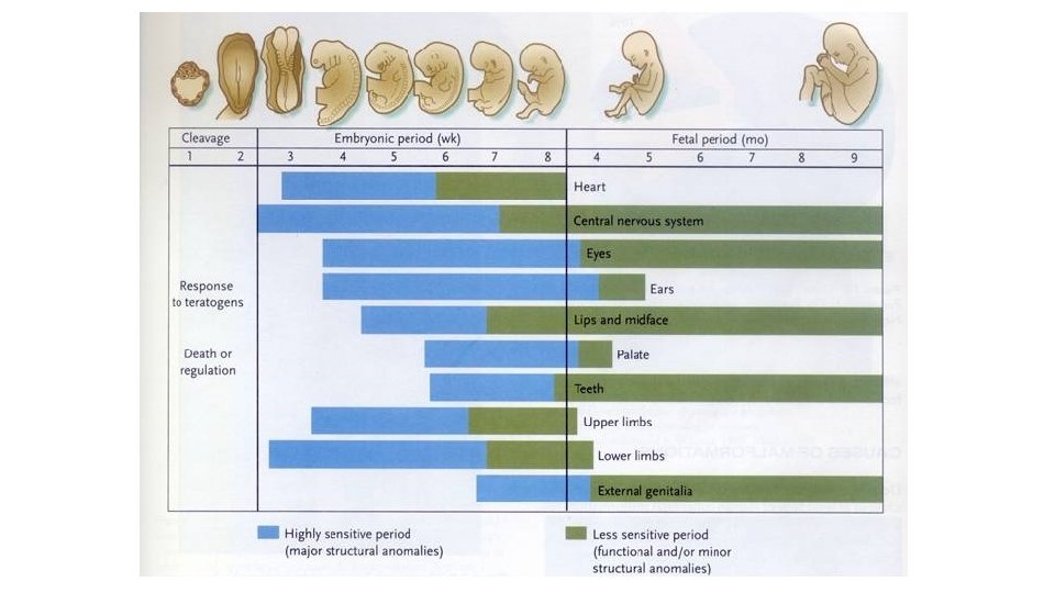
The first principle of teratology is that an embryonic structure is usually susceptible to teratogens only during specific critical sensitive periods, which usually correspond to periods of active differentiation and morphogenesis. Thus, a potent teratogen may have no effect on the development of an embryonic structure if it is administered before or after the critical period during which that structure is susceptible to its action. Because the major events of organogenesis take place during the first 8 weeks of development, that is the period during which the fetus is most vulnerable to teratogens.

A second principle of teratology is that an embryonic structure is susceptible to a critical dose of teratogen during its specific critical sensitive period. Thus, in teratologic studies a dose response curve is constructed for a suspected teratogen in which lowest dose has no effect and the highest dose is lethal to the embryo. A third principle of teratology is that susceptibility to a teratogen depends on the genetic constitution of the developing embryo or fetus. For example, if two embryos of the same age are exposed to the same dose of teratogen, one may develop severe cardiac malformations whereas the other may remain unaffected. The molecular basis for this difference in susceptibility might, for instance, be a genetic difference in the rate at which the enzyme systems of the two embryos detoxify the teratogen. Thus, there is a gene-environmental interaction underlying susceptibility to birth defects that varies from embryo to embryo.
: „to")
Critical periods during the pregnancy Pre-embryonic period: • 0 -3. embryonic weeks (EW): „to be or not to be”, the teratogen kills the whole embryo (abortion), or only some of the early cells and these cells will be substituted (no congen. malf. ) Embryonic peroid: • 3 -8. EW: most vulnerable period: time of the development of the most serious (major) anomalies Fetal period: • 8. EW: minor morphological or functional defects (e. g. mental retardation).

• The sensitivity of the different organs are different in the time: some organs are sensitive in early period (heart) the others are sensitive in later periods (genitals) and some of them are sensitive all along (nervous system). Diagram of sensitivity period

Incidence of abnormalities in Europe: 2 -3% of all live born children at the birth, -(congenital) but in the first year: 4 -6%. Range of CONGENITAL ANOMALIES is very vide from the severe morfological defects till enzime defects (no morfological picture)

Major malformations drastic differences from normality they cause prenatal or perinatal death require surgical or medical care soon after birth gravely physically handicapping extreme cosmetic burden Minor malformations minor physical features absence of associated major congenital malformations little or no medical or cosmetic consequence

Major congenital malformations by system Neural ~9. 3% Cardiovascular GIT/Respiratory ~ 7 % anencephalus spina bifida encephalocele microcephalus hydrocephalus transposition of great vessels ventricular septal defect hypoplastic left heart coarctaction of aorta cleft palate cleft lip oesophageal atresia or stenosis small intestine atresia or stenosis anorectal atresia or stenosis Urinary ~20 % ~6. 4 % hypospadias renal agenesis and dysgenesis cystic kidney disease obstructive defects of renal pelvis, ureter Genital organs ~ 12 % Skeletal/Muscular congenital dislocation of the hip limb reduction defects diaphragmatic hernia exomphalos gastroschisis ~24 % Chromosomal ~ 9. 5% trisomy 21 (Down syndrome) trisomy 18 (Edwards syndrome) unicornuate, bicornuate uterus cryptorchidism About 3% of newborns have a "major physical anomaly", meaning a physical anomaly that has cosmetic or functional significance Infant mortality: over 20% subsequent upon malformation

Minor Congenital Malformations Cardiovascular system patent ductus arteriosus or foramen ovale (gestational age <37 weeks or birthweight <2500 g) mild, trivial, or physiological valvular regurgitation… http: //bestpractice. bmj. com/best-practice/ images/bp/en-gb/766 -1 -hlight_default. jpg Skin skin cysts noncavernous single small hemangioma (<10 cm dia. ) nevus flammeus…. . https: //i. ytimg. com/vi/g_z. QEQBLv_g/hqdefault. jpg Gastrointestinal system Hepatomegaly, splenomegaly Meckel's diverticuluum naevus flammeus consists of superficial and deep dilated capillaries in the skin https: //encrypted-tbn 0. gstatic. com/ images? q=tbn: ANd 9 Gc. Sb. U 199 kxnp 3 k 1 T 4 Mrf. Lj 1 q. Ptoj_ OWECkknq 07 YFc. TS_FYSf. OQP http: //thehealthscience. com/ thsattachs/939954/111918441287277. jpg

Major Congenital Malformations

Major malformations: drastic differences from normality

Causes

Classification by causes of malformations Genetic Environmental. cause of more than half of all congenital malformations is not known!!!!! Large terra incognita…. . „Description is infinite and easy: explanation is limited and difficult” Hippocrates The Human Genome Project (HGP) -international scientific research project -to determine the sequence of chemical base pairs which make up DNA -identifying and mapping the approximately 20, 000– 25, 000 genes of the human genom -in May 2006, the sequence of the last chromosome was published in the journal Nature

environment 7%

Genetic Abnormal chromosome number Trisomy 21 – Down syndrome Trisomy 13 Trisomy 18 Abnormal chromosome structure dominant or recessive autosomes or sex chromosomes Deletion Duchenne muscular dystrophy Xchromosome Deletion of the short arm of chromosome 5 Cri du chat syndrome Monosomy of sex chromosome 45 X 0 – Turner syndrome female fenotype sterile Genetic mutation Duplication autosomal recessive polycystic kidney disease (perinatal type) autosomal dominant polycystic kidney disease (adult onset) 1: 400– 1: 1, 000 individuals X-linked recessive hemophilia Reciprocal translocation

http: //www. ncbi. nlm. nih. gov/pubmed/

 Homologues are not separated (non-disjunction)")
Chromosomal mutations Aneuploidy: in meiosis (first metaphase) Homologues are not separated (non-disjunction)

Down syndrome is one of the most common chromosome abnormalities in humans Down syndrome: chromosome 21 trisomie It is typically associated with physical growth delays, characteristic facial features and mild to moderate intellectual disasbility. Single crease of the palm.

21 st chromosome - smallest chromosome Only 200 to 250 genes Genes that may have input into Down syndrome include: Down syndrome Overexpression!! COL 6 A 1 -- overexpression may be the cause of heart defects ETS 2 -- overexpression may be the cause of skeletal abnormalities CAF 1 A -- overexpression may be detrimental to DNA synthesis DYRK -- overexpression may be the cause of mental retardation CRYA 1 -- overexpression may be the cause of cataracts GART -- overexpression may disrupt DNA synthesis and repair IFNAR -- the gene for expression of Interferon, overexpression may interfere with the immune system In 1959, Jérôme Lejeune discovered that Down syndrome resulted from an extra chromosome. Mosaicism! -% of trisomic cells in the muscle may differ from that in the brain, or in the blood or skin -presence of cells with normal number of chromosomes (46) may result in a less severe Down syndrome Sottish award-winning film and TV actress Paula Sage

Abnormal chromosome number Down syndrome – chromosome 21 - trisomy 21 Non-disjunction - 88% coming from non-disjunction in the maternal gamete - 8% coming from non-disjunction in the paternal gamete 1 per 800 to 1, 000 births (earlier data) 1 in every 700 babies Between 1979 and 2003, the number of babies born with Down syndrome increased by about 30%

Why these signs result from the trisomy is unknown

Other aneuploidies Trisomy of 13. Trisomy 13, also called Patau syndrome, is a chromosomal condition associated with severe intellectual disability and physical abnormalities in many parts of the body. Individuals with trisomy 13 often have heart defects, brain or spinal cord abnormalities, very small or poorly developed eyes (microphthalmia), extra fingers or toes, an opening in the lip (a cleft lip) with or without an opening in the roof of the mouth (a cleft palate), and weak muscle tone (hypotonia). Due to the presence of several life-threatening medical problems, many infants with trisomy 13 die within their first days or weeks of life. Only five percent to 10 percent of children with this condition live past their first year. Trisomy of 18. (rocker bottom feet) Trisomy 18, also called Edwards syndrome, is a chromosomal condition associated with abnormalities in many parts of the body. Individuals with trisomy 18 often have slow growth before birth (intrauterine growth retardation) and a low birth weight. Affected individuals may have heart defects and abnormalities of other organs that develop before birth. Other features of trisomy 18 include a small, abnormally shaped head; a small jaw and mouth; and clenched fists with overlapping fingers. Due to the presence of several life-threatening medical problems, many individuals with trisomy 18 die before birth or within their first month. Five to 10 percent of children with this condition live past their first year, and these children often have severe intellectual disability.
: female but")
Aneuploidy of the sex chromosomes: Turner syndrome: 45 X, O (X monosomy): female but weak fenotype, steril Others: XXY: Klienefelter syndrome: male, but small testis, steril, long limbs XYY: seems to be normal male, high figure, but violent (choleric) behavior XXX: „super women”: seems to be normal feminin habit, but mental retardation occurs

MONOGENIC DEFECTS

Marfan syndrome Genetic disorder of the connective tissue. The degree to which people are affected varies. People with Marfan tend to be tall, and thin, with long arms, legs, fingers, and toes. . They also typically have flexible joints and scoliosis. The most serious complications involve the heart and aorta with an increased risk of mitral valve prolapse and aortic aneurysm. Other commonly affected areas include the lungs, eyes, bones, and the covering of the spinal cord. Abraham Lincoln had this disorder. Autosomal dominant disorder. About 75% of the time the condition is inherited from a parent while 25% of the time it is a new mutation. It involves a mutation to the gene that makes fibrillin which results in abnormal connective tissue.

Polysyndactylia: Hox. D 13 pointmutation Syndaktilia: az ujjak összeolvadása. Ebben a humán esetben a HOXD 13 gén pontmutációja a gén kieséséhez vezetett. Az üres csillagokkal jelölt képletek kézközépcsontok, a két kicsi csillag normálisan nem létező carpalis csontokat jelöl.
 phocomelia - heart")
Holt-Oram syndrome -TBX 5 mutation -1: 100. 000 -limb bud (100%) phocomelia - heart defects (67%) Anthony Bagliano is a kicker for North High School North kicker on Holt-Oram syndrome - 'I'm always happy. I try to look at the positives in life'
 genetic sources Developmental abnormalities")
CAUSES OF CONGENITAL ANOMALIES Environmental sources (teratogens) genetic sources Developmental abnormalities

Maternal derived abnormalities lifestyle, environment and nutrition directly affect embryonic development some effects are more subtle and relate to later life health events low birth weight data later health outcomes of same babies weight named as "fetal origins" or "programming” diabetes

Chromosome mutation 10% unknown Gene mutation 8% 50% multifactorial 25% ? ? : - age of the parents - different ethnical groups - familiar background (genetical) - seasons - social and geographical differences
")
AGE Incidence of Down syndrome is dependent on the age of mother (left figure) but the Apert syndrome is on the age of father (right blue line) Apert syndrome: turricephalus – tower head

Apert syndrome

Apert syndrome: turricephalus – tower head FGFR 2 mutation: craniosynostosis, syndaktylia

RACE Incidence of cleft palate is different in different ethnical groups far-east > (2 x) caucasian > (2 x) afro-american

Jaxon Buell: Presents pile up for 'miracle baby' who made national news SEASON Anencephaly is abundant in the spring: it is caused by the deficit of the mother’s folic acid level. Given folic acid to the mothers cut down drastically the number of these fetuses. he was born Anencephaly = cranioschisis – anterior part of the neural tube (neuroporus anterior) is not closed the brain will not be developed. Aug. 27, 2014.

Frequency of anencephaly in different countries
 2. drugs/chemicals 3. mechanical agents")
Environmental factors 1. biotic agents (bacteria, viruses, fungi) 2. drugs/chemicals 3. mechanical agents

1. Infectious agents as teratogens • Rubella • German or Three-Day Measles • First trimester – most serious ED 0 - ED 60: cataracta (blindness )and heart problems ED 0 - ED 120: deafness • Cytomegalovirus (CMV) • Herpes Simplex Virus (HSV) • Varicella • Human Immunodeficiency Virus (HIV) • Congenital Syphilis • Toxoplasmosis

Different organs different sensitive periods
 :")
Different organs different sensitive periods Tetraciklin: - after the 120. embrionic day (ED) : milk-teeth and adult teeth become coloured -after ED 250: only the adult teeth become coloured Rubella infection: ED 0 - ED 60: cataracta (blindness )and heart problems ED 0 - ED 120: deafness https: //drmarthaszabolcs. com/2017/03/06/firstblog-post/#jp-carousel-140

2. Mechanical factors as teratogens • Mechanical forces • • Restrict the mobility of the fetus Cause prolonged compression in an abnormal posture E. g. congenital dislocation of hip and clubfoot Malformed uterus

Mechanical factors as teratogens • Amniotic fluid • Absorb mechanical forces • Oligohydramnios • Significantly reduced fluid-quantity • Mechanically induced deformations of the limbs • Knee hyper-extends • Amniotic bands • Rings formed from amnion rupture • Local constriction during fetal growth • Intrauterine amputations
: causes problems in the brain development :")
3. Drugs/chemicals Etil-alcohol: Fetale Alcohol Syndrome (FAS): causes problems in the brain development : holoprosencephalia. Prosencephalon is the most anterior part of the neural tube : hemispheres, hypothalamus, hypophysis , eye developed from this part Thalidomid (Contergan): tranquillizier Many therapeutic drugs are known to be teratogenic; these include retinoids (vitamin A and analogs), the anticoagulant warfarin, the anticonvulsants valproic acid and phenytoin, and a number of chemotherapeutic agents used to treat cancer. Most teratogenic drugs exert their main effects during the embryonic period. Although, as stated above, most care must be exercised in administering certain anesthetics and other drugs even late in pregnancy or at term, because they may endanger the health of the fetus.
: This disorder affects 2 in 1000 live-born infants (Fig. 5")
Fetale Alcohol Syndrome (FAS): This disorder affects 2 in 1000 live-born infants (Fig. 5 -2). Consumption of amounts of alcohol as low as 80 g per day (i. e. , between two and three shots of a grain liquor such as rum) during the 1 st month of pregnancy can cause significant defects, and it has been suggested that even a single binge may be teratogenic. Common components of the disorder include defects of brain and face development, namely, microcephaly (small head), short palpebral fissures (eye openings), epicanthal folds (folds overeye lids), a low nasal bridge with a short nose, flat midface, minor external ear anomalies, and jaw anomalies including a thin upper lip with indistinct philtrum and micrognathia (small jaw). Chronic consumption of even quite small amounts of alcohol later in pregnancy can result in other, less-destructive effects, such as some degree of growth retardation and minor physical defects.

FAS: holoprosencephaly and craniofacial CA. Prosencephalon regulate not only the development of the brain but the head and face also B: cebocephalia C: no nose, hypotelorismus D: cyclopia

Some recreational drugs are also teratogenic; these include tobacco, alcohol, and cocaine. Cocaine, used by alarming numbers of pregnant women (the drug affected 300, 000 to 400, 000 newborns in 1990 in the United States), readily crosses the placenta and may cause addiction in the developing fetus. In some of the major cities of the United States, as many as 20% of babies are born to mothers who abuse cocaine. Unfortunately, fetal cocaine addiction may have permanent effects on the individual, although studies suggest that early intervention with intensive emotional and educational support in the first few years of life may be helpful. Two mechanisms have been proposed by which cocaine could cause preterm labor: cocaine, a potent constrictor of blood vessels, may cause abruption of the placental membranes (premature separation of the placenta from the uterus) by partly shutting off the flow of blood to the placenta; or as there is evidence that cocaine directly affects the contractility of the uterine myometrium (muscle layer), it perhaps makes the myometrium hypersensitive to signals that initiate labor. high frequency of preterm labor. https: //www. youtube. com/watch? v=QYICa. Ho 6 t. WQ
, often called small for gestational age (SGA), is a condition")
Intrauterine growth restriction (IUGR), often called small for gestational age (SGA), is a condition in which fetal growth in markedly retarded. IUGR carries a higher risk of perinatal mortality and morbidity, so IUGR is a life-threatening birth defect. A newborn is considered to be SGA if he/she weighs less than 2500 grams at term or falls below the 10 th percentile for gestational age. CAUSES: teratogen exposure such as congenital viral or bacterial infections, fetal chromosomal anomalies (e. g. , Down syndrome), maternal factors (such as preeclampsia, a condition affecting about 5% of pregnancies characterized by high blood pressure and protein in the urine), placental factors (such as placenta previa, or ‘‘low-lying’’ placenta, a condition in which the blastocyst implants near the uterine cervix and the placenta covers part of the opening of the cervix). involves the entire fetus
: tranquillizier thousands of pregnants used as a sleeping -pill")
Contergan scandal • Thalidomid (Contergan): tranquillizier thousands of pregnants used as a sleeping -pill in the 60’s • About 40 thousands person had got periferal nerve inflammation; • 8 -12 thousands baby born with phocomelia • among them 5 thousands grown up…

Thalidomid-case: phocomelia: part of the limbs are missing the diagram shows the amount of sold Contergan (Thalidomid), empty columns: in kilogramm!!!; stripped colums : number of phocomelia patient , in Germany between 1956 and 1964

In 1964 the drug was given to patients with leprosy as the last possibility for sleeping. And the drug dissolved the distress, gave good sleeping periods and mollified the leprosy. It seems to be very bid fair: it seems to be usable not only in leprosy but in some autoimmune deseases also Today we know that it has antiangiogenetic effect , that caused the problem in the developing embryo

Both maternal diabetes and maternal obesity during pregnancy constitute risk factors for birth defects of the fetus neural tube defects and heart defects

Maternal obesity (defined in the United States as a body mass index greater than 30 kg/meter squared) is also a risk factor for birth defects. Fetuses born to obese women are 2 to 3. 5 times more likely than those born to average-weight women to have neural tube defects, heart defects, and omphalocele.

Thank You for attention!

List of important malformations • Gamete production, meiosis: trisomy 21, trisomy 18, Klinefelter- (47, XXY), Turner- (45, X) syndrome 22 q 11 Deletion ectopic or extrauterin pregnancy • gastrulation: situs inversus • neurulation: anencephalia, spina bifida • ectopias (ectopia cordis) • somites: lumbalisation, sacralisation, preatlas • placenta: preeclampsia • strangulations • oligo- and polyhydramnion • Skull: hydrocephalus, craniosynostosis

References T. W. Sadler: Langman’s Medical Embryology, 7 th edition, 1995, Baltimore, Maryland, USA – képek Carlson’s embryology book Lectures of Dr. Ágnes Csáki, Dr. Attila Magyar, Dr. Nándor Nagy, Dr. Ágnes Nemeskéri
- Slides: 65