Developmental Dysplasia of the Hip DDH Dr M
 Dr M. Abu Hilal Ass. Prof Mutah University")


: more adduction Risk factors (5 F’s) First")


- Slides: 28
Developmental Dysplasia of the Hip (DDH) Dr M. Abu Hilal Ass. Prof Mutah University
Normal hip development depends on 1 -well formed acetabulum by triradiat cartilage 2 -concentrically located femoral head.
Mechanism 1 -The instability : imperfect seating of the femoral head 2 - acetabular dysplasia: underdeveloped acetabulum
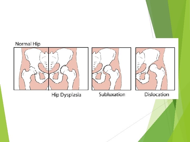
DDH a spectrum of disease that includes dysplasia : shallow or underdeveloped acetabulum subluxation : displacement of the joint with some contact remaining between the articular surfaces dislocation : complete displacement of the joint with no contact between the articular surfaces
CDH Teratologic or congenital hip dislocated in utero irreducible on neonatal exam presents with a pseudoacetabulum associated with neuromuscular conditions and genetic disorders commonly seen with arthrogryposis, myelomeningocele, Larsen's syndrome, Ehlers -Danlos
Epidemiology of DDH Incidence of neonate hip instability is 1: 1000 Girls much more commonly than boys 7: 1 Left more affected than Right (60 -70%) due to the most common intrauterine position being left occiput anterior (left hip is adducted against the mother's lumbrosacral spine) bilateral in 20% Culture increases or decreases the risk? ?
Etiology Genetic factors heritable features which could predispose to hip instability, generalized joint laxity and shallow acetabulum (Both more common in females) Hormonal factors high levels of maternal oestrogen, progesterone and relaxin in the last few weeks of pregnancy) may aggravate ligamentous laxity in the infant. This explains the rare instability in premature babies
Family history: genetic Foot first( breech position): more adduction Risk factors (5 F’s) First born : small uterus Female: more laxity and more affected by Elastin Fluid abnormality: Oligohydramnios (less movement)
Post natal factor: Swaddling
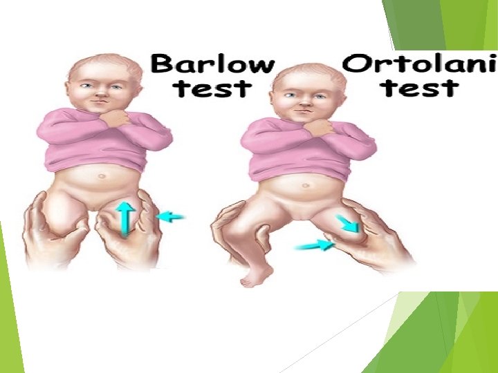
Clinical features Usually not very obvious Decreased abduction or asymmetrical abduction Clicky hip although most clicky hips are found to be normal Asymmetrical thigh creases Leg length discrepancy : Galeazzi’s sign Ortolani test: while hips flexed, move one hip from adduction to abduction to reduce the dislocated hip ( Ortolani: hip In) Barlow test: while hips flexed, move one hip from abduction to adduction to dislocate the hip Ortolani and Barlow are none specific and maybe false negative Positive if Click or clunk is heard
Galeazzi’s sign
Asymmetrical creases
Imaging Ultrasonography Plain x-rays No radiation Soft tissue and bone evaluation Bone only Static and dynamic Static only Can be done as early as 6 weeks Not before 4 months Operator dependent Not operator dependent Less available Available More coast Less coast
Ultrasound Base line : line pass through the ilium Alpa Angle : AA measures bony Acetabulum Normal more then 60 degrees Ø Beta Angle: BB la. Brum depth Normal less than 55 degrees BBB: Big Beta Bad
Ultrasound
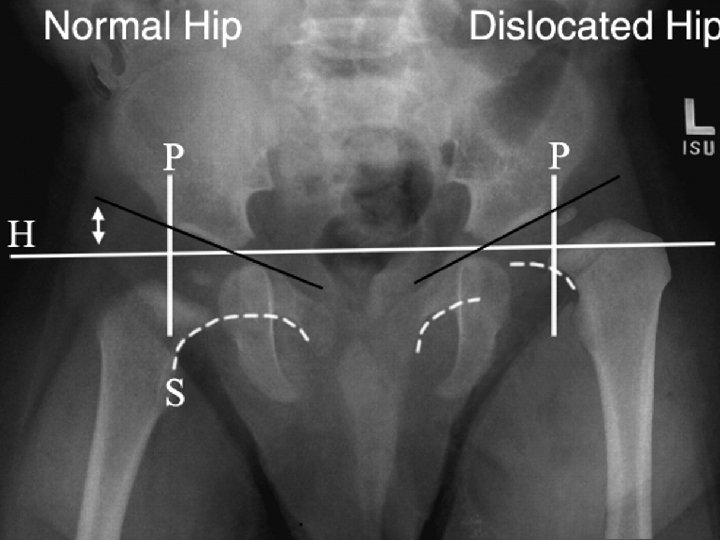
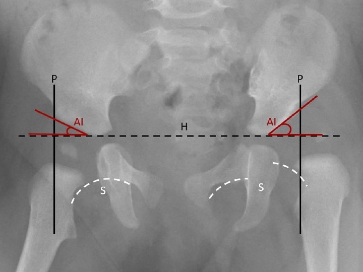
Plain Radiography From AP radiograph of the hip Hilgenreiner line : Horizontal line is drawn between each triradiate cartilage Perkin line: Lines Perpendicular to H line are drawn through the outer edge of the acetabulum (Perkin line), dividing the hip into 4 quadrants - Normally femur head lies in the lower inner quadrant Acetabular index or angle : H line and second line which extend from inner to outer edge of acetabulum -Normally less than 30 degree after age 6 months Shenton’s line : imaginary curved line drawn along the inferior border of the superior pubic ramus and along the inferomedial border of the neck of the femur. - Normally it’s smooth semicircular with no interruption
Management Depend on the AGE and THE DEGREE OF INSTABILITY 1 -Pavlik harness or abduction splint (often used as initial treatment of hip dysplasia in newborn and infants under 6 -9 months of age for 6 -12 weeks) 2 - Close reduction + Spica cast -Failed Pavlik Harness or older child (9 -12 months ) -performed under GA with an arthrogram to confirm a concentric reduction 3 - Open reduction + spica cast +- pelvic or femoral osteotomy -failed closed reduction -older than one year
Pavlik harness or abduction splint Pavlic Harness is dynamic brace that maintains the hip in flexion and abduction, less stiffness , enhance deepening of acetabulum, and less AVN more time to apply and needs co-operative parents Abduction splint: static splint that maintains the hip in flexion and abduction , no movement so more stiffness, less deepening of acetabulum and more AVN easy to apply
Pavlik harness and abduction splint
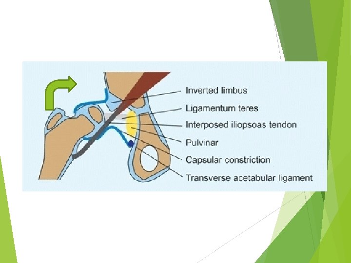
Factors that prevent reduction 1 -Redundant capsule but constricted ? ? ? 2 -elongated ligamentum teres 3 -Tight abductor muscle 4 -fibrouse tissue in the socket (Pulvinar) 5 -inverted labrum (limbus) 6 -Transverse Acetabular ligament 7 -interposed iliopsoas tendon
Hip spica after closed reduction or open reduction
Complications Late diagnosis and difficult treatment AVN : more with static abduction and multiple surgeries Recurrence : 10% even with proper treatment Stiffness Early osteoarthritis Transient femoral nerve palsy seen with excessive flexion during Pavlik bracing
Thank you