Development Policies and Procedures Manual Examples of table

Development Policies and Procedures Manual Examples of table of contents: Example 1: demonstrates a organization numbering system Example 2: demonstrates a service-specific numbering system
 FAMILY and COMMUNITY MEDICINE")
DATA INTERPRETATION Dr. HUSSEIN SAAD Assistant Professor and Consultant, MRCP(UK) FAMILY and COMMUNITY MEDICINE College of Medicine King Saud University

A 37 - year- old lady, presents with 3 months H/O dizziness and easy fatigue. The following CBC is shown below: WBC. . . 7. 0 4 – 11 x 10. e 9/L RBC. . . 3. 68 L 4. 2 – 5. 5 x 10. e 12/L HGB. . . 87 L 120 – 160 g/L HCT. . . 27. 1 L 42 – 52 % MCV. . . 73. 6 L 80 – 94 fl MCH. . . 23. 6 L 27 – 32 pg MCHC. . . . . 321 320 – 360 g/L RDW. . . 15. 5 H 11. 5 – 14. 5 % PLT. . . 445 H 140 – 450 x 10. e 9/L Diagnosis: Hypochromic Microcytic Anaemia (IDA) On systemic enquiry, she added that she has menorrhagia for the last 4 months. ◘ Mention one investigation of importance to reach the diagnosis. TSH : FT 4: 89 8. 6 m. IU/L pmol/l (0. 25 – 5) (10. 3— 25. 8)

A 16 -year-old girl presents with 2 m H/O dizziness, palpitation and recurrent faints. The following CBC is shown below: WBC. . . 8. 1 4 – 11 x 10. e 9/L RBC. . . 1. 42 L 4. 2 – 5. 5 x 10. e 12/L HGB. . . 24 L 120 – 160 g/L HCT. . . 8. 0 L 37 – 47 % MCV. . . 56 L 80 – 94 fl MCH. . . 16. 6 L 27 – 32 pg MCHC. . . 295 L 320– 360 g/L RDW. . . 22. 9 H 11. 5 – 14. 5 % PLT. . . 181 140 – 450 x 10. e 9/L Retic. Count ………. 3. 5 H 0. 2 - 2. 0 % HOW ARE YOU GOING TO MANAGE THIS PATIENT? Blood Transfusion, Admission, Treat the cause beside Iron and Folic A.

A 17 year old lady presents with dizziness and bouts of fall. WBC …………. 7. 4 x 10. e 9/L 4 -11 RBC …………. 3. 57 x 10. e 12/L 4. 2 - 5. 5 HGB …………. 57 g/L 120 -160 HCT …………. 20. 1 % 37 - 47 MCV ………… 56. 2 fl 80 - 94 MCH ………… 15. 9 pg 27 - 32 MCHC ……… 282 g/L 320 - 360 RDW ………. 25. 0 % 11. 5 - 14. 5 PLT ………. . 578 x 10. e 9/L 140 - 450 Iron ………. . ………………. . 1. 0 umol/L Total Iron-Binding cap …. . 89. 6 umol/L 9 - 30 44. 8 - 80. 6 Transfused (one pint of blood) and Put on : ferrous sulphate and folic acid

Cont. A 17 year old lady with low Hb, after 6 weeks. WBC …………. 8. 4 x 10. e 9/L 4 -11 RBC …………. 4. 71 x 10. e 12/L 4. 2 - 5. 5 HGB …………. 105 g/L 120 -160 HCT …………. 32. 5 % 37 - 47 MCV ………… 68. 9 fl 80 - 94 MCH ………… 22. 3 pg 27 - 32 MCHC ……… 324 g/L 320 - 360 RDW ………. 35. 7 % 11. 5 - 14. 5 PLT ………. . 296 x 10. e 9/L 140 - 450 Ferritin. . 6. 77 ug/L 13 -150 Gluc. 6 phosphate dehydrog NORMAL IU/10^9 100 - 200 Hemoglobin A 2 2. 3 % 2. 0 - 3. 5 Hemoglobin F 0. 0 % 0 - 2. 0 Hemoglobin A 97. 7 % 95 - 99 Hemoglobin S 0. 0

Microcytosis: low MCV Serum Iron Ferritin ◘ IDA Low ◘ Thalassaemia Minor Normal ◘ Sideroplastic Anaemia High Uncommon, defect in heme synthesis and ringed sideroplasts in bone marrow. ◘ RDW: Red Cell Distribution Width When increased reflect, heterogeneity in cell size. Also indicating low serum iron level

Iron Defeciency Anaemia Oral iron therapy, characterized by a modest reticulocytosis beginning in about five to seven days, followed by an increase in hemoglobin at a rate of about 2 to 4 g/d. L every three weeks until the hemoglobin concentration returns to normal. The serum or plasma ferritin concentration is an excellent indicator of iron stores.

A 55 year old man, who is a known case of hypertension controlled on 25 mg hydrochlorthiazide. He is a smoker of 20 cig. per day for >20 years. He came for routine follow up. ◦ WBC……. . 6. 5 4— 11 x 10. e 9/L ◦ RBC……. . . 7. 1 H 4. 7— 6. 1 x 10. e 12/L ◦ HB………. . 197 H 130— 180 g/L ◦ HCT……… 56. 3 H 42— 52 % ◦ MCV……. . . 88 80 - 94 fl ◦ MCH……. . . 30. 3 27 - 32 pg ◦ PLT………. 305 140 - 450 x 10. e 9/L ◦ ESR 0 - 10 mm/hr 4 What is the differential diagnosis? 1) 2 nd Polycythemia (mostly 2 nd Polycythaemia due to smoking) 2) Polythycaemia rubra vera How are you going to manage this patient? Blood Donation, Stop smoking, Aspirin, shift to another anti. HTN
 ◘ Relative Polycythaemia: ( Gais.")
Polycythaemia ◘ Absolute Polycythaemia (Red Cell mass ↑ ) ◘ Relative Polycythaemia: ( Gais. Bock’s ) - Normal Red Cell Mass - Decrease in plasma volume - Obese, middle aged men with anxiety and hypertension. Absolute: ◘ Primary Polycythaemia Rubra Vera (↑ RBC, WBC and Platelets) (Increase in RBCs with ↑in WBCs or ↑Platelets or both) ◘ Secondary Polycythaemia: - Smoking - COPD - High altitude - Cyanotic Cong. H. D - Renal Cysts - Uterine Fibromyoma - Hypernephroma - Adrenal adenoma - Hepatoma - Phaeochromocytoma

CONT. Polycythaemia What is the role of erythropoietin? If the erythropoietin level is high: secondary polycythaemia If the erythropoietin level is low: polycythaemia rubra vera Lap. Features of Polycythaemia Rubra Vera: Increased in HB Increased in WBC (>12. 000) Increased platelets (> 400. 000) could be within normal level Increased uric acid Increased LAP (Leukocyte Alkaline Phosphatase) Score Increased serum Vit B 12 Bone Marrow Examin. Hypercelularity
 Major Criteria: Elevated cell mass Normal arterial oxygen concent.")
Contin. Polycythaemia vera (Diagnostic criteria) Major Criteria: Elevated cell mass Normal arterial oxygen concent. (≥ 92%) Splenomegally Minor Criteria: Platelet count > 400. 000 WBC count >12. 000 ↑ LAP Score ↑ B 12 level

A 25 year old man came for pre-marital checkup. The following CBC is shown below: WBC. . . 6. 6 RBC. . . 5. 87 HGB. . . 121 HCT. . . 38. 1 MCV. . . 64. 0 MCH. . . 20. 6 MCHC. . . . . 318 RDW. . . 14. 3 PLT. . . 271 L L L 4 - 11 x 10. e 9/ L 4. 7 – 6. 1 x 10. e 12/L 130 – 180 g/L 42 - 52 % 80 – 94 fl 27 – 32 pg 320 – 360 g/L 11. 5 – 14. 5 % 140 - 450 x 10. e 9/L Interpret this data. Low HB (slight), RBCs are high normal and not matching with HB. The decrease in MCV is more and is disproportionate to the HB level

Cont. A 25 year old man Haemoglobin Electrophoresis Hemoglobin A Hemoglobin F Hemoglobin A 2 Hemoglobin S Hemoglobin E Hemoglobin C 94. 5 (95 -99 %) 0. 6 (0 - 2. 0 %) 4. 9 H (2. 0 -3. 5 %) 0. 0

A 34 -year-old man came to check some of results because of being have IBS. # Test Result Unit Range EDTA Whole Blood - SAMPLE: 1 1 2 3 4 5 6 7 8 9 WBC RBC HGB HCT MCV MCHC RDW PLT # Test 7. 75 6. 83 135. 0 43. 0 63. 0 19. 8 314. 0 16. 20 175 x 10. e 9/L x 10. e 12/L g/L % fl pg g/L % x 10. e 9/L Result Unit 4 4. 7 130 42 80 27 320 11. 5 140 - 11 6. 1 180 52 94 32 360 14. 5 450 Range Venous Blood - SAMPLE: 1 1 2 3 4 5 6 7 Hemoglobin A 2 Hemoglobin F Hemoglobin A Hemoglobin S Hemoglobin C Hemoglobin E Hemoglobin O 2. 5 0. 50 97. 0 0 0 % % % % 2. 0 0 95 - 3. 5 2. 0 99 Thalassemia Trait mostly “alpha Thalassemia”

Thalassaemia Minor Microcytosis is much more profound, and the anemia much milder, than that seen in iron deficiency anemia. Patients with beta thalassemia minor/trait also tend to have total red blood cell counts higher than normal, often into the "polycythaemic" range. The RDW in patients with thalassemia trait tends to be normal, since virtually all cells are hypochromic and microcytic.

Thalassaemia Minor ◘ MCV usually < 70 f. L ◘ The decrease in MCV is disproportionate to the HB level. ◘ Mentzer Index: MCV / RBC is < 13 ◘ If RDW is high, Correct Iron level first before proceeding to HB electrophoresis, otherwise giving a false negative result. ◘ If HB A 2 > 3. 5 → B-Thalassaemia Minor If HB A 2 is normal → alpha Thalassaemia Minor

A 31 -year-old man presents with heart burn and known to have IBS. The following CBC is shown below. Result # Test Unit Range EDTA Whole Blood - SAMPLE: 1 1 2 3 4 5 6 7 8 9 10 13. 6 4. 94 106 33. 1 67. 1 21. 4 319 19. 7 0. 0 375 WBC RBC HGB HCT MCV MCHC RDW HDW PLT Result # Test x 10. e 9/L x 10. e 12/L g/L % fl pg g/L % g/L x 10. e 9/L 4 4. 7 130 42 80 27 320 11. 5 0 140 - 11 6. 1 180 52 94 32 360 14. 5 0 450 Unit Range % % % % 2. 0 0 95 Venous Blood - SAMPLE: 1 1 2 3 4 5 6 7 Hemoglobin A 2 Hemoglobin F Hemoglobin A Hemoglobin S Hemoglobin C Hemoglobin E Hemoglobin O 7. 3 5. 2 0. 0 87. 5 0. 0 What is your diagnosis? SCA and Beta Thalassaemia Trait - 3. 5 2. 0 99

A 49 -year-old woman presents with weakness and easy tiredness. The following investigations are shown: WBC. . . 7. 8 4 – 11 x 10. e 9/L RBC. . . 4. 16 4. 2 – 5. 5 x 10. e 12/L HGB. . . 76 L 120 – 160 g/L HCT. . . 25. 2 L 37 – 47 % MCV. . . 60. 6 L 80 – 94 fl MCH. . . 18. 3 L 27 – 32 pg MCHC. . . . . 303 L 320– 360 g/L RDW. . . 19. 2 H 11. 5 – 14. 5 % PLT. . . 383 140 – 450 x 10. e 9/L Iron ………………… ……. 2. 0 umol/L ( 9 - 30 ) Ferritin ………… 4. 57 ug/L ( 13 - 150 ) Total Iron-Binding cap … 89. 3 umol/L (44. 8 - 80. 6 ) What is your diagnosis? Iron def. anaemia + Thalassaemia trait

41 yo SF preop screening 45 yo Indian male preemployment 52 yo Filipino Normal male HTN Anemia Microcytic RBC 3. 40 5. 87 4. 98 4. 7 -6. 1 x 10. e 12/L Hb 89 126 119 130 – 180 g/L MCV 70. 9 63. 3 70. 8 80 -94 fl S. Iron 2. 6 13 34 9 -30 mol/L Ferritin 3. 39 266. 7 691 30 -400 g/L Hemogl. A 2 2. 1 5. 4 2. 2 2. 0 -3. 5 Hemogl F 0 <0. 5 0 0 -2. 0 Hemogl A 97. 9 >94 97. 8 95 -99 Hemogl S 0 0 0 - Hemogl C 0 0 0 - IDA B Th. Trait

A 44 year old man, who is a known case of HCV positive. ◦ ◦ ◦ ◦ WBC……. . 2. 0 RBC……. . . 2. 95 HB………. . 110 HCT……… 31. 9 MCV……. . . 108. 1 MCH……. . . 37. 3 RDW ……. 19. 5 PLT………. 92 L L H H % L 4— 11 x 10. e 9/L 4. 7— 6. 1 x 10. e 12/L 130— 180 g/L 42— 52 % 80 - 94 fl 27 - 32 pg 11. 5 – 14. 5 140 - 450 x 10. e 9/L HEPATITIS C RNA QUALITATIVE ………… Positive HEPATITIS C RNA QUANTITATIVE ………. . 389744 IU/ML What is your diagnosis? Pancytopenia 2 nd to therapy Like interferon.

A 70 -year-old man, presents with 2 month H/0 easy fatigue and tiredness. PMH: unremarkable The following CBC is shown below: WBC. . . RBC. . . HGB. . . HCT. . . MCV. . . MCHC. . . RDW. . . PLT. . . 7. 8 2. 26 69 20. 2 89. 3 30. 6 343 15. 8 179 L L L H 4 – 4. 7 – 130 – 42 – 80 – 27 – 320 – 11. 5 – 140 – 11 x 10. e 9/L 6. 1 x 10. e 12/L 180 g/L 52 % 94 fl 32 pg 360 g/L 14. 5 % 450 x 10. e 9/L What is your diagnosis? Normocytic Normochromic Anaemia D. D. Hypothyroidism, Chronic Diseases, Malignancy

Normocytic Normochromic Anaemia of chronic diseases characterized by: Serum Iron Low Ferritin Normal or High RDW Normal or High Causes: ◘ Acute blood loss ◘ Hypothyroidism ◘ Chronic Diseases ◘ Malignancy

A 70 -year-old man, known diabetic, admitted because of abdominal pain. The following investigations are shown below: Result # Test Unit Range EDTA Whole Blood - SAMPLE: 1 WBC 2 RBC 3 HGB 4 HCT 5 MCV 6 MCH 7 MCHC 8 RDW 9 HDW 10 PLT 1 # Test 7. 0 10. e 9/L 10. e 12/L g/L % fl pg g/L % g/L 10. e 9/L 4 4. 7 130 42 80 27 320 11. 5 140 - ug/L PM/L 30 - 400 145 - 637 Result Unit Range 9. 4 umol/L 11 - 31 3. 38 101 30. 0 88. 8 29. 9 336 17. 8 0 246. 0 Result Unit 11 6. 1 180 52 94 32 360 14. 5 450 Range Serum - SAMPLE: 1 Ferritin 2 Vitamin B 12 1 # Test 1583. 000 630. 600 Serum - SAMPLE: 1 1 Iron Interpret the results. normocytic normochromic anaemia, due to chronic disease, malignancy, hypothyroidism

Cont. A 70 -year-old man, known diabetic, admitted because of abdominal pain. Test Result Unit Range 1 Urea 21. 0 mmol/L 2. 9 - 7. 5 2 Serum Creatinine 330 umol/L 62 - 115 3 Sodium mmol/L 135 - 145 mmol/L g/L mml/L 3. 5 3. 9 30 2. 1 12 Inorganic Phosphorus 128 4. 2 8. 6 37 2. 4 1. 68 mmol/L 0. 74 - 1. 3 13 Total Bilirubin 58 umol/L 3 - 17 14 Direct Bilirubin 42 umol/L 0 - 5 15 Total Proteins 84 g/L 60 - 80 16 Alkaline Phosphatase 189 U/L 50 - 136 17 Alanine Aminotransferase 72 U/L 20 - 65 18 Aspartate Aminotransfer. 62 U/L 12 - 37 19 Gamma G T 142 U/L 15 - 85 21 Globulins 47. 0 g/L 20 - 40 U/L 39 - 308 mmol/L 0. 7 - 1. 1 U/L 25 - 125 U/L 0 - 200 4 7 10 11 Potassium Random Blood Sugar Albumin Corrected Calcium 23 Creatine Kinase 24 Magnesium 25 Amylase 26 Lipase 6 0. 8 168 1414. 0 - 5. 1 9 50 2. 55

A 57 year old man presents with 5 weeks H/O numbness and weakness of the lower limbs. He was looked pale with signs of peripheral neuropathy. The following CBC is shown below: WBC. . . 3. 20 L 4 – RBC. . . HGB. . . HCT. . . MCV. . . MCHC. . . . . RDW. . . PLT. . . 1. 90 53 15 118 40 134 24. 6 39 L L L H H L 4. 7 130 42 80 27 320 11. 5 140 11 x 10. e 9/L – – – – 6. 1 x 10. e 12/L 180 g/L 52 % 94 fl 32 pg 360 g/L 14. 5 % 450 x 10. e 9/L Blood film : Hypersegmentation of neutrophils. WHAT IS THE MOST LIKELY DIAGNOSIS? Vitamin B 12 Deficiency / Pernicious Anaemia

Cont. A 57 year old man with numbness Mention three investigations necessary for this patient? 1 - Vitamin B 12 level 67 PM/L (145 – 637) 2 - Bone Marrow Aspiration 3 - Gastroscopy

A 64 -year-old man presents with 3 month H/O Dizziness and headache. His PMH: unremarkable O/E: plethoric and tip of the spleen is palpable. The following CBC is shown below. WBC. . . 21. 8 RBC. . . 8. 59 HGB. . . 213 HCT. . . 66. 6 MCV. . . 81 MCH. . . 28. 3 MCHC. . . 324 RDW. . . 14. 3 PLT. . . 350 LAP SCORE 237 4 – 4. 7 – 130 – 42 – 80 – 27 – 320 – 11. 5 – 14. 5 140 – 11 6. 1 180 52 94 32 360 % 450 20 – 80 x 10. e 9/L x 10. e 12/L g/L % fl pg g/L x 10. e 9/L What is your diagnosis and action taken? Polycythaemia Rubra Vera Referral to Haematology, Bone marrow aspiration

A 53 -year-old man booked for control of high blood pressure. He used to smoke 20 – 40 cig. per day and cheesha. The following CBC is shown below: # Test EDTA Whole Blood - SAMPLE: 1 1 WBC 2 RBC 3 HGB 4 HCT 5 MCV 6 MCH 7 MCHC 8 RDW 9 HDW 10 PLT Result 3. 9 7. 18 224 66. 6 92. 7 31. 3 337 13. 7 0 163. 0 What is your diagnosis? 2 nd Polycythemia Think in causes: Smoking, COPD, ……. Unit 10. e 9/L 10. e 12/L g/L % fl pg g/L % g/L 10. e 9/L Range 4 4. 7 130 42 80 27 320 11. 5 140 - 11 6. 1 180 52 94 32 360 14. 5 450

A 63 year old woman presents with a 2 months' H/0 tiredness and easy bruising. 0/E cervical lymph nodes are felt and her spleen is palpable 4 cm below the costal margin. The following investigations are shown below: WBC. . . 42. 7 4 – RBC. . . 2. 6 L 4. 7 – HGB. . . 83 L 130 – HCT. . . 30. 2 L 42 – MCV. . . 102 H 80 – MCH. . . 36. 4 H 27 – PLT. . . 52 L 140 – Differential NEUT. . . 8. 5% LYMP. . . 89% RETIC…………. 5. 3% Immunoglobulins IGG…………………. 3. 5 8 - 18 IGM………………… 0. 1 0. 6 - 2. 5 IGA…………………. 0. 1 0. 9 - 4. 5 Interpret the results and what complications 11 6. 1 180 52 94 32 450 x 10. e 9/L x 10. e 12/L g/L % fl pg x 10. e 9/L 40 – 75 20 – 45 0. 2 - 2 g/L g/L are seen? % % %

Cont. A 63 year old woman presents with a 2 months' H/0 tiredness and easy bruising. Interpretations: High WBCs with mainly lymphocytes predominant Lymphadenopathy and splenomegally Diagnosis: chronic lymphocytic leukaemia Complications: Autoimmune Haemolytic Anaemia based on: Low Hb and high reticulocytes Thrombocytopenia (bone marrow filteration) Hypogammaglobulinaemia

A 49 - year- old lady, presents with 2 months H/O weakness and lethargy. The following CBC is shown below: WBC. . . RBC. . . HGB. . . HCT. . . MCV. . . MCHC. . . . . RDW. . . PLT. . . 7. 8 4. 1 75 24. 4 59. 2 18. 2 308 20. 0 530 L L L H H 4 – 4. 2 – 120 – 42 80 27 – 320 11. 5 140 – 11 x 10. e 9/L 5. 5 x 10. e 12/L 160 g/L – 52 % – 94 fl 32 pg – 360 g/L – 14. 5 % 450 x 10. e 9/L ◘ What is your diagnosis? Hypochromic Microcytic Anaemia (IDA) and Th. Trait

A 12 -year-old boy presented with two days H/O of lethargy. His mother has noted him to be jaundiced. He was usually well. His PMH is unremarkable. O/E, he was pale and obviously jaundiced, no hepatomegally. The following investigations are shown below: HB. . . . . 76 L 130 – 180 g/L WBC. . . . . 6. 90 4 – 11 x 10. e 9/L PLT. . . . . 413 140 - 450 xl 0. e 9/L Retic. ……………. 5. 4 % H Total bilirubin…………………. 94 H (3 - 17 umol/L) Direct bilirubin ………. . ……… 5 Alanine aminotransferase …. . 35 (20 -65 u/L) Urine urobilinogen : …………… +ve 1 - What is the most likely diagnosis? G 6 PD deficiency 2 - What additional details in history and further investigations? ● H/O exposure to Fava Beans / Drugs ● Screening test for G 6 PD, when haemolysis is not present.

A 15 year old girl presents with 6 months H/O hair fall. The following investigations are shown. Hb ……………… 111 Ferritin ………. 4. 7 Vit D …………. … 11. 2 TSH …………… 3. 2 Zinc …………… 10. 2 g/L ng/ml nmol/L m. IU/L umol/L (120 – 160 ) (13 – 150) (75 – 250) (0. 25 – 5) (7. 65 – 22. 95) What is your management? Ferrous fumerate and folic acid to restore Ferritin level Vitamin D 3

A 62 -year-old lady, known case of IHD presents with one week H/O black stools which is documented to be melena on PR. She was pale and abdomen is soft. Investigations revealed: HGB. . . 96 PLT. . . 260 120 – 160 g/L 140 – 450 x 10. e 9/L What is the most common cause could be responsible for this condition? Aspirin The most appropriate next step to do is: A- Start her on ferrous sulphate B- Start her on H 2 blocker C- Start her on proton pump inhibitor D- Refer her for gastroscopy Answer D

A 24 year old man presents with 2 days H/O loose motions, 3 – 5 times per day with blood and mucous. He gave H/O URTI and a course of antibiotic. Stool analysis: Mucous ++ Management: RBCs 30 – 40 /HPF WBCs 10 – 20 /HPF §Discontinue Antibiotic §Oral fluids C/S: No growth §Metronidazole Mention two differential diagnosis. § In severe cases, 1. Acute dysentery e. g. Shigella / Amoebic Vancomycin 2. Pseudo Membranous Colitis What is the most appropriate diagnosis based on the scenario? Pseudo Membranous Colitis Mention three drugs responsible for that picture. 1. Clindamycin 2. Ciprofloxacin 3. Amoxicillin What is the causative agent? Clostridium Difficile

A 42 year old lady presented with 2 days H/O lower abdominal pain and vomiting. Result Unit Range URINE - SAMPLE: 1 NITRITE …………. POSITIVE PH ……………. . . . . 8. 5 PROTEIN …………………. . 1+ GLUCOSE ………………. . NIL KETONE ………… TRACE BLOOD …………. . 3+ HEMOGLOBIN …………… 3+ WHITEBLOODCELLS …. . 467 cmm REDBLOODCELLS ……. 968 cmm CAST …………… NIL CRYSTAL …………………. . NIL OTHERS …………. BACTERIA ++ SPECIFICGRAVITY ………. . 1. 025 What is your diagnosis? Lower UTI, Cystitis

A 14 year-old boy presents with one month H/O puffiness of eye lids mainly by morning. The following urine analysis is shown below. NITRITE negative PH 5. 8 PROTEIN 4+ WBC 10 / CMM RBC 10 / CMM CASTS NIL ANTIBACTERIAL ACTIVITY NIL HEMOGLOBIN NIL CULTURE NO GROWTH INTERPRET THE RESULTS Proteinuria and mostly Nephrotic syndrome

A 32 year old man who is a known case of IBS for the last 3 years, has the stool analysis shown below. OCCULT BLOOD: OVA, CYST & PARASITE: NEGATIVE NO OVA CYST or PARASITE SEEN CULTURE: …………SALMONELLA SEROGROUP C 1 How are you going to manage this patient? Self limiting and no need for antibiotic

Components of Liver Chemistry Tests • Alanine amino Transferase ALT • Aspartate amino Transferase AST • Alkaline phosphatase • γ-Glutamyl-transpeptidase • Bilirubin (mainly Direct) • Serum albumin • Prothrombin time / INR Hepatocyte integrity Cholestasis “true” liver function

A 40 year old man, came for routine medical check up. The following LFT is shown below: Total bilirubin …………………. 10 (3 - 17 umol/L) Total protein ………… 73 (60 -80 g/L) Albumin …………… 38 (35 -50 g/L) Alkaline phosphatase ………… 116 (50 -136 u/L) Alanine aminotransferase …… 55 (20 -65 u/L) Aspartate aminotransferase. . 27 (10 -31 u/L) G. G. Transferase ………………. 198 H (5 -55 u/L) Mention two causes for the abnormality? Drugs like anti-epileptics e. g. Carbamazepine Alcohol Fatty liver

A 32 year old man referred from PHC Center because of Jaundice. Liver function test Profile Total Bilirubin ………………… 57 H 3 – 17 Direct Bilirubin ………………… 6 Total Protein ………… 78 Albumin ……………. . . 47 Alkaline phosphatase …………. . . 69 Alanine Aminotransferase ……. . . 63 Asparate Aminotransferase ……. 31 Gamma Glutamyl transferase …. . 25 mmol/L 0– 5 umol/L 60 – 80 g/L 30 – 50 g/L 50 – 136 u/L 20 – 65 u/L 12 – 37 u/L 15 – 85 u/L How are you going to deal with this gentleman? Request CBC and Reticulocytes to R/O haemolytic anaemia If normal so it is mostly due to Gilbert Syndrome

A 25 year old man on 4 drug anti-tuberculous treatment. On 2 months follow up visit, he presents with mildly elevated transaminases. Physical examination is unremarkable. Total bilirubin …………………. 10 Total protein ………… 71 Albumin …………… 37 Alkaline phosphatase ………… 126 Alanine aminotransferase …… 99 H Aspartate aminotransferase. . 65 H G. G. Transferase ………………. 98 H (3 - 17 umol/L) (60 -80 g/L) (35 -50 g/L) (50 -136 u/L) (20 -65 u/L) (10 -31 u/L) (5 -55 u/L) What is the most likely diagnosis? Drug induced Hepatitis, mostly due to Isoniazide.

A 58 year old asymptomatic woman presents with elevated liver enzymes on routine screening. Her past medical history is significant for HTN, DM 2 and dyslipidaemia. On examination, her BMI is 38 and there is significant acanthosis nigricans on her neck. CBC …. . . Normal U&E ………… Normal Total bilirubin …………………. 10 (3 - 17 umol/L) Total protein ………… 69 (60 -80 g/L) Albumin …………… 38 (35 -50 g/L) Alkaline phosphatase ………… 146 H (50 -136 u/L) Alanine aminotransferase …… 112 H (20 -65 u/L) Aspartate aminotransferase. . 61 H (10 -31 u/L) G. G. Transferase ………………. 126 H (5 -55 u/L) T. chol. …. . 6. 1 Trig. . . 3. 2 INR …… 1. 2 (Normal) Mention two investigations of significance? Viral serology B & C (Negative) U/S liver (increased echogenicity) What is the most likely diagnosis? NAFLD (non-alcoholic fatty liver disease)

A 19 year old girl presents with new onset fatigue, jaundice and mild pruritus. Her past medical history is significant for acne, which is being treated with minocycline for the past 2 months. There is no history of travel or contact with patients with viral hepatitis. On examination there is mild icterus, no organomegaly. Total bilirubin …………………. 58 H (3 - 17 umol/L) Indirect bilirubin ………………. 5 Albumin …………… 38 (35 -50 g/L) Alkaline phosphatase ………… 346 H (50 -136 u/L) Alanine aminotransferase …… 116 H (20 -65 u/L) Aspartate aminotransferase. . 91 H (10 -31 u/L) Viral serology for B and C is Negative U/S is within normal What is the most likely diagnosis? Drug induced cholestasis- secondary to minocycline. Symptoms resolve within 2 weeks of drug discontinuation Liver profile normalize within 8 weeks.

A 38 -year-old lady presented with 2 weeks H/O yellowish discouloration of sclera together with weakness. The following investigations are shown below: Total bilirubin …………………. 98 H (3 - 17 umol/L) Indirect bilirubin ………………. 43 Albumin …………… 36 Alkaline phosphatase ………… 356 H Alanine aminotransferase …… 316 H Aspartate aminotransferase. . 291 H G. G. Transferase ………………. 286 H INR ………………. . normal (35 -50 g/L) (50 -136 u/L) (20 -65 u/L) (10 -31 u/L) (5 -55 u/L) What are the possible DD? What are essential investigations needed to help to reach diagnosis?

Cont. A 38 -year-old lady presented with 2 weeks H/O yellowish discouloration Differential Diagnosis: v Viral Hepatitis v Autoimmune Hepatitis v Primary biliary cirrhosis v Alcoholic hepatitis v Drug induced Investigations: v Viral markers (screening) for B, C and A v Ultrasound liver v Autoimmune antibodies (ANA, Anti mitoch. Ab and Anti smooth musc. Ab) v Liver biopsy

A 62 -year-old man is a known case of HCV +ve. The following investigations are shown below: Total bilirubin …………………. 6 Indirect bilirubin ……………. 3 Albumin …………… 23 (3 - 17 umol/L) L (35 -50 g/L) Alanine aminotransferase … 71 H (20 -65 u/L) G. G. Transferase …………… 111 H Alkaline phosphatase ……… 180 H Aspartate aminotransferase. . 77 INR ……………… RBC. . . HGB. . . HCT. . . MCV. . . MCH. . . H (50 -136 u/L) (10 -31 u/L) (5 -55 u/L) 1. 36 H (0. 8 – 3. 08 L 4. 2 – 88 L 120 – 26. 7 L 42 86. 7 80 28. 5 27 – What is your diagnosis? 1. 2) 5. 5 x 10. e 12/L 160 g/L – 52 % – 94 fl 32 pg Chronic liver disease (CLD), uncompensated, post HC virus. Normocytic Normochromic Anaemia due to CLD.

A 53 -year-old man known case of dyslipidaemia. As a routine investigation: FPG: 6. 2 mmol/L 5. 9 mmol/L What is your diagnosis? Impaired FPG OGTT is requested (FPG and 2 hr post 75 gm glucose) FPG: 6. 9 mmol/L 2 hr: 13. 4 mmol/l What is your diagnosis? Diabetes

Diagnosis of Diabetes: FPG ≤ 5. 5 mmol/L = normal FPG ≥ 5. 6 mmol/L to 6. 9 mmol/L= IFG ( If OGTT is requested) 2 -h post 75 gm glucose < 7. 8 mmol/L = normal GTT 2 -h post 75 gm glucose ≥ 7. 8 mmol/L and < 11. 1 mmol/L = impaired GTT 2 -h post 75 gm glucose ≥ 11. 1 mmol/L = DM

A 70 -year-old blind man known case of hypothyroidism, vitiligo and left ventric. dysfunction presents with 2 m H/O SOB, bouts of dry and irritating cough, loss of appetite, hoarseness of voice and low mood. TSH: 0. 288 miu/L …………(0. 25 – 5) T 4: 20. 5 pmol/L ………(10. 3 – 25. 8) Ca. 1. 4 mmol/L ………(2. 10 – 2. 55) Ph. 1. 67 mmol/L ………(0. 74 – 1. 30) Alb. 35 gm/L ………. (30 – 50 ) Alk. Ph. 86 u/l …………. . (50 – 136) What is your diagnosis? Primary hypoparathyroidism

Contin. A 70 -year-old blind man known case of hupothyroidism, vitiligo What is the next investigation of choice? Parathyroid hormone 0. 353 pmol/L ……. . (1. 65 – 6. 9) What is your management? Vitamin D Oral Calcium What other organs or diseases you may screen for? Diabetes (FPG) Adrenal gland (Cortisol level)

A 14 -year-old girl presents with 1 year H/O pain in lower limbs. O/E: unremarkable The following results are shown: Calcium …………………. Corrected calcium ………… Inorganic Phosphorus …… Albumin …………… Alkaline phosphatase ……. . 1. 62 1. 6 1. 13 39 1191 Vit D …………………. . 4. 0 [ Defeciency <25 Suffecient 75 – 250 See attached X-Ray L L H nmol/L Insuffeciency Toxicity 2. 10 – 2. 55 0. 87 – 1. 45 35 – 50 195 – 476 25 – 75 >250 ] What is your diagnosis and management? mmol/L g/L u/L

Widened growth plate with fraying, splaying And cupping of the Metaphysis Involving both distal both Femurs and proximal Tibias and fibulas suggestive of Rickets.

Cont. A 14 -year-old girl presents with 1 year H/O pain in lower limbs. She was put on Vit. D 3 45000 U for 2 months. The results are shown below: Calcium …………………. Corrected calcium ………… Inorganic Phosphorus …… Albumin …………… Alkaline phosphatase ……. . /week and calcium carbonate 600 mg BID 2. 27 2. 30 2. 00 39 687 H H 2. 10 – 2. 55 mmol/L 0. 87 – 1. 45 mmol/L 35 – 50 g/L 195 – 476 u/L

Rickets / Osteomalacia Hypoparathyroidism v. Low calcium v. Low or Normal phosphate v. High alkaline phosphatase v. Normal alkaline phosphatase

A 15 -year-old girl referred to obesity clinic. BMI 34 The following investigations are shown below: Test Result Unit Range Serum - SAMPLE: 1 165. 900 3. 150 1. 550 9. 020 13. 040 3. 860 27. 870 103. 500 194. 000 277. 800 97. 350 25. 670 Prolactin 2 Lutenizing Hormone 3 Follicle Stimulating Horm 4 Para Thyroid Hormone 5 FT 4 6 Thyroid Stimulating Hormo 7 VITAMIN D - T 8 Insulin 9 Cortisol 10 Vitamin B 12 11 Ferritin 12 Folate 1 # Test MIUL IU/L PM/L MIU/L nmol/L MIU/L NM/L PM/L ug/L NML Result Unit 3. 560 NM/L 102 1. 65 10. 3 0. 25 75 2. 6 193 145 13 4. 5 - 496 6. 9 25. 8 5 250 24. 9 690 637 150 20. 7 Range Serum - SAMPLE: 1 1 C-PEPTIDE 2 Fasting Sugar 4. 3 mmol/L Interpret the results. ü ü Hyperparathyroidism 2 nd to Vit. D defeciency Insulin resistance 0. 37 - 1. 47 3. 3 5. 5

A 50 year- old man presents to your office with 6 month H/O of fatigue and weakness. . O/E: no objective positive findings. ◦ TSH: 12. 2 miu/l (0. 25— 5) ◦ FT 4: 11. 6 pmol/l (10. 3— 25. 8) What is your diagnosis? a- Primary Hypothyroidism b- Subclinical Hyperthyroidism c- Subacute Thyroiditis d- Subclinical Hypothyroidism e- Secondary Hypothyroidism Answer D

Subclinical Hypothyroidism Indication of treatment: Clinical symptoms Presence of goiter TSH > 10 miu/l High positive antithyroid antibodies If TSH < 10 and asymptomatic: Repeat TSH after 6 – 12 months Request thyroid antibodies, if high +ve then treat. To treat, start with Thyroxin 25 ugm OD

A 19 -year-old lady presents with 3 weeks H/O a neck swelling discovered incidentally. The swelling move with deglutition and related to left lobe of thyroid and no L N swellings. She is euthyroid. What is the most appropriate first step in management? A- TSH and T 4 B- Ultrasound Thyroid C- Thyroglobulin antibodies D- Fine needle aspiration under U/S guide E- Technetium thyroid scan Answer D (Note: U/S is requested to see if there is one nodule or more and also to localize the nodule for biopsy)

A 19 -year-old lady presents with 3 weeks H/O a neck swelling Technetium-99 m pertechnetate thyroid scan is shown. Cold nodule of left lobe of thyroid.

A 32 -year-old lady, nurse, single presented with one month H/O palpitation and loss of weight. O/E: pulse 116 / min Bp 140 / 70 Apart from fine tremors nothing was significant. The following investigations are shown: WBC : ……… 8. 4 ESR : …. . 4 TSH: < 0. 01 miu/l (0. 25— 5) FT 4: 92. 6 pmol/l (10. 3— 25. 8) Thyroid scan: Reduced iodine uptake ◦ Mention three causes of reduced iodine uptake. 1 - Subacute thyroiditis 2 - Post-partum thyroiditis 3 - Factitious thyroiditis

A 42 -year-old man booked recently in the clinic. Followed in a private psychiatry clinic because of depression mainly insomnia, weakness and fatigue, on 40 mg Paroxetine. Still not improving, so another antipsychotic drug was added. The patient has good insight and very cooperative. Mention one investigation of importance for this patient. TSH : FT 4: 329. 0 2. 87 Cholesterol: 9. 86 Trig. : ………. . 3. 12 H L m. IU/L pmol/L mmol/L (0. 25 – 5) (10. 3 - 25. 8)

A 27 -year-old man presents with 3 months H/O weakness and tendency to sleep. The following investigation is shown. # Test Result Unit Range Serum - SAMPLE: 1 1 FT 4 2 Thyroid Stimulating Hormo 0. 87 PM/L 10. 3 - 25. 8 1653. 00 MIU/L 0. 25 - 5 3 FT 3 4 Lutenizing Hormone 1. 69 PM/L 3. 96 - 6. 8 2. 10 IU/L - 5 Follicle Stimulating Horm 5. 81 IU/L - 2 months later 0/12/2010 # Test Result Unit Range Serum - SAMPLE: 1 1 FT 4 14. 69 PM/L 10. 3 - 25. 8 2 Thyroid Stimulating Hormo 1549. 00 MIU/L 0. 25 - 5 3 FT 3 1. 75 PM/L 3. 96 - 6. 8 4 Prolactin 549. 20 MIUL 5 Cortisol ACTH 476. 40 NM/L 8. 63 PM/L Result Unit 86 - 324 193 - 690 3 months later # Test Range Serum - SAMPLE: 1 1 FT 4 2 Thyroid Stimulating Hormo 3 Prolactin 13. 63 PM/L 10. 3 - 25. 8 0. 59 MIU/L 0. 25 - 5 334. 80 MIUL 86 - 324

A 30 -year-old lady with menstrual irregularities. ◦ TSH: …… 44. 58 miu/l (0. 25 - 5) ◦ FT 4: …… 5. 58 pmol/l (10. 3 - 25. 8) ◦ Prolactin. . 1499 miu/l (102 - 496) 3 months later: (after 100 micgm thyroxin) ◦ TSH: …… 7. 37 ◦ FT 4: …… 10. 68 miu/l pmol/l (0. 25 - 5) (10. 3 - 25. 8) ◦ Prolactin. . 1161 miu/l (102 - 496) 3 months later: (after 125 micgm thyroxin) ◦ TSH: …… 2. 59 ◦ FT 4: …… 12. 58 ◦ Prolactin. . 1557 miu/l pmol/l miu/l (0. 25 - 5) (10. 3 - 25. 8) (102 - 496) MRI sella turcica: No significant Macro or Microadenoma. Cabergoline (dopamine agonist) was started 0. 5 mg once weekly.

A 27 -year-old woman presents with one month H/O weight loss, sweating and tremors. She has diffuse neck swelling. Pulse: 124 bpm CBC: ◦ TSH: ◦ FT 4: normal ESR: <0. 001 miu/l 139. 2 pmol/l 12 mm/h (0. 25 -5) (10. 3 -25. 8) What are the differential diagnosis? 1 - Graves’ disease 2 - Subacute thyroiditis 3 - Multinodular toxic goiter 4 - Toxic nodule /adenoma Mention one appropriate investigation to reach the diagnosis. 1. Thyroid Scan

A 28 year old woman presents to your office with 10 days H/O palpitation, sweating and neck discomfort. O/E: Wet hands and neck tenderness pulse: 116/m CBC: ◦ TSH: ◦ FT 4: normal temp. 37. 7 ESR: <0. 01 miu/l 89. 2 pmol/l 82 mm/h (0. 25 -5) (10. 3 -25. 8) What is the most likely diagnosis? A- Graves’ disease B- Subacute thyroiditis C- Hashimotos thyroiditis D- Multinodular toxic goiter Answer B

Cont. A 28 year old woman with neck discomfort. Select one investigation to confirm your diagnosis. A- Ultrasound neck B- Thyroid antibodies C- Free T 3 level D- Radioactive Iodine thyroid uptake E- Fine needle aspiration Answer D What is the treatment? Choose one or more. A- L- Thyroxin B- B Blockers C- NSAID D- Iodine therapy E- Carbimazole Answer B and C

A 19 -year-old lady, presents with 2 months H/O generalized aches and inability to stand from sitting position. She gave H/O passing 1 – 3 motions of bulky stools. She lost 5 Kg. The following investigations are given below. Stool analysis: Fat cells, undigested food particles No RBC, No WBC, NO ova and NO cysts HGB. . . . . 98 Serum Iron …………………. . 7 Calcium …………………. Corrected calcium ………… Inorganic Phosphorus …… Albumin …………… Alkaline phosphatase ……. . 1. 97 1. 954 0. 85 33 525 What is your provisional diagnosis? L L H 120 – 160 g/L 11. 0 – 31. 0 umol /L 2. 10 – 2. 55 0. 87 – 1. 45 35 – 50 60 – 190 Malabsorption syndrome / Coeliac disease What further investigations are you going to do? Coeliac antibodies / upper endoscopy for biopsy mmol/L g/L u/L

A 52 - year- old woman presents to your office with 6 month H/O polyuria and lethargy. O/E: looks dehydrated and has a neck swelling (she has the swelling for years and informed to be a simple goitre) ◦ Ca: ……… 3. 4 mmol/L (2. 1 - 2. 6) ◦ Ph: ……. . 0. 62 mmol/L (0. 8 - 1. 4) ◦ Urea: …. . 9. 2 mmol/L (2. 6 - 6. 6) ◦ Chloride: . . 113 mmol/L (95 - 105) What is your diagnosis? Hyperparathyroidism due to parathyroid adenoma

A 48 year old woman presents with 5 month H/O difficulty in raising from sitting position. The following investigation is shown below: Calcium Phosph. Alk. Phos. Albumen 1. 65 mmol/L 1. 52 mmol/L 134 mmol/L 38 g/L What is your diagnosis? Hypoparathyroidism (2. 1 – 2. 6) (0. 8 – 1. 4) (43 – 154) (35 – 50)
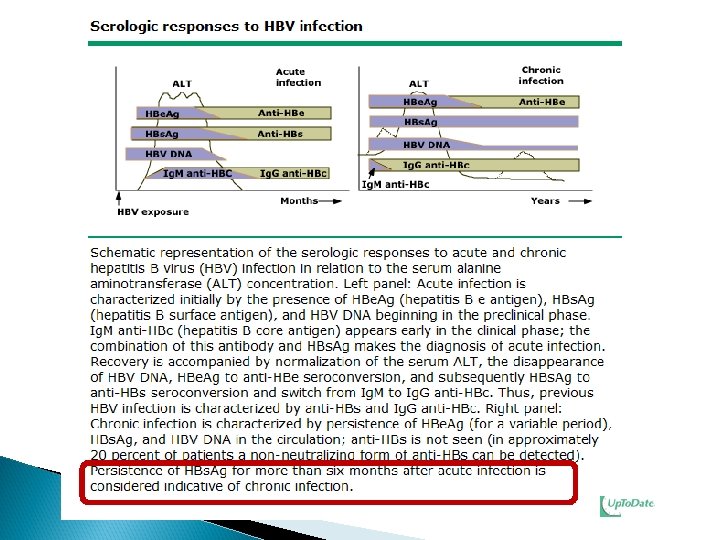

A 28 year old man, referred from Blood Bank because of being HBs. Ag positive. The following HB markers are shown below: ◦ Hepatitis B S antigen…………. . Positive ◦ Anti-Hepa B Core Ig. G ………… Positive ◦ Hep-B e Antigen …………… Negative ◦ Anti- Hepa B e Antigen ………. . Positive ◦ Anti- Hepa B Surface …………. Negative ■ What is your next step? LFT, U/S liver, PCR, HEPATITIS B DNA QUALITATIVE ………… Positive HEPATITIS B DNA QUANTITATIVE ………. . 889796 IU/ML ■ How are you going to deal with patient? Measure for Family Contacts, screen and vaccinate the negative ones Referral to hepatologist, No blood donation

A 35 year old man came to the clinic for screening, as one member in his family is HBV positive. The following HB markers are shown below: ◦ Hepatitis B S antigen ……. . ◦ Anti-Hepa B Core Ig. G …… Negative Positive ◦ Hep B e Antigen …………. Negative ◦ Anti- Hepa B e Antigen … Negative ◦ Anti- Hepa B Surface …… Positive ● What is your diagnosis? Immune post exposure to HB virus ● How are you going to deal with patient? • Reassurance, No further actions could be taken

A 23 -year-medical student came to the clinic for screening. The following HB markers are shown below: ◦ Hepatitis B S antigen ……. . ◦ Anti-Hepa B Core Ig. G …… Negative ◦ Hep B e Antigen …………. Negative ◦ Anti- Hepa B e Antigen … Negative ◦ Anti- Hepa B Surface …… Positive ● What is your diagnosis? Immune post Vaccination

A 32 -year old man presents to your clinic for routine check up. The following viral markers are shown below: ◦ ◦ ◦ Hepatitis B S antigen …… Negative Anti-Hepa B Core lg. G …… Positive Hep- B e Antigen ………. . Negative Anti- Hepa B e Antigen … Negative Anti-Hepa B Surface … Negative Interpret the results. H/O chronic exposure to HB virus see Explanations /Options in next slide

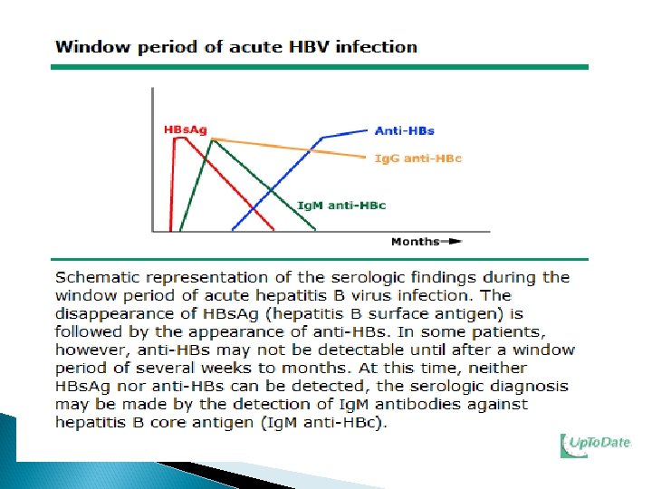
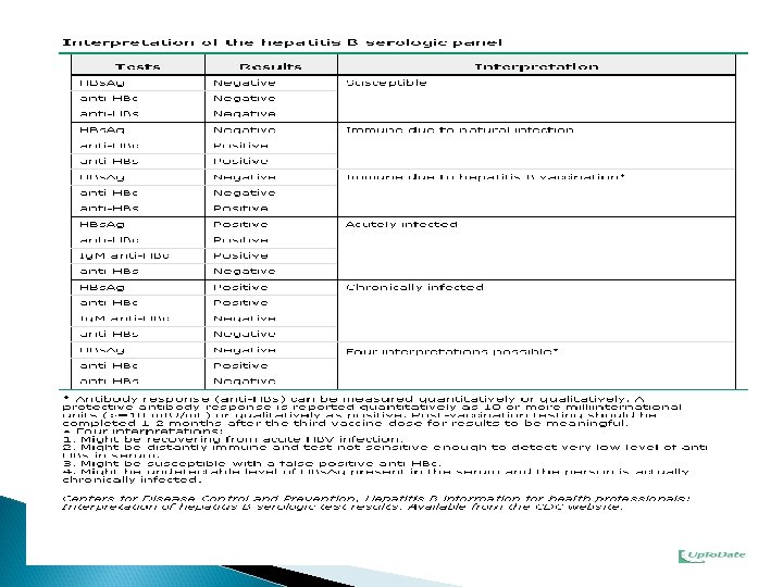
Cont. anti-HBc positive 1 - May be recovering from acute HBV infection ( window period ) 2 - May be distantly immune and test is not sensitive enough to detect very low level of anti-HBs in serum. 3 - May be undetectable level of HBs. Ag present in the serum and the person is actually a carrier. 4 - May be a false positive anti-HBc.

Cont. A 32 -year old man presents to your clinic for routine check up. HEPATITIS B DNA QUALITATIVE Positive HEPATITIS B DNA QUANTITATIVE <20 IU/ML Actions: Measures to Contacts No blood donation Not candidate for treatment by e. g. Interferon

A 26 -year-old female came for premarital check up. The following hepatitis B markers are shown: ◦ ◦ ◦ Hepatitis B S antigen……. Anti-Hepa B Core lg. G……. Hep- B e Antigen ……. Anti- Hepa B e Antigen … Anti-Hepa B Surface……. Positive Negative HEPATITIS B DNA QUALITATIVE Positive HEPATITIS B DNA QUANTITATIVE >110 million IU/ML Total bilirubin …………………. 15 Albumin …………… 39 Alkaline phosphatase ………… 225 Alanine aminotransferase …… 960 Aspartate aminotransferase. . 296 G. G. Transferase ………………. 235 (3 - 17 umol/L) (35 -50 g/L) (50 -136 u/L) (20 -65 u/L) (10 -31 u/L) (5 -55 u/L) ◦ What is your diagnosis and What actions are you going to do? ◦ Chronic viral Hepatitis with active replication and highly infectious (e antigen is positive)

Cont. A 26 -year-old female came for premarital check up. After one and half year of treatment. Result Unit Range # Test Serum - SAMPLE: 1 Positive 0 31 IU/ML 1 HEPATITISBDNAQUALITATIVE 2 HEPATITISBDNAQUANTITATIVE # Test Serum - SAMPLE: 1 1 Urea 2 Serum. Creatinine 3 Sodium 4 Potassium 5 Chloride 6 Carbon. Dioxide 7 Total. Bilirubin 8 Total. Proteins 9 Albumin 10 Alkaline. Phosphatase 11 Alanine. Aminotransferase 12 Aspartate. Aminotransfer. 13 Calcium 14 Inorganic. Phosphorus 15 Albumin 16 Alkaline. Phosphatase 17 Corrected. Calcium Result 4. 6 75 138 4. 4 102 29. 2 10 74 42 94 52 27 2. 26 1. 15 42 94 2. 2 Unit mmol/L umol/L mmol/L umol/L g/L U/L U/L mmol/L g/L U/L mml/L - - Range 2. 5 62 135 3. 5 98 22 3 60 30 50 20 12 2. 1 0. 87 30 50 2. 1 - 6. 4 115 145 5. 1 107 32 17 80 50 136 65 37 2. 55 1. 45 50 136 2. 55

- Slides: 89