Development of the Nervous System Matt Velkey matt


- Slides: 14

Development of the Nervous System Matt Velkey matt. velkey@duke. edu

Neural Induction: In addition to patterning the forming mesoderm, the primitive node also sets up the neural plate • • Ectoderm exposed to BMP-4 (from endoderm and mesoderm below), develops into skin However, the node secretes BMP-4 antagonists: (e. g. noggin, chordin, & follistatin) that allow a region of the ectoderm to develop into nerve tissue.

Neurulation: folding of the neural plate 1. Median hinge point forms (probably due to signaling from notochord) –columnar cells adopt triangular morphology (apical actin constriction, like a purse string) 2. Lateral hinge point forms by a similar mechanism (probably due to signaling from nearby mesoderm). 3. As neural folds close, neural crest delaminates and migrates away (more on that later…) 4. Closure happens first in middle of the tube and then zips rostrally and caudally.

Neurulation: folding and closure of the neural plate • Folding and closure of the neural tube occurs first in the cervical region. • The neural tube then “zips” up toward the head and toward the tail, leaving two openings which are the anterior and posterior neuropores. • The anterior neuropore closes around day 25. • The posterior neuropore closes around day 28.

Major divisions of the developing brain: 3 parts, then 5 parts • 3 parts: Prosen-, Mesen-, and Rhomencephalon • 5 parts: Prosencephalon divides into telen- and diencephalon, Rhombencephalon divides into met- and myelencephalon

Major divisions of the developing brain: 3 parts, then 5 parts

The developing neural tube is divided into 4 plates • Roof plate: signaling center (BMPs and Wnts) • Alar plate: sensory • Basal plate: motor • Floor plate: signaling center (Shh)

Early signaling establishes the neural crest, alar plate, and basal plate • • • Neural crest established in the intermediate zone between Shh from the notochord and BMPs from the skin After neural tube closure, BMPs from skin induce expression of BMPs in roof plate, which largely patterns the alar plate. Shh from the notochord induces expression of Shh in floor plate, which largely patterns the basal plate.

Development of the myelencephalon • Alar plate develops into 4 sensory tracts: – Olivary nucleus (cerebellar input) – Somatic afferent (general sensation from the face, via CN V, external ear, auditory meatus, and eardrum via CN IX, X) – Special visceral afferent (taste, CN IX, X) – General visceral afferent (autonomic input, CN IX, X) • Basal plate develops into 3 motor tracts: – General visceral efferent (autonomic output, CN IX, X) – Special visceral efferent (innervation of pharyngeal arch muscles of the larynx & pharynx, CN IX, X, XI) – aka “branchiomotor” – Somatic efferent (innervation of skeletal muscles of the tongue, CN XII) (covered by ependymal cells that produce CSF) Note: the final position of the SVE column is generally ventral (and slightly lateral) to the GSE column

Development of the metencephalon • Rhombic lip develops into cerebellum • Alar plate develops into 4 sensory tracts: – – Pontine nuclei (cerebellar input) Somatic afferent (general sensation from the face, tongue, and extraocular muscles, CN V, VII) Special visceral afferent (taste, CN VII) General visceral afferent (autonomic input from soft palate & pharynx, CN VII) • Basal plate develops into 3 motor tracts: – General visceral efferent (autonomic output to salivary and lacrimal glands, CN VII) – Special visceral efferent (innervation of muscles of the face, via CN VII, and of mastication, via CN V) – Somatic efferent (innervation of lateral rectus muscle of the eye, via CN VI) Note: the final position of the SVE column is generally ventral (and slightly lateral) to the GSE column

Anatomy of the developing cerebellum

Development of the mesencephalon • Alar plate generates superior and inferior colliculi – Inferior colliculus: auditory relay – Superior colliculus: visual relay • Basal plate generates 4 motor tracts: – – Somatic efferent (motor output to extraocular muscles, CN III, IV) Visceral efferent (motor output to ciliary ganglion of the eye, CN III) Red nucleus (primarily a cerebellar relay in humans, but some motor to upper limb flexors) Substantia nigra (dopaminergic output to the basal ganglia of the telencephalon)

Development of the diencephalon • Epithalamus (habenula, stria medullaris, pineal gland): circadian behaviors, limbic and basal ganglia circuitry • Thalamus: major relay of sensory input to cerebral cortex • Hypothalamus: master regulatory center (autonomic and endocrine) and also limbic system (emotion & behavior) • Hypophysis/infundibulum: posterior pituitary gland, secretes ADH and oxytocin • Optic cup: retina of eye

Development of the telencephalon • Generates cerebral cortex: – Neocortex: 6 -layered cortex of frontal, temporal, parietal, and occipital lobes – Archicortex: 3 -layered cortex of hippocampus and dentate gyrus – Paleocortex: olfactory cortex • Generates basal ganglia: – – Caudate nucleus Globus palladus Putamen Amygdala
 Which neuron is rare
 Sensory input and motor output
 Neuronal processes
 Nervous system and digestive system
 Endocrine system and nervous system
 General mechanism of hormone action
 Adh function
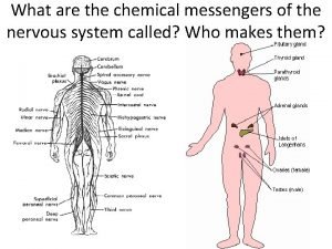
 Chemical messengers of the nervous system
 Segmentation of platyhelminthes
 The nervous system is made up of
 What are the three basic functions of the nervous system
 Learning objectives of nervous system
 Bipolar neuron function
 Stimulus in nervous system
 Nervous system objectives
