Development of the Endocrine Glands DEVELOPMENT OF THE


- Slides: 40
Development of the Endocrine Glands
DEVELOPMENT OF THE PITUITARY GLAND
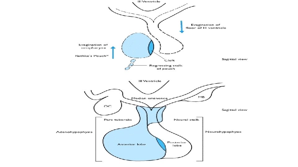
-it develops from: 1 - Rathke’s pouch is an upward ectodermal diverticulum from roof of stomodeum 2 - Infundibulum is a downward ectodermal diverticulum from floor of diencephalon -During 2 nd month Rathke’s pouch come into contact with ant. Surface of infundibulum. - Then, loses connection with stomodeum
-Anterior wall of pouch increases to form pars distalis - An upward extension around infundibulum forms pars tuberalis -Posterior wall of pouch forms pars intermedia - Infundibulum forms Pit. Stalk and pars nervosa
During 3 rd and 4 th months, the cells of pars distalis differentiate into: -Chromophobe cells -Acidophil cells -Basophil cells
Congenital anomalies of the pituitary gland Craniopharyngioma arises from remnants of Rathke’s pouch
Pharyngeal hypophysis due to persistence of small portion of Rathke’s pouch in the wall of the pharynx
DEVELOPMENT OF THE thyroid GLAND
-During 3 rd week a median endodermal thickening appears at junction of ant. 2/3 and post. 1/3 of tongue at the foramen coecum The thickening forms diverticulum grows into the mesenchyme called thyroglossal duct. -The duct elongates and its distal end becomes bilobed to form thyroid gland.
Then, thyroid gland descends in front of hyoid bone and laryngeal cartilages. -Finally, it reaches final position in front of trachea at 7 th week. - Meanwhile, the cord connecting thyroid gland to tongue degenerates
Histological differentiation of thyroid gland - At first thyroid gland consists of solid mass of cells -Later vascular mesenchyme invades them and breaks them into clusters of cells
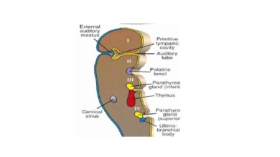
-By 3 rd month colloid starts to accumulate in the center of each cluster to form follicles -Ultimobranchial bodies form parafollicular cells
Congenital Anomalies of the Thyroid Gland
1 - Agenesis of the thyroid gland results in cretinism
2 - Incomplete descent of the thyroid gland Lingual thyroid is the commonest form
3 - Ectopic thyroid tissue: -in the thorax -at bronchi -at the oesophagus
4 - Thyroglossal cyst: -Due to persistence of part of thyroglossal duct. The common site is below hyoid bone
Localization of thyroglossal cysts
5 - Thyroglossal fistula: Due to rupture of thyroglossal cyst
DEVELOPMENT OF THE suprarenal GLAND
Suprarenal gland develops from two components: 1 -Cortex is mesodermal from coelomic epithelium 2 -Medulla is ectodermal from sympathetic ganglion
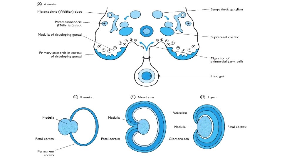
Neural crest cells -During 5 th week mesodermal cells of coelomic epithelium form foetal (primitive) cortex - Neural crest cells differentiate into sympathetic ganglion then migrate and invade the medial aspect of cortex to form suprarenal medulla
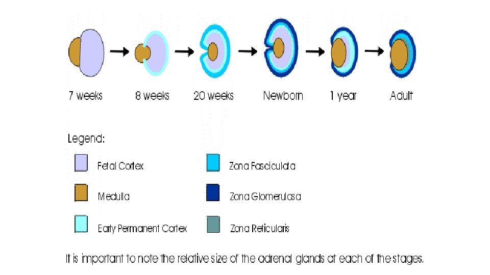
- Figure A shows Foetal cortex and the chromaffin cells migrating to form the medulla- A second series of cells arise from coelomic epithelium, and enclose fetal cortex to form definitive (permanent) cortex - Differentiation of the cortex begins with zona glomerulosa and zona fasciculata which are present at birth - Zona reticularis appears at 3 rd year and it may be a remnant of foetal cortex
Congenital anomalies of the Suprarenal Gland
Phiochromocytoma It is a tumor of chromaffin cells of suprarenal medulla
Development of the Pancreas • It develops from ventral and dorsal buds originating from endodermal lining of duodenum. • Dorsal bud is located short distance above ventral bud. • Ventral bud arises in common with hepatic bud close to junction of foregut with midgut.
• As the duodenum rotates to right and becomes C- shaped the ventral pancreatic bud migrates dorsally. • It lies immediately below and behind dorsal pancreatic bud. • Then, parenchyma and duct system of ventral and dorsal buds fuse together. • Ventral bud forms uncinate process and inferior part of head of pancreas. • Dorsal bud forms the remaining part of pancreas.
• The main pancreatic duct is formed by distal part of dorsal pancreatic duct and the whole ventral pancreatic duct.
• Proximal part of dorsal pancreatic duct is obliterated or persists as accessory pancreatic duct.
• Main pancreatic duct and common bile duct enters duodenum at major papilla. • Accessory pancreatic duct enters it at site of minor papilla.
• Islets of Langerhans are proliferated epithelial cells which become separated from duct system.
Congenital anomalies of the Pancreas • Annular pancreas: • Ventral pancreatic bud is formed of two parts. • Right portion migrate normally. • Left portion migrates in opposite direction • Thus duodenum is surrounded by pancreatic tissue and may be obstructed.
Accessory pancreatic duct: Develops from proximal part of dorsal pancreatic duct.
Accessory pancreatic tissue: May be found in mucosa of stomach, gall bladder and spleen.