Development of Spinal Cord And Vertebral Column 2

Development of Spinal Cord And Vertebral Column ﴾ ﴿ ﺍ ﺍ ﺍﻹﻧﺴﺎﻥ ﺍ ﻳﺍ ﻳﺍ : 2]. ]ﺍﻹﻧﺴﺎﻥ Embryology 436 MEDICINE KING SAUD UNIVERSITY Important Dr. notes Explanation - We recommend you to study anatomy of spinal cord lecture
 (middle) (inner)")
The Three Germ Layers: 1 - Ectoderm 2 -Mesoderm 3 -Endoderm (outer) (middle) (inner) - Neural Tube is a derivative of the ectoderm. It is dorsal to notochord. Amniotic cavity - Notochord stimulates neural tube formation which in turn stimulates development of the vertebral column. Yolk sac Development of Neural Tube : - Ectodermal cells dorsal to notochord thicken to form the neural plate*. A longitudinal groove, neural groove, develops in the neural plate. - The margins of the neural plate (neural folds) approach to each other and fuse to form the neural tube. * That mean the notochord stimulates the ectoderm to increase in size and number to form neural tube. Neural plate embryo

Development of the Spinal Cord: The spinal cord develops from the caudal 2/3 of the neural tube. While the superior 1/3 will form the brain. ﺃﻤﺎ ﺍﻟﺜﻠﺜﻴﻦ ﺍﻷﺨﻴﺮﺓ ﺇﻟﻲ ﻓﻲ ﺍﻟﺬﻳﻞ ﺣﺘﻜﻮﻥ brain *ﺍﻟﺜﻠﺚ ﺍﻷﻮﻝ ﻣﻦ ﺍﻟﻨﻴﻮﺭﺍﻝ ﺗﻴﻮﺏ ﺣﻴﻜﻮﻥ ﺍﻝ spinal cord ﺃﻮﻝ ﻃﺒﻘﺔ ﻣﺎﻟﻨﺎ ﻓﻴﻬﺎ ﻷﻨﻪ ﻓﻴﻬﺎ ﺧﻼﻳﺎ ﻏﻴﺮ ﻣﻌﺮﻭﻓﺔ ﺑﺲ ، ﻃﺒﻘﺎﺕ 3 *ﺍﻟﺨﻼﻳﺎ ﻓﻲ ﺍﻟﻨﻴﻮﺭﺍﻝ ﺗﻴﻮﺏ ﺗﺘﻘﺴﻢ ﺇﻟﻰ ﺍﻟﻄﺒﻘﺘﻴﻦ ﺍﻟﺜﺎﻧﻴﺔ ﻫﻲ ﺍﻟﻤﻬﻤﺔ The cells of the neural tube arranged in three layers: 1 - An inner ventricular zone of undifferentiated cells. 2 - A middle mantle zone of cell bodies of neurons (future grey matter). 3 - An outer marginal zone of nerve fibers or axons of neurons (future white matter). Mantle Layer of Spinal Cord: Neurons of mantle layer differentiate into: 1 -A dorsal alar plate (future dorsal horn) containing sensory neurons - The 2 areas are separated by a longitudinal groove (sulcus limitans). 2 - A ventral basal plate (future ventral horn) containing motor neurons ” ﺇﻟﻲ ﺑﺎﻟﻠﻮﻥ ﺍﻷﺤﻤﺮ mantle” *ﺃﻮﻝ ﺷﻴﺀ ﻧﺘﻜﻠﻢ ﻋﻦ ﺍﻟﻄﺒﻘﺔ ﺍﻟﺜﺎﻧﻴﺔ ﻫﺬﻱ ﺍﻟﻄﺒﻘﺔ ﻫﻲ ﺇﻟﻲ ﻣﺴﺘﻘﺒﻼ ﺣﺘﻜﻮﻥ ﻗﺮﻱ ﻣﺎﺗﺮ ﻟﺬﻟﻚ ﺗﻨﻘﺴﻢ ، ﻭﺍﻷﺰﺭﻕ 2 - basal. . (Dorsal horn ) ﻭﻫﻮﺍ ﺣﻴﻜﻮﻥ 1 - alar plate ﻟﻘﺴﻤﻴﻦ ﻭ ﺍﻟﻤﻨﻄﻘﻪ ﺍﻟﻴﻤﻴﻦ ﺍﻟﻤﻔﺼﻮﻟﺔ. . (Ventral horn ) ﻭﻫﻮ ﺣﻴﻜﻮﻥ plate sulcus limitans ﻋﻦ ﺍﻟﻴﺴﺎﺭ ﺑـ

Count. ﺣﻴﺼﻴﺮ ﻓﻲ ﺗﻜﺎﺛﺮ ﻭﺗﺮﺍﻛﻢ ﻟﻠﻄﺒﻘﺔ ﻋﺸﺎﻥ ﺍﻟﺠﻬﺔ ﺍﻟﻴﻤﻴﻦ ﺗﻘﺮﺏ ﻣﻦ ﺍﻟﺠﻬﺔ ﺍﻟﻴﺴﺎﺭ ﻭﻳﻜﺘﻤﻞ ﺍﻟﻘﺮﻱ ، mantle *ﺑﺮﺿﻮ ﻧﺘﻜﻠﻢ ﻋﻦ ﺍﻟﻄﺒﻘﺔ ﺍﻟﺜﺎﻧﻴﺔ 2 - dorsal median septum 3 - ventral ﺳﻴﻨﺘﺮﺍﻝ ﻛﻨﺎﻝ ﺇﻟﻲ ﻓﻲ ﺍﻟﻮﺳﻂ : 1 - ﺃﺸﻴﺎﺀ 3 ﻭﺑﺴﺒﺐ ﻫﺬﺍ ﺍﻟﺘﻘﺎﺭﺏ ﺣﺘﺘﻜﻮﻥ ، ﻣﺎﺗﺮ ﻣﺴﺘﻘﺒﻼ median fissure Proliferation and bulging of both alar and basal plates result in: 1 -Formation of dorsal median septum. 2 Formation of ventral median fissure. 3 -Narrowing of the lumen of the neural tube to form a small central canal. ، ﺣﺘﻜﺒﺮ ﻭﻳﺰﻳﺪ ﺣﺠﻤﻬﺎ ﺑﺴﺒﺐ ﺯﻳﺎﺩﺓ ﺍﻟﻔﺎﻳﺒﺮﺯ ، « ﺇﻟﻲ ﺣﺘﻜﻮﻥ ﺍﻟﻮﺍﻳﺖ ﻣﺎﺗﺮ ﻓﻲ ﺍﻟﻤﺴﺘﻘﺒﻞ marginal» *ﻧﺠﻲ ﻟﻠﻄﺒﻘﺔ ﺍﻟﺜﺎﻟﺜﺔ ﻓﻴﻨﺘﺮﺍﻝ ﻭﺩﻭﺭﺳﺎﻝ ﻭﻻﺗﻴﺮﺍﻝ : ﺃﻌﻤﺪﺓ ﺃﻮ ﻓﺎﻧﻴﻜﻴﻮﻻﻱ 3 ﻭﺗﻨﻘﺴﻢ ﺇﻟﻰ -The marginal layer (future white matter) increases in size due to addition of ascending, descending and intersegmental nerve fibers. - marginal layer (future white matter) is divided into: 1 - dorsal funiculi 2 - lateral funicli 3 - ventral funiculi (white column). Dorsal funiculus Lateral funiculus ﻭﺍﻟﻤﻮﺗﺮ ﻓﺎﻳﺒﺮ ، *ﺍﻟﻤﺎﻳﻠﻨﻴﺸﻦ ﻳﺒﺪﺃ ﻣﻦ ﺍﻟﺸﻬﺮ ﺍﻟﺮﺍﺑﻊ ﻟﻠﺠﻨﻴﻦ ﻭﻳﻜﻤﻞ ﺇﻟﻰ ﺃﻮﻝ ﺳﻨﺔ ﺑﻌﺪ ﺍﻟﻮﻻﺩﺓ ﻳﺼﻴﺮ ﻟﻬﺎ ﻣﺎﻳﻠﻨﻴﺸﻦ ﻗﺒﻞ ﺍﻟﺴﻴﻨﺴﻮﺭﻱ ﻓﺎﻳﺒﺮ -Myelination of nerve fibers starts at 4 th month and continues during the 1 st postnatal year. Ventral funiculus - Motor fibers myelinate before sensory fibers: (So, After a nerve injury, both motor and sensory axons have the ability to regenerate and, given a proper pathway). Only in girl’s slide

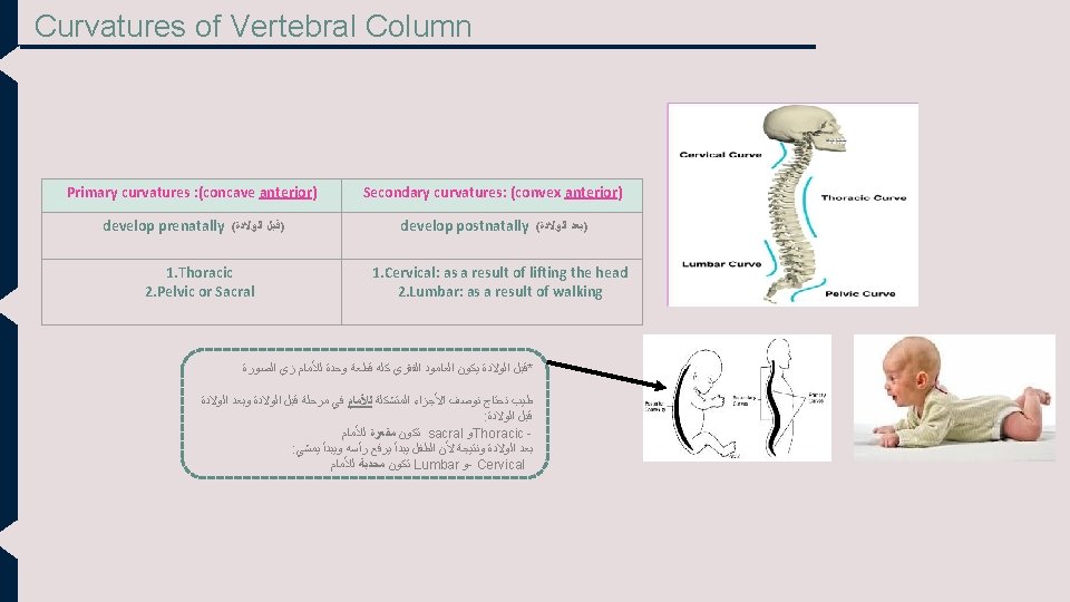
Development Of Spinal Meninges - This slide is very important ♥ Meninges : these are 3 membranes covering the neural tube: 1 - dura mater 2 - arachnoid mater 3 - pia mater. Dura mater (Outer thick layer) Arachnoid mater (Middle layer) MESODERMAL in origin Pia mater (Inner thin layer) ECTODERMAL in origin - A cavity appears between the arachnoid and the pia mater is (subarachnoid space) and becomes filled with cerebrospinal fluid (CSF). Positional Changes of Spinal Cord: * very important* - Initially, (at week 8) the spinal cord occupies the whole length of the vertebral canal. -As a result a faster growth of vertebral column, the caudal end of spinal cord (conus medullaris) shifts gradually to a higher level. -The spinal cord in Adult between L 1 and L 2 and new born in L 3*. *because in new borns their vertebral column is shorter ﺑﻌﺪ ﻛﺪﺍ ﻓﻘﺮﺍﺕ ﺍﻟﻈﻬﺮ ﺗﻨﻤﻮ ﺑﺴﺮﻋﺔ ﻭﺗﺼﻴﺮ ، *ﻓﻲ ﺑﺪﺍﻳﺔ ﺗﻜﻮﻥ ﺍﻟﺠﻨﻴﻦ ﻓﻘﺮﺍﺕ ﺍﻟﻈﻬﺮ ﺗﻜﻮﻥ ﻋﻠﻰ ﻧﻔﺲ ﻣﺴﺘﻮﻯ ﺍﻟﺴﺒﺎﻳﻨﺎﻝ ﻛﻮﺭﺩ » ﺃﻄﻮﻝ ﻣﻦ ﺍﻟﺴﺒﺎﻳﻨﺎﻝ ﻛﻮﺭﺩ ﻭﺧﻼﺹ ﻣﺎ ﻳﺼﻴﺮﻭ ﻓﻲ ﻧﻔﺲ ﺍﻟﻤﺴﺘﻮﻯ » ﺍﻟﻔﻘﺮﺍﺕ ﺗﻜﻤﻞ ﻟﺘﺤﺖ ﺃﻤﺎ ﻧﻬﺎﻳﺔ ﺍﻟﺴﺒﺎﻳﻨﺎﻝ ﻛﻮﺭﺩ ﺗﻄﻠﻊ ﻟﻔﻮﻕ

Development of the Vertebral Column Recall: - Notochord stimulates neural tube formation which in turn stimulates development of the vertebral column. -The vertebral column develops from the ventromedial parts (sclerotomes) of the somites. *ﺑﻌﺪ ﻣﺎ ﻋﺮﻓﻨﺎ ﻛﻴﻒ ﺃﺘﻜﻮﻥ ﺍﻟﺴﺒﺎﻳﻨﺎﻝ ﻛﻮﺭﺩ ﻧﺠﻲ ﻟﻌﻈﺎﻡ ﺟﻴﺮﻡ ﻻﻳﺮﺯ 3 ﻧﺮﺟﻊ ﻟﻞ ، « ﻓﻘﺮﺍﺕ ﺍﻟﻈﻬﺮ » ﺍﻟﻌﺎﻣﻮﺩ ﺍﻟﻔﻘﺮﻱ ﺍﻟﻄﺒﻘﺔ ﺇﻟﻲ ﻓﻲ ﺍﻟﻮﺳﻂ ﻭﻫﻲ mesoderm ﻭﻧﺘﻜﻠﻢ ﻋﻨـ ﻫﺬﻱ ﺍﻟﻄﺒﻘﺔ ،notochord ﻣﻘﺴﻮﻣﺔ ﻟﻴﻤﻴﻦ ﻭﻳﺴﺎﺭ ﺑﺴﺒﺐ ﺍﻝ ﻣﻨﺎﻃﻖ ﻓﻲ ﻛﻞ ﺟﻬﺔ ﻭﻣﻦ ﺍﻟﻤﻨﻄﻘﺔ ﺍﻷﻮﻟﻰ 3 ﻣﺘﻘﺴﻤﺔ ﺇﻟﻰ ﺃﺸﻴﺎﺀ ﺩﻳﺮﻣﺎﺗﻮﻡ ﻭﻣﺎﻳﻮﺗﻮﻡ 3 ﻓﻴﻬﺎ Paraxial mesoderm ﺗﻨﺸﺄ ﻋﻈﺎﻡ ﻓﻘﺮﺍﺕ Sclerotome ﻭﻣﻦ ﺍﻝ ، ﻭﺳﻜﻠﻴﺮﻭﺗﻮﻡ ﺍﻟﻈﻬﺮ ﺃﻮ ﺑﺎﻟﻤﻌﻨﻰ ﺍﻷﺼﺢ ﺍﻟﻌﺎﻣﻮﺩ ﺍﻟﻔﻘﺮﻱ - The somites develop from the para-axial mesoderm. Intraembryonic Mesoderm - Located between Ectoderm and Endoderm EXCEPT in the central axis of embryo where notochord is found. -Differentiates into 3 parts: 1. Paraxial mesoderm 2. Intermediate mesoderm 3. Lateral mesoderm notochord Paraxial mesoderm divides into segments called “somites” each somtie divides into 3 parts : 1 NT 2 1. Dermatome 2. Myotome 3. Sclerotome N 3 GUT somites Extra Picture

DEVELOPMENT OF VERTEBRA Only in male’s slide Notochord Neural tube Sclerotome 1 - Sclerotome around neural tube: forms vertebral (neural) arch. 1 2 2 - Sclerotome around notochord: forms body of vertebra. 3 3 3 -Sclerotome in body wall near to neural tube and notochord : forms costal process (gives ribs in thoracic region only ) 1 - Loosely arranged cells 2 - densely packed cells

Formation of Body of Vertebra ، *ﻓﻲ ﺍﻷﺴﺒﻮﻉ ﺍﻟﺮﺍﺑﻊ ﺍﻟﺴﻜﻠﻴﺮﻭﺗﻮﻡ ﺗﻨﻘﺴﻢ ﻟﺠﺰﺋﻴﻴﻦ ، ﺟﺰﺀ ﻭﺭﺩﻱ ﻭﻫﻮ ﺍﻟﺮﺃﺲ ﻭﺟﺰﺀ ﻓﻮﺷﻲ ﻭﻫﻮ ﺍﻟﺬﻳﻞ ﺑﻌﺪ ﻛﺪﺍ ﻛﻞ ﺟﺰﺀ ﻓﻮﺷﻲ ﺣﻴﻨﺪﻣﺞ ﻣﻊ ﺍﻟﺠﺰﺀ ﺍﻟﻮﺭﺩﻱ ﺇﻟﻲ ﺗﺤﺘﻪ ﻭﺑﻜﺪﺍ ﻳﺘﻜﻮﻥ ﺟﺴﻢ ﺍﻟﻔﻘﺮﺓ ﺍﻟﻮﺍﺣﺪﺓ ﻭﺍﺳﻤﻪ Centrum -At 4 th week, each sclerotome becomes subvidided into two parts : -A cranial part, consisting of loosely arranged cells -A caudal part, of more condensed tissue. -The caudal part of each somite fuses with the cranial part of the consecutive somite, around the notochord to form the body of the vertebra, called the centrum (body primordium). - Thus each centrum develops from 2 adjacent sclerotomes The fused sclerotomes grow dorsally around the neural tube and form the vertebral (neural) arch. Ventrolaterally, costal processes develop that give rise to ribs in thoracic region. Pictures only in girl’s slide centrum -Fate of Notochord : - In the region of the bodies of vertebrae: notochord degenerates. -Between bodies of vertebrae: It forms the central part, ’nucleus pulposus’ of the intervertebral discs - Annulus fibrosus part of the intervertebral discs is formed by the mesoderm surrounding the notochord. *ﺍﻟﺴﻜﻠﻴﺮﻭﺗﻮﻡ ﻟﻤﻦ ﺗﻠﺘﺤﻢ ﻋﺸﺎﻥ ﺗﻜﻥ ﺍﻟﻔﻘﺮﺓ ﻛﺎﻣﻠﺔ ﻳﻜﻮﻥ ﺇﻟﺘﺤﺎﻣﻬﺎ ﺣﻮﻝ ﺍﻝ ( ﻭﺑﻜﺪﺍ vertebral neural arch) « ﻭﻧﻘﻄﺔ ﺍﻹﻟﺘﺤﺎﻡ ﺍﺳﻤﻬﺎ neural tube » ﻳﻜﻮﻥ ﺑﻴﻦ ﺍﻟﻔﻘﺮﺍﺕ ﻓﺮﺍﻍ ﻟﻠﺴﺒﺎﻳﻨﺎﻝ ﻛﻮﺭﺩ - The fused sclerotomes grow dorsally around the neural tube and form the vertebral (neural) arch. - ventrolaterally, costal processes develop that give rise of ribs in thoracic region. ﺍﻟﻨﻮﺗﻮﻛﻮﺭﺩ ﺇﻟﻲ ﻭﺳﻂ ﺟﺴﻢ ﺍﻟﻔﻘﺮﺓ ﻳﺨﺘﻔﻲ ﺃﻤﺎ : *ﻣﺼﻴﺮ ﺍﻟﻨﻮﺗﻮﻛﻮﺭﺩ ﺇﻟﻲ ﺑﻴﻦ ﻛﻞ ﻓﻘﺮﺓ ﻭﻓﻘﺮﺓ ﺣﻴﻜﻮﻥ ﺟﺰﺀ ﻣﻦ nucleus ﻋﻦ ﻃﺮﻳﻖ intervertebral discs ﺍﻝ pulposus)) ﺇﻟﻲ ﻫﻮﺍ intervertebral discs ﺍﻟﺠﺰﺀ ﺍﻟﺘﺎﻧﻲ ﻣﻦ ﺍﻝ (ﺃﺘﻜﻮﻥ ﻣﻦ ﺍﻟﻤﻴﺰﻭﺩﻳﺮﻡ ﺍﻟﻲ ﺣﻮﻝ Annulus fibrosus) !!!! ﺍﻟﻨﻮﺗﻮﻛﻮﺭﺩ ﻣﻮ ﻣﻦ ﺍﻟﻨﻮﺗﻮﻛﻮﺭﺩ ﻧﻔﺴﻪ

Vertebral Development Mesenchymal Stage Chondrification Stage Primary Ossification Stage Dr. sanaa Notes This picture represents the changes that occurs into 2 stages : chondrotification stage and ossification stage. • • • The chondrotification centers appeare at 6 th week (cartilage): (Pic. B) And by the (at the end of 8 th week)the 3 primary ossification centers appears (bone): (Pic. c) And The 5 secondary ossification centers appear at puberty. Fusion of bony halves of vertebral arch occurs at 3 -5 years (pic. D). and Fusion of centrum with vertebral arch occurs at 3 -6 years (pic. D). Stage of Fusion *Primary ossification: before birth *second ossification: after birth Stage of Secondary Ossification - All centers unite around 25 years *ﻣﺮﺍﺣﻞ ﺗﺤﻮﻝ ﻧﺴﻴﺞ ﺍﻟﻤﺴﺰﻳﻨﻜﺎﻳﻤﻞ ﺇﺍﻟﻰ ﻏﻀﺮﻭﻑ ﺛﻢ ﺇﻟﻰ ﻣﺮﺣﻠﺔ ﺃﻮﻟﻴﺔ ﻟﻠﺘﻌﻈﻢ ﺛﻢ . ﺇﻟﻰ ﻣﺮﺣﻠﺔ ﺛﺎﻧﻮﻳﺔ ﻟﻠﺘﻌﻈﻢ SO, Ossification starts at the end of embryonic period ( end of 8 th week) and ends at adult age 25 years. Team 435 ♥. .

Spina Bifida -Cause: Failure of fusion of the halves of vertebral arches -Incidence: 0. 04 -0. 15% -Sex: more frequent in females -Types: vertebral neural arch *ﺗﺤﺪﺙ ﻫﺬﻩ ﺍﻟﺘﺸﻮﻫﺎﺕ ﺑﺴﺒﺐ ﺍﻧﻪ ﺍﻝ ﻣﻜﺸﻮﻑ ﻭﻣﺎ ﻋﺎﺩ ﻓﻲ spinal cord ﻓﺸﻞ ﻓﻲ ﺍﻻﻟﺘﻔﺎﻑ ﻭﺍﻻﻟﺘﺤﺎﻡ ﻓﺎﻟـ ﺷﻴﺀ ﻳﻐﻄﻴﻪ * ﻭﻫﺬﻩ ﺍﻟﺘﺸﻮﻫﺎﺕ ﻧﻮﻋﻴﻦ 1 - Occulta. . ( )ﻣﻘﻔﻞ 2 -Cystica ( )ﻣﻔﺘﻮﺡ 1. Spina bifida occulta (20%) • • • The closed type Only one vertebra is affected No clinical symptoms Skin overlying it is intact. Sometimes covered by a tuft of hair. Usually does not involve underlying neural tissue. ﻣﻌﻨﻰ ﻛﺬﺍ ﺍﻥ ﺍﻟﻌﻤﻮﺩ ﺍﻟﻔﻘﺮﻱ ﺗﻀﺮﺭ ﻟﻜﻦ ﺍﻟﺴﺎﻳﻨﺎﻝ ﻛﻮﺭﺩ ﺍﻭ ﺃﻲ ﻧﻴﻮﺭﺍﻝ ﺗﻴﺸﻮ ﺳﻠﻴﻤﺔ ﻣﺎﺗﺄﺜﺮﻭﺍ ﻭﺻﺎﺭﻟﻬﻢ ﺿﺮﺭ 2. Spin bifida cystica (80%) • The open type • Neurological symptoms are present • • • A portion of the nerves and the spinal cord are exposed outside the body Cystica is the most severe and complex form of spina bifida It usually involves serious or fatal neurological problems Subdivided into: Meningo = meninges , myelo =spinal cord , ocoele = sac contains fluid or cysist , schisis= opening 1. Spina bifida with meningocoele. 2. Spina bifida with meningomyelocoele. 3. Spina bifida with myeloschisis. ﻣﺎﻟﻜﻢ ﺣﺠﺔ ﻋﺸﺎﻥ ﻣﺤﺪ ﻳﺤﻔﻆ ﺷﻜﻞ : p ﺍﻟﻜﻠﻤﺔ ﺑﺲ

Count. ☻√ ﺍﻷﻨﻮﺍﻉ ﺍﻟﺠﺎﻳﺔ ﺍﺳﺘﻨﺘﺠﻮﺍ ﺗﻌﺮﻳﻔﻬﺎ ﻣﻦ ﺍﺳﻤﻬﺎ - 1. Spina bifida with meningocoele protrusion of sac containing meninges and cerebrospinal fluid. *ﻻ ﻳﻮﺟﺪ ﻋﻈﺎﻡ ﺍﻟﻔﻘﺮﺓ ﺗﻐﻄﻲ ﺍﻟﺴﺒﺎﻳﻨﺎﻝ ﻛﻮﺭﺩ ﻭﻟﻜﻦ ﻓﻲ ﺯﻱ ﻛﻴﺲ ﺩﺍﺧﻠﻪ ﻃﺒﻘﺎﺕ ﺍﻟﻤﻨﻨﺠﻴﺰ CSF ﻭﺍﻝ 2. Spina bifida with meningomyelocoele protrusion of sac containing meninges with spinal cord and/or nerve roots. spinal cord and/or *ﻧﻔﺲ ﺇﻟﻲ ﻗﺒﻞ ﺑﺲ ﺯﻳﺎﺩﺓ ﻓﻴﻪ nerve roots 3. Spina bifida with myeloschisis spinal cord is open due to failure of fusion of neural folds. *ﻫﺬﺍ ﺍﻟﻨﻮﻉ ﻣﺮﻩ ﺳﻴﺀ !! ﻣﺎﻓﻲ ﻭﻻ ﺷﻴﺀ ﻳﻐﻄﻲ ﺍﻟﺴﺒﺎﻳﻨﺎﻝ ﻛﻮﺭﺩ Very rare

SUMMARY Structure Origin Neural tube Ectoderm. Spinal cord Caudal 23 of the neural tube. Grey matter Mantle layer. White matter Marginal layer. arachnoid mater and pia mater Ectoderm. Dura matter mesoderm Vertebral column ventromedial parts (sclerotomes) of the somites. Somaits Paraaxial mesoderm. nucleus pulposus Notochord between the bodies of vertebrae. Annulus fibrosus Mesoderm.
 Three germ cell layers. 4 th week")
SUMMARY Time Changes 3 rd week (early) Three germ cell layers. 4 th week Each sclerotome becomes subdivided into cranial and caudal part. 6 th week Chondrification centers appear. End of 8 th week 3 primary ossification centers appear. 4 th month Starting of myelination of nerve fibers. During 1 st postnatal year Continuation of the myelination of nerve fibers. 3 -5 years Fusion occurs (fusion of 2 vertebral arches) 4 -6 years Fusion of centrum with vertebral arch. At puberty 5 secondary ossification centers appear. 25 years All centers unite. During development the end of spinal cord shifts its position: at 24 weeks (level of S 1), at birth (level of L 3), adult position (level of L 1 -L 2).

MCQ’S
. Embryology team")
References ANY SUGGESTION OR ISSUE • • Dr. slides (male and female). Embryology team 435. USEFUL VIDEOS https: //www. youtube. com/watch? v=4 Swn 8_Jnlss&t=60 s https: //www. youtube. com/watch? v=E-bq. LIQCbq. E Embryology @Embryology 436@gmail. com Editing file Your Suggestion here

TEAM MEMBERS § TEAM LEADERS : ▪ Yazeed Al-mutairi Nehal Beyari. • Abdulrahman Al. Omrani • Saqr Altamimi ▪ EDITING By: MUHANNED ALZAHRANI BOYS : GIRLS : • Razan Alotaibi • Thikrayat Omar • Do’aa Walid • Ohood Abdullah • Nouf Aloqili
- Slides: 18