DEVELOPMENT OF SKELETAL MUSCLES Paraxial mesoderm Gives rise


")


")


- Slides: 49
DEVELOPMENT OF SKELETAL MUSCLES
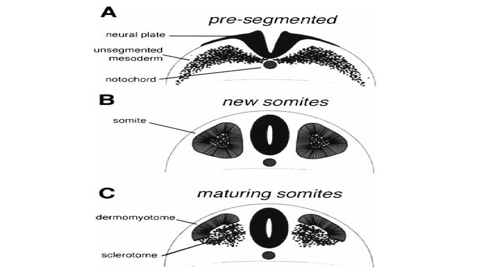
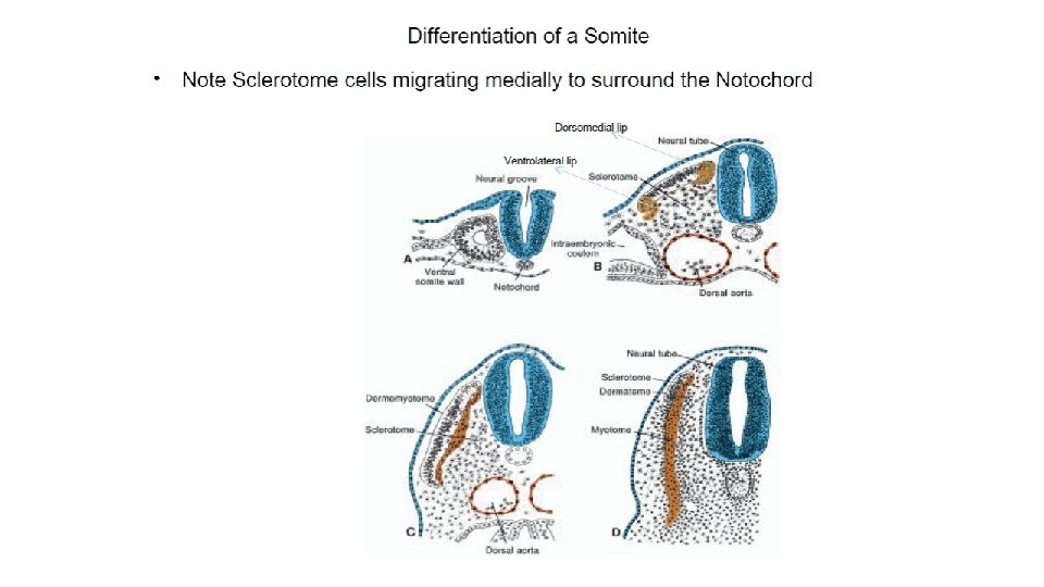
Paraxial mesoderm • Gives rise to somitomeres in the head region • Gives rise to somites in postcranial region Somite differentiate into Scelerotome (ventromedially) forms bones of axial sceleton Dermomyotome (dorsolaterally) dermatomte (dermis) myotome (skeletal muscles)
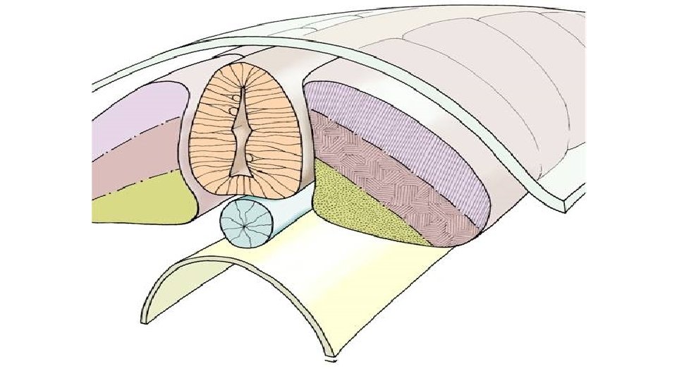
Differentiation of the intraembryonic mesoderm (transverse section)
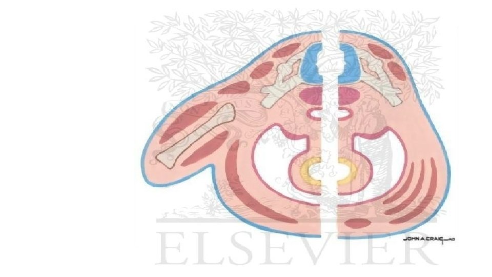
By the end of the fifth week , each myotome is divided into : 1 - Epimere the extensor muscle column of the back. 2 - Hypomere in the thorax inter costal muscles in the abdomen muscles of the anterior abdominal wall. .
From the ventral tip of the hypomers a ventral longitudinal column arises: -In the cervical region infrahyoid muscles. -In the abdominal region rectus abdominis -In the thorax except sternocostalis muscle. -
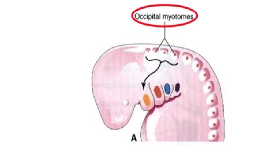
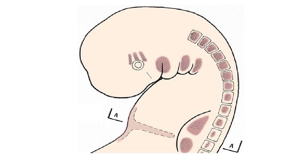
Head musculature -Muscles of the tongue : 4 pairs of occipital somites but the first pair soon disappears. -Muscles of the eye : From the 3 preotic myotome. -The remaning muscles of the head and neck: Develop from the mesoderm of the branchial arches
Branchial Apparatus (Pharyngeal Arches)
• During 5 th week, the mesoderm on each side shows an anteroposterior segmentation into 6 segments called pharyngeal arches.
• Each pharyngeal arch consists of a core of mesoderm (derived from neural crest cells) and covered on outside by ectoderm and on inside by endoderm. • Each pharyngeal arch has its blood and nerve supply.
Mesodermal Derivatives of pharyngeal arches
• The muscle derivatives of 1 st arch are: 1. 2. 3. 4. 5. Muscles of mastication Anterior belly of digastric Mylohyoid Tensor palati Tensor tympani The nerve of 1 st arch is mandibular nerve The artery is 1 st aortic arch which disappears except a small portion forming maxilllary artery.
The second pharyngeal arch: The cartilage is called Reichert’s cartilage
• Muscle derivatives of 2 nd arch are: 1. 2. 3. 4. 5. 6. 7. Muscles of expression Occipitofrontalis Muscles of the auricle Platysma Posterior belly of digastric Stylohyoid Stapedius of middle ear • Nerve of 2 nd arch is facial nerve • The artery is 2 nd aortic arch which disappears except a small portion forming stapedial and hyoid arteries.
The third pharyngeal arch: Its cartilage forms lower part of body and greater horn of hyoid bone.
• Stylopharyngeus is the only muscle develops from 3 rd arch. • Nerve of 3 rd arch is glossopharyngeal nerve. • Artery is 3 rd aortic arch that persists to form common carotid artery and stem of internal carotid artery.
The fourth pharyngeal arch Its cartilage forms thyroid cartilage. Cricothyroid is the only muscle. Nerve supply is superior laryngeal nerve. Its external branch supplies cricothyroid. Artery is 4 th aortic arch that persists: 1. On left side forms main part of arch of aorta 2. On right side forms stem of right subclavian artery.
The sixth pharyngeal arch: Its cartilage forms cricoid, arytenoid, corniculate and cuniform cartilages. Its muscles are intrinsic muscles of larynx. Nerve supply is recurrent laryngeal branch of vagus nerve.
Development of the Tongue • At 4 th week 3 swellings appears from 1 st pharyngeal arches: 1 - 2 lateral lingual swellings 2 - 1 median swelling called tuberculum impar. • Another swelling called copula appears in floor of pharynx at pharyngeal arches 2, 3, 4.
Muscles of tongue develop from occipital myotomes. They migrate around the side of pharynx to reach their position under mucosa of tongue They carry their nerve supply (hypoglossal nerve) with them.
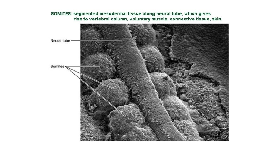
Somite Scelerotome Myotome Dermatome
Development of the vertebral column
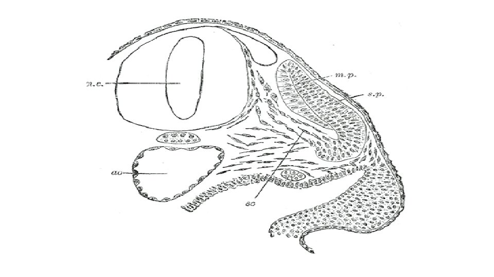
-each scelerotome is related laterally to myotome and its segmental nerve And separated from its fellow by intersegmental artery
-Each scelerotome is formed of loosely arranged cells cranially and densely backed cells caudally due to cellular condensation. -Densely backed cells of scelerotome above fuse with loosely arranged cells of scelerotome below resulting in: 1 - formation of mesenchymal centrum (body) of vertebra which is intersegmental in position 2 -Alternation of vertebral body with myotomes
3 - Some densely backed cells move cranially to form intervertebral disc 4 - degeneration of notochord except between vertebrae it is enlarged to form nucleus pulposus 5 - entrance of intersegmental artery into middle of body and exit of segmental nerve at disc towards myotome
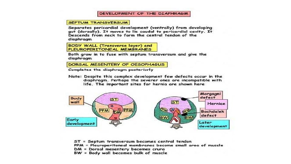
Development of the Diaphragm
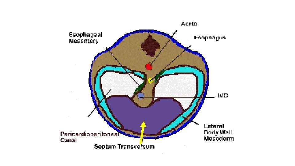
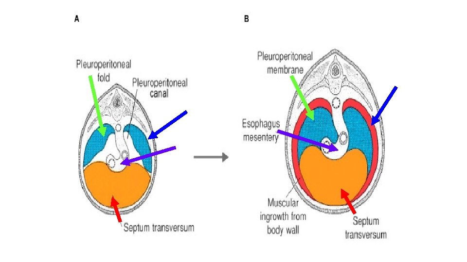
Diaphragm is derived from the following structures: 1 - The septum transversum: Forms central tendon 2 - Pleuro- peritoneal membranes: Form dorsilateral parts 3 - The mesoderm of the thoracic wall: Forms peripheral muscular part 4 - The mesentery of the esophagus: Forms crura of diaphragm
- Septum transversum is mass of mesoderm lies between pericardial cavity cranially and vitello-intestinal duct caudally - It is formed in the neck by 3 rd, 4 th and 5 th cervical somites
With descent of heart to thorax, the septum transersum is pushed caudally pulling phrenic nerve with it Phrenic nerve
Pleuro-peritoneal membranes grow medially from body wall they encroach on pleuroperitoneal canals till fuse with septum transversum anterior to esophagus And the esophageal mesentery posterior to it
The peripheral muscular part is derived from mesoderm of body wall
Congenital Anomalies of the Diaphragm
Congenital diaphragmatic hernia Herniation of abdominal contents into pleural cavity due to failure of development of pleuroperitoneal membrane
Esophageal hiatal hernia Due to congenital shortening of esophagus where cardiac opening and upper part of stomach lie in the thorax
Parasternal Hernia Due to failure of formation of part of muscles of diaphragm between sternal and costal parts
Thank you