Development of kidney ureter The kidneys developed from


 • It develops from the cranial part of the intermediate")
 • The middle part of the intermediate")
 of mesonephros - By the end of the 5 th week")
 of mesonephros - In female embryo: 1 - The mesonephric tubules:")
 Before the disappearance of mesonephros (by")

 * Dorstal end lies in contact with")


- Slides: 23
Development of kidney & ureter - The kidneys developed from the intermediate mesoderm (urogenital mesoderm). - The kidneys pass into 3 successive stages of development (overlap each other) 2
The first stage {Pronephros) • It develops from the cranial part of the intermediate mesoderm. • It is divided into 7 or 8 mesodermal masses called nephrotomes. • Each nephrotome gets a small cavity changing it into nephrocele. • The nephroceles elongated and form the pronephric tubules. • Each tubule has dorsal and ventral ends. 1)Ventral ends open into the intraembryonic coelom. 2) Dorsal ends join each other forming the pronephric Duct. • The pronephric duct elongates caudally and opens into the cloaca. • ** Function, it has no excretory function (no glomeruli). • ** Fate of pronephros: 1 - The pronephric tubules: disappear completely 2 - The pronephric duct: remain to be used as a mesonephric duct.
• Second stage • {Mesonophros} (WOLLFIAN) • The middle part of the intermediate mesoderm becomes segmented into 70 -80 masses called nephrotomes. • There is a small cavity transforming it to nephrocele. • Each nephrocele elongates forming S-shape mesonephric tubule. • Each tubule has ventral and dorsal ends. • a- Dorsal end of each mesonephric tubule opens into mesonephric duct. • b- Ventral end of each tubule enlarged and invaginated by a branch from dorsal aorta forming a transient glomerulus. • So; the mesonephros has an excretory function.
** Fate (derivatives) of mesonephros - By the end of the 5 th week of development shows the following changes: - In male embryo: 1 - Mesonephric tubules: - Cranial part forms appendix of Epididymis. - Middle part will form vasa efferentia. - Caudal part forms Paradidymis. 2 - Mesonephric (Wolffian) duct: - It forms epididymis, vas difference, seminal vesicle and ejaculatory duct. - Trigone of urinary bladder - Ureteric bud
** Fate (derivatives) of mesonephros - In female embryo: 1 - The mesonephric tubules: - Cranial part forms the Epoophron. - Caudal part forms Paroophron. 2 - The mesonephric (Wolffian) ducts: - Gartner's cyst in the vaginal wall. - Trigone of urinary bladder - Ureteric bud
• Third stage (The Metanephros, Permanent Kidney) Before the disappearance of mesonephros (by the 5 th week), the metanephros starts its development: - Ureteric bud from mesonephric duct. b) This bud grows upward and backward till invading caudal part of intermediate mesoderm that called metanephric cap or blastema (opposite the lower lumbar and sacral somites).
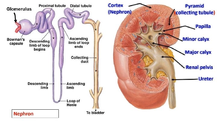
- Changes in the metanephric cap (blastema) * Dorstal end lies in contact with collecting tubule but without canalization. * Ventral end invaginated by branch from internal iliac artery forming glomerulus and Bowman's capsule. • This tubule will elongate forming proximal convoluted tubules, loop of Henle and distal convoluted tubule. • Later on distal convoluted tubule will be canalized with the collecting tubule.
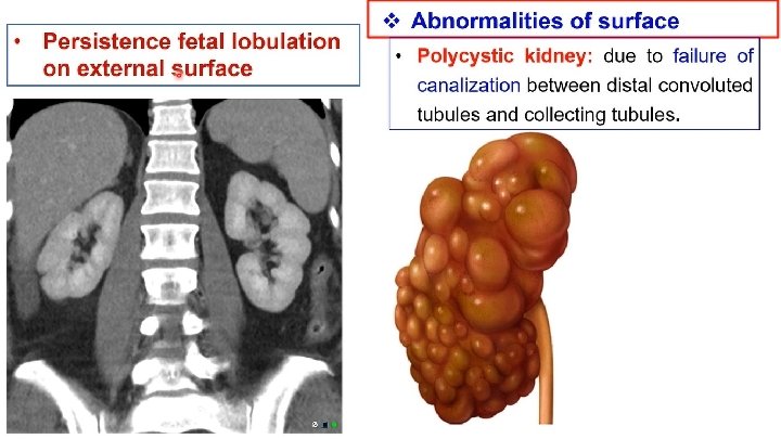
Post-developmental changes of the kidneys 1. Change in surface; disappear of the fetal lobulation by the capsule. 2. Change in position; ascend upward to the lumbar region. 3. Medial rotation 90 degree, Hilum becomes medially after rotation. 4. Change in blood supply; a) In the pelvis, it is supplied from the internal iliac artery. b) During its ascent, it is supplied by the common iliac artery. c) At its normal position, it is supplied by the abdominal aorta. v Definitive nephrons secret urine in the 2 nd half of pregnancy.
Congenital anomalies of kidney & ureter 12
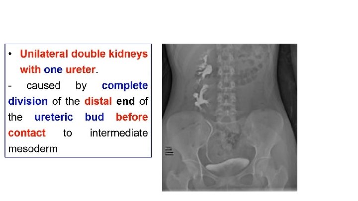
v. Agenesis - Causes: 1 - Failure of development of the ureteric bud (no ureter and kidney). 2 - Failure of contact of the ureteric bud and intermediate mesoderm (ureter and no kidney). - It may be - Unilateral agenesis, It may be noticed until problems occur in the solitary kidney. - Bilateral agenesis the amount of amniotic fluid decreased (oligohydramnios) and the fetus die within few days after birth.
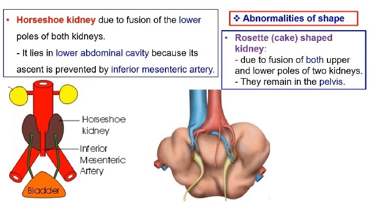
v. Abnormalities of the position a. Pelvic kidney: failure of the ascent of one or both kidneys to their normal positions. b. Incomplete ascent: it ascends but not reaches its terminal position. c. Ectopic kidney due to abnormal ascent. d. Mobile (floating) kidney: Not fixated to posterior abdominal wall, The kidney is movable with changes of body position. This lead to torsion of renal artery or ureter (Dietl's disease).
Th ank you Qu est ion s 23