Development of CNS Central nervous system Development of


 Ependymal layer: the most inner layer which contains")


- Slides: 20
Development of CNS Central nervous system
Development of the neural tube: - The neural tube develops as neural plate from the ectoderm dorsal to the notochord. - A neural pit that forming the neural groove. - The edges of the neural groove fused forming the neural tube that later on separated from the ectoderm. - Neural tube lies opposite developed somites: a- The part cranial to the 4 th somite is dilated and forms the brain. b- The part caudal to the 4 th somite remains narrow and forms the spinal cord.
**Cyto-differentiation of the neural tube: (1) Ependymal layer: the most inner layer which contains 2 types of cells; a- Ependymal cells line the cavity of the neural tube. b- Germinal cells divide repeatedly and migrated peripherally to form the mantle layer. (2) Mantle layer: the middle layer; It forms the gray matter of the spinal cord and is formed of two types of cells: a- Neuroblasts (nerve cells or neurons), the axons of these cells extend to the marginal layer. b- Neuroglial cells (supporting cells). (3) Marginal layer: the outer layer containing ascending and descending tracts (white matter of the spinal cord).
** At first, the neural tube has thick lateral wall, thin roof plate and floor plate, and a narrow slit-like lumen. - The ventral and dorsal parts of the lateral wall become thick by proliferation of the cells in the mantle zone. As a result, it is divided by lateral sulcus called sulcus limitans into ventral part (basal lamina) and dorsal part (alar lamina) and the cavity becomes rhomboidal in shape.
A- Ventral parts: contain motor cells, form the anterior and the lateral horns of the spinal cord. • The anterior horns are found in all segments. • The lateral horns are found in; 1) All the thoracic and upper 2 -3 lumbar segments (sympathetic). 2) In the 2 nd, 3 rd and 4 th sacral segments (parasympathetic). B- Dorsal parts: contain the sensory cells, form the posterior horns of the spinal cord. C- The cavity becomes reduced in size (narrow) to form the central canal. D- Marginal layer: the outer layer containing ascending and descending tracts (white matter of the spinal cord anterior lateral and posterior columns.
§ Termination of the spinal cord varies with the age: 1 - At the 3 rd month of intrauterine life, tip of coccyx. 2 - At birth, intervertebral disc of L 3/L 4. 3 - Adult; at the level of intervertebral disc of L 1/L 2. • below this level, vertebral canal contains roots of lumbar, sacral, and coccygeal nerves around filum terminale which form a bundle called Cauda Equina (L 2 -C 1) • The lower nerve roots are longer & more oblique because the spinal cord is shorter than the vertebral canal
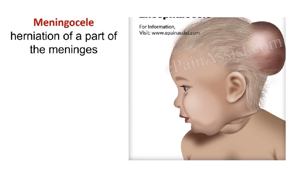
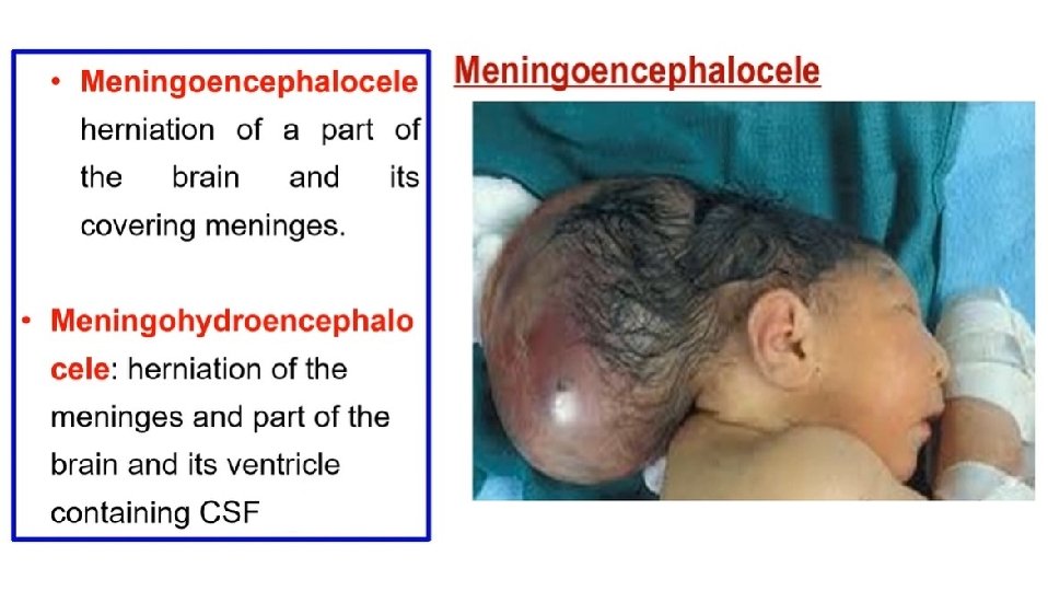
Congenital anomalies of the spinal cord 1 - Spina bifida: the cord is exposed directly to the skin due to failure of fusion of neural arch of vertebra. **Types of spina bifida: a- Spina bifida occulta: bifid spines of the vertebra with normal spinal cord. b- Meningocele; bulge of the meninges through the spina bifida. c- Meningo-myelocele; bulge of the meninges and spinal cord through the spina bifida. d- Myelocele; the spinal cord is exposed directly to the spina bifida.
Craniorachisis: Failure of closure of the neural tube leading to longitudinal cleft in the back of the head & vertebral column.
Development of Brain
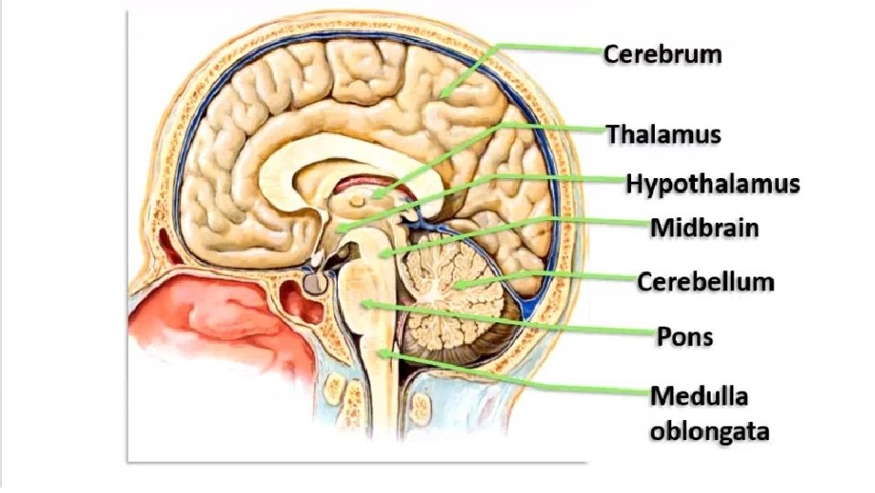
DEVELOPMENT OF THE BRAIN - The brain develops from the part of the neural tube cranial to the 4 th somite. - This part shows 3 dilatations, forebrain, midbrain and hindbrain vesicles. • Differentiation of the brain vesicle 1 - Forebrain vesicle (prosencephalon): divided into 2 parts: 1 - A median part (diencephalon) gives rise to; - Thalamus and 3 rd ventricle. - Hypothalamus - Epithalamus. - Metathalamus. 2 - Two lateral parts (telencephalon) gives rise cerebrum and lateral ventricles. 2 - Midbrain vesicle (mesencephalon) Gives rise to the midbrain. 3 - Hindbrain vesicle (Rhombencephalon): divided into 2 parts: - Cranial part or metencephalon gives pons and cerebellum and 4 th ventricle. - Caudal part or myelencephalon gives medulla oblongata.
• Anencephaly: Failure of development of greater part of the brain & vault of the skull due to failure of cephalic part of the neural tube to close
DERIVATIVES OF THE NEURAL CRESTS Ø A special neuro-ectodermal cells dorsolateral to the neural tube. 1 - Sensory Cells of the para-sympathetic ganglia ((Ciliary of 3 rd, pterygopalatine and submandibular of 7 th, Otic of 9 th and enteric ganglia of 10 th cranial nerves)) 2 - Sensory Cells of the sympathetic ganglia 3 - Sensory Cells of the dorsal root ganglia of the spinal nerves 4 - Pia and arachnoid matters of the meninges (dura matter mesodermal in origin). 5 - Schwan cells that form the myelin sheath 6 - Chromaffin cells of the suprarenal medulla 7 - Pigment cells in the skin, iris and retina.
Th ank Qu you est ion s I/Azzam - 2004