DETERMINANTS OF CELL ORGAN AND TISSUE TROPISM One


- Slides: 29
DETERMINANTS OF CELL, ORGAN, AND TISSUE TROPISM One of the most important concepts for understanding pathogenesis is the concept of cell, organ, and species tropism. Tropism is determined by many factors in both the virus and the host, including how the virus enters the host, how the virus spreads within the host (lymphatic, neural, or hematogenous spread), the permissiveness of specific cell types for the virus (as defined by receptors, cellular differentiation, and intrinsic cellular resistance to infection), the nature of innate and adaptive immune responses, and specific properties of tissues such as accessibility and effectiveness of the immune system (immunoprivilege). Each of these factors can play a determining role during viral infection and must be considered when defining mechanisms of viral pathogenesis.
Entry into the Host A virus must access permissive cells to establish infection and therefore must overcome a series of anatomic and innate immune barriers to enter the host. The route of entry and the mechanisms of spread are therefore important determinants of viral tropism. The route and tissue through which a virus infects the host may be clear from epidemiologic studies. For example, both measles virus and VZV spread by the respiratory route, and polioviruses and noroviruses spread fecal–orally. However, even when the route of infection is known, the precise events involved in entry into the host are mostly unknown. There are six primary portals of entry for viruses, each used by a variety of viruses. Five of these are epithelial surfaces: skin, conjunctiva, respiratory tract, gastrointestinal tract, and genitourinary tract. The sixth is the unique interface between the mother and the germ cell or the developing fetus.
Penetration Through Epithelial Barriers The body is covered by epithelia, presenting a large surface for viruses to access. However, epithelia share several properties that inhibit viral entry. For example, epithelial cells are constantly turning over and being replenished; thus, cells that are contacted by a virus are shed continuously. The skin has an added protective mechanism, being many cells deep with the surface comprised of metabolically inactive cells that cannot support viral replication. In addition, barriers may be protected by low p. H or secretions, including mucus. Epithelial tissues are highly active immune organs. In all epithelia, DCs (e. g. , Langerhans cells in the skin) serve as sentinels for invasion, having the capacity to, when activated, move to lymph nodes to induce immune responses. These sentinel cells play a dual role in infection, both as critical for induction of immunity and as cells targeted by viruses as an initial site of infection. For example, when VEE is inoculated into mice, the first cells infected are DCs; these cells rapidly move to draining lymph nodes, providing the virus access to the
Viruses cross epithelial barriers through mechanical breaches (e. g. , vectorborne delivery of arboviruses or bite wound delivery of rabies) or by accessing specialized cells
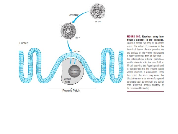
within the epithelium. An excellent example of this latter situation is provided by reovirus, which accesses the Peyer’s patch through specialized epithelial cells called M cells. Upon entry into the intestine, the reovirus virion is proteolyzed, triggering a remarkable structural transition with cleavage and loss Many viruses infect either the immature fetus or the newborn during the birth—a process referred to as vertical transmission. There are two mechanisms for entry into the developing fetus. The first is via placental penetration, as when a virus enters the fetus after invasion of the fetal circulation or amniotic fluid. Viruses such as HCMV and rubella can spread transplacentally, with devastating consequences for the developing fetus. Viruses such as human endogenous retroviruses (HERVs) can also be vertically transmitted via the germ line and constitute a significant fraction of the human genome. HERVs continue to proliferate within the genome and are likely to exert both beneficial and detrimental effects on their hosts.
Systemic Spread of Virus Infection Viruses spread via three host systems that can provide access to a large number of tissues and cells: blood, lymphatics, and nerves. Although the blood is a major highway for spread of viruses through the host, many viruses use nerves or a combination of hematogenous and neural spread to access host tissues. The level of viremia has been correlated with the severity of acute viral disease, the prognosis of chronic viral disease (as in HIV), the extent of viral dissemination, and the efficiency of viral spread between hosts. The level of viremia is a function of viral access to the blood, viral clearance from the blood, and the vehicle (plasma vs. cell associated) that the virus uses to travel through the blood.
The classical example of this strategy is rabies virus, which spreads along nerves from the area of inoculation to the CNS. The time between inoculation and development of signs and symptoms of rabies encephalitis depends on the length of the nerves between the site of inoculation and the CNS. The virus travels up nerves toward the CNS at a rate of 50 to 100 mm/day, and the disease can be cured by surgical removal of the infected limb as long as the virus has not entered the CNS. For example, if the initial bite is on a lower extremity, there is a longer time within which vaccination and passive transfer of rabies-immune antibody can be effective than if the bite is on the face. Importantly, the immune system can also modulate neural spread. For example, antibodies can interrupt many of the steps in neural spread of reoviruses to and within the CNS.
The Role of Viral Receptors in Cell, Tissue, and Species Tropism An important step in viral infection, and a primary determinant of the distribution of virus between and within tissues, is the interaction of a virus with specific receptors on permissive cells. Receptor expression plays a major role in determining the tropism of several viruses, including poliovirus and measles virus, and the use of molecular tools, including transgenic mice, expressing the virus-specific receptors provides key insights into the role of receptors in regulating viral tissue tropism and pathogenesis.
Innate Immunity and Intrinsic Cellular Resistance to Infection Determine Tropism Factors in addition to specific receptors play a major role in determining viral tropism. Tropism can be determined by proteins responsible for cell-intrinsic defense against viral infection, transcription factors, cell cycle regulators, and micro. RNAs. For example, the liver-specific host micro. RNA mi. R-122 is essential for robust HCV replication, 137 and inhibition of mi. R-122 in nonhuman primates limits HCV replication. Once a virus has bound to a cell and delivered its genome or capsid to the cytoplasm, a series of events occur that can have a profound effect on the viral cell tropism. These events likely explain why receptor expression does not always explain the cell and tissue tropism of a virus. The host cell expresses on its cell surface and in its cytoplasm molecules that can either directly inhibit viral replication (intrinsic cellular resistance to infection) or can induce a signaling cascade that in turn generates antiviral molecules (innate immune responses). Together, these molecules and pathways are determinants of both the permissiveness of cells for viral replication and species tropism.
Immunoprivilege. For example, in studies of LCMV infection of mice, 32, 38 many organs including liver, spleen, lung, and pancreas are cleared relatively efficiently within 30 days of transfer of immune cells. However, virus persists in the CNS for up to 90 days and in the kidney and genitourinary system for more than 200 days, indicating that both the CNS and the genitourinary tract are immunoprivileged. These data also show that immunoprivilege is a relative term, with the efficacy of the immune system varying depending on when infection is analyzed. Analysis of the mice at a time point more than 90 days after transfer of immune cells identifies only the genitourinary tract as immunoprivileged (Fig. 10. 9). It is likely that the selective inability of the immune system to clear virus infection from certain tissues is owing to a combination of two interrelated factors. The first is intrinsic limitations of immune system function in certain tissues or to limited capacity to address infection of certain cell types. For example, CD 8 T cells may be more effective at eliminating infection from major histocompatibility complex (MHC) class I–expressing hepatocytes than from neurons that do not express MHC
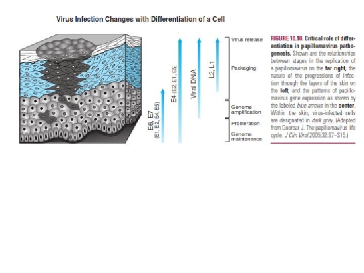
Cellular Differentiation as a Determinant of Viral Tropism and Pathogenesis Another important determinant of viral tissue distribution is cellular differentiation. Thus, cells at different stages of differentiation may have specific properties that favor or disfavor viral infection or replication. This is especially true in a tissue responding to virus-induced damage. For example, when hepatocytes are damaged during chronic HBV or HCV infection, the liver regenerates, providing viruses with access to cells in differentiation states. Many viruses take advantage of differentiated functions of the cells that they target. For example, HSV establishes latency in fully differentiated neurons, and EBV latently infects memory B cells; in each case, the reservoir for chronic infection is a particularly long-lived cell type. Papillomavirus infection of the skin provides an outstanding example of the impact of cellular differentiation on pathogenesis (Fig. 10). In normal skin, basal stem cells give rise to ever more differentiated cells that move toward the skin surface, lose their nucleus, become cornfield, and are finally shed.
Papillomaviruses infect basal stem cells of the skin, where they can persist in a latent state for prolonged periods. Activation of a program of gene expression initially involving the expression of E 6 and E 7 proteins results in cell cycle progression, inhibition of apoptosis, and viral replication. As infected cells differentiate and move toward the skin surface, they become permissive for expression of genes involved in viral replication and assembly. The normal differentiation process is subverted by the virus, resulting in retention of the nucleus and synthesis of proteins required for viral DNA and protein synthesis. The virus assembles and is released from the skin in shed cells. The principle that cellular differentiation is a determinant of viral pathogenesis is very important and general. When tissues are damaged, stem cells are activated to generate new cells with consequent proliferation of somatic cells and differentiation into cells such as hepatocytes, endothelial cells or epithelial cells. These processes are plausibly subverted by several viruses, many of which grow optimally in replicating cells.
FATE OF THE INFECTED CELL, TISSUE, AND HOST The presence of avirus in atissue may or may not result in damage. The variables that determine the fate of the infected tissue and host are complex and involve the interplay between the virus and its cytopathic potential; the age, sex, and nutritional status of the host; the regenerative capacities of different primary cell types; whether the immune system damages the tissue as part of the protective response; whether the virus induces autoimmune responses; and perhaps most importantly, whether the virus is ever cleared from the tissue and, if not, the nature of residual virus infection. Determinants of tissue damage may be local in nature, such as the death of infected cells owing to viral cytopathicity or killing by immune cells, or operative over longer distances, as in the case of vascular damage leading to ischemia, systemic hormonal and cytokine responses, induction of fever or cachexia, cytokine induction of the death of uninfected cells, metastatic tumors, or virus-induced autoimmunity. Age as a Determinant of Susceptibility to Viral Infection Many viral infections are far more severe in young than older hosts—an increase in severity that often correlates with increased replication and dissemination of virus. For example,
This observation extends to many other viruses, including poliovirus, where 2 -week-old poliovirus receptor transgenic mice are 10, 000 fold more susceptible to paralytic disease than adult mice. . It is commonly considered that maturation of the immune system explains increased resistance to infections seen in older hosts. However, this has not been rigorously demonstrated as the reason for age-dependent susceptibility to viral infection. Other age-dependent processes, such as cellular differentiation and proliferation, may play a role as large as that of the immune system in age-related susceptibility. For example, the genes expressed in the nervous system during infection of younger versus older hosts differ, indicating that there are fundamental differences in how older and younger tissues respond to viral infection.
Fate of Infected and Uninfected Cells in Tissues The consequences of viral infection range from rapid destruction of tissue cells, as seen with variola or Ebola viruses, to continuous replication in the absence of severe cytopathology, as seen with LCMV, to the establishment of latency, as seen with herpesviruses.
Direct Killing of Cells by Viruses Cytopathic effect is defined as the destructive consequences of virus infection on cells. Direct killing of cells by viruses may be attributable to viral subversion of cell metabolism for replication or to cell-intrinsic programmed cell death pathways such as necrosis, apoptosis, or nonnecrotic and non apoptotic cell death. The term cytopathic effect is most commonly used to describe the consequences of infection in cultured cells. Cell death that occurs in a cultured cell may or may not reflect the process of cell death that occurs in vivo. An advantage of studying cell death and cytopathicity in cultured cells is that the interaction between the virus and the cell occurs without cell-extrinsic factors, with exception of those owing to autocrine stimulation (e. g. , the expression of interferons by infected cells). A disadvantage of this approach to studying cell death is that events in cultured cells may have little relationship to what happens in primary cells in an infected tissue.
. It is important to remember that the presence of inflammation does not necessarily indicate the participation of inflammatory cells in the killing of infected cells. Inflammatory cells may be beneficial or present as a host response to killed cells and tissue damage rather than being participants in the death of infected cells.
Killing of Cells by Cell-Extrinsic Effects of the Immune System Not all cell death in an infected tissue is a result of direct viral effects or cell-intrinsic mechanisms of programmed cell death triggered by the presence of the virus. The host has many cell-extrinsic mechanisms for initiating apoptosis or lysis of infected cells via cytokines, serum proteins such as complement, and proteins such as the granzymes that are injected via perforin into infected cells by leukocytes. The role of cell killing by the lytic activity of granzymes is important because perforin- and/or granzyme-deficient mice are susceptible to coronaviruses, West Nile virus, LCMV, ectromelia virus, HSV, Theiler’s virus, MCMV, and murine g. HV. The complement cascade is a two-edged sword important for antiviral immunity but can also contribute to virus-induced tissue damage. Complement proteins are involved in induction of antibody and T-cell responses, trapping of viral antigen on antigenpresenting cells, induction of chemotactic and vascular permeability changes in infected tissues, and activation of leukocytes. In addition to being immunoregulatory, complement can lyse both virus-infected cells and lyse or neutralize virions.
Clearance of Virus Infection and Chronic Viral Infection A fundamentally important variable in viral pathogenesis is whether the immune system can clear a virus from the body. Some viruses are readily cleared but can do significant damage to the host during their brief time of residence in the body. An example is variola virus, which causes smallpox. This pattern of acute infection (see Figs. 10. 1 and 10. 3) is associated with development of sterilizing immunity and effective resistance to reinfection. Many viruses for which effective vaccines are available fall into this category. Other viruses are less amenable to elimination by the immune system, resulting in latent, chronic, or progressive infection (see Fig. 10. 1). Examples of chronic viral infections of great medical significance are those caused by HIV, HCV, and HBV. Other chronic infections are less consistently harmful to their hosts but do cause significant disease. Examples include herpesviruses such as EBV, VZV, HCMV, and HSV, each of which permanently infects most human beings. It is possible that such chronic infection can, in addition to causing disease in rare cases, provide a symbiotic benefit to the host via chronic stimulation of the innate immune system, including macrophages and NK cells.
Tissue Damage During Beneficial Immune Responses Is a Necessary Evil Although there are clear examples in which the immune system is primarily responsible for virus-induced tissue pathology, and thus disease is immunopathologic, the situation is seldom that simple. The antiviral immune response has two faces: a beneficial face as the host system responsible for curing infection and a harmful face via induction of tissue pathology and systemic toxicity. This sets the stage for consideration of one of the most complex topics in viral pathogenesis: the relationship between immunemediated damage and clearance of infection. It is not always true that the function of the immune system causes pathology. For example, protective effects of antibody that do not damage tissues are seen in most if not all viral infections.
Immunopathology Occurs When the Immune System Goes Too Far In some cases, the balance between the protective effects of immunity and the harmful effects of immunity clearly shifts to immunity being the primary cause of tissue pathology and even death of the host. These processes can be driven by overactive innate immune responses, which result in inflammatory responses that drive virus-induced tissue pathology and disease, nonspecific cell killing, or even induction of autoimmunity by the host adaptive immune response. The balance between acceptable tissue damage associated with viral clearance and immunopathology is determined by the effectiveness of the immune system in clearing the infection. For example, even severe hepatitis would be an acceptable outcome of HCV infection if the virus could be cleared; however, the immune response to HCV infection is often ineffective. Immunopathology can be attributable to either T- or B-cell responses or to innate immunity and may be the consequence of virus-induced immune responses to either viral antigens or, in the case of induced autoimmunity, host antigens.
Virus-Induced Autoimmunity A variant mechanism of immunopathology is the induction of autoimmunity by virus infection. The contributions of autoimmunity to tissue pathology associated with viral infection, or putatively associated with viral vaccination, remain quite controversial. The role of autoimmunity in virus infection and post infection syndromes has been studied in several systems, and viruses have at one time or another been candidate etiologic agents for many autoimmune or inflammatory diseases in humans, including rheumatoid arthritis, multiple sclerosis, Crohn’s disease, myocarditis, uveitis, hepatitis, Sjögren’s syndrome, and type 1 diabetes. A proposed mechanism for the induction of autoimmunity is molecular mimicry between virus and host antigens leading to a breakdown of self-tolerance. This might plausibly occur in the setting of virus-induced tissue damage and inflammation.
VIRAL DETERMINANTS OF VIRULENCE Mutation and Selection of Viral Variants An excellent example of a mutation contributing to the pathogenesis of viral infection is the emergence of immune escape mutants. The principle that viral escape mutants can evade CD 8 responses was first identified by study of murine LCMV infection. 227 In this study, investigators created T-cell receptor transgenic (LCMV TCRtg) mice containing a large number of T cells specific for the epitope containing amino acids 3242 of the LCMV glycoprotein. Within 8 days of infection, many LCMV isolates were no longer recognized by TCRtg T cells and contained mutations in the 3242 epitope. 227 Importantly, mutations in other T-cell epitopes that were not recognized by TCRtg T cells were not selected, indicating specificity of the selective pressure.
Virus-Induced Immunosuppression Virus infection is often associated with the induction of an immunosuppressed state. Immunosuppression is defined as a virus infection-associated decrease in the capacity of the immune system to respond to antigen. The phenomenon of immunosuppression was first noted as inhibition of tuberculin skin test reactivity during measles virus infection. That initial observation has been followed by a deluge of reports of altered immune reactivity in virus-infected hosts.
An immunosuppressed state may benefit a virus by inhibiting antiviral responses; however, it is important to distinguish between immunosuppression and viral immune evasion that occurs without inducing a general deficiency in immune function. It is also important not to confuse immunosuppression with the normal down-regulation of virus-specific responses that occurs as viral infection is cleared. There are three categories of immunosuppressive mechanisms. In the first, virus kills significant numbers of critical immune system cells; an example of this is HIV-associated depletion of CD 4 T cells. Likewise, LCMV clone 13 causes immunodeficiency owing to destruction of DCs that are critical antigen-presenting cells. In this case, activated CD 8 T cells that recognize viral antigens expressed by infected DCs damage the immune system. A second potential mechanism involves alterations in cytokine secretion by infected cells or the induction of secreted molecules that inhibit immune responses. This mechanism, however, does not provide a clear explanation for a case in which immunosuppression persists long after infection is thought to be cleared. A third but largely speculative potential mechanism only now being explored is viral induction of regulatory T
Viral Evasion and Subversion of Host Cytokine Responses Host molecules present in the extracellular fluid including cytokines, prostaglandins, steroid hormones, peptide hormones, growth factors, and serum components such as complement provide important targets for viral evasion and subversion strategies. Viruses encode molecules that evade or subvert host hormonal and cytokine responses that regulate both innate and adaptive immunity, such as interferons, TNF, IL 18, IL 6, IL 10, complement, and chemokines