Describe the Pathophysiology of Burns Akshay Kapoor Asst

Describe the Pathophysiology of Burns Akshay Kapoor Asst. Professor Burns & Plastic Surgery

Introduction • • • 66% of burn injuries occur at home Fatalities at extremes of age Flame and Scald most common cause Scald burn victims commonly< 5 years Survival rate for all burns 94. 6%

Burn Classification Causes • Flame • Scald • Contact • Chemical • Electricity

Classification. .

Depth of Burn 1 st Degree • Burns involving only the epidermis. • Erythematous and very painful but do not form blisters. • Sunburns fit this category of superficial, epidermal injury. • Within 3– 4 days, the dead epidermis sloughs and is replaced by regenerating keratinocytes.
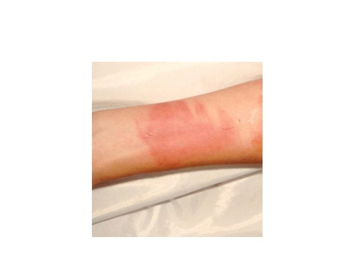
 • Extend into the papillary dermis and characteristically")
2 nd degree (Superficial dermal burns) • Extend into the papillary dermis and characteristically form blisters. • Appearance is pink, wet and hypersensitive to touch. • Painful as uncovering the wound allows currents of air to pass over it. • These wounds blanch with pressure as the blood flow to the dermis is increased due to vasodilation. • Superficial dermal burns usually heal within 2– 3 weeks without risk of scarring and therefore do not require operation.
 • Extend into the reticular dermis and generally")
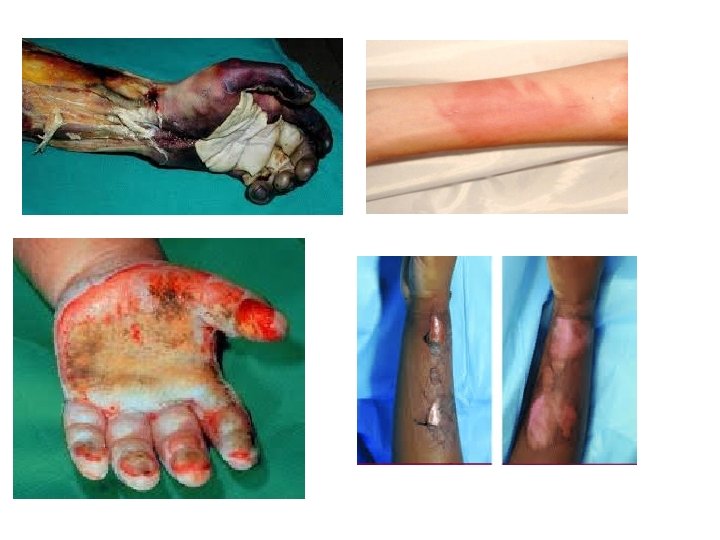
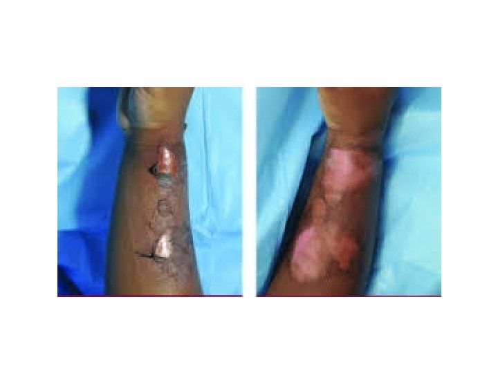
3 rd degree (Deep Dermal Burns) • Extend into the reticular dermis and generally will take 3 or more weeks to heal. • They also blister, but the wound surface appears mottled pink and white • The patient complains of discomfort and pressure rather than pain. • When pressure is applied to the burn, capillaries refill slowly • Partial-thickness burns that are predicted not to heal by 3 weeks should be excised and grafted.
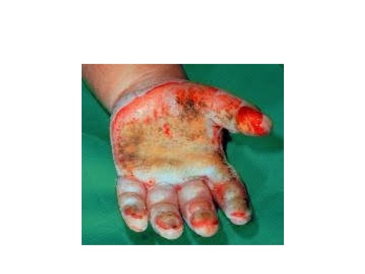
 • Full-thickness burns involve the entire dermis and extend")
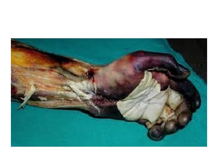
4 th Degree (Full Thickness) • Full-thickness burns involve the entire dermis and extend into subcutaneous tissue. • Their appearance may be charred, leathery, firm, and depressed when compared to adjoining normal skin. • These wounds are insensitive to light touch and pinprick. • Non-charred full-thickness burns can be deceptive as they may have a mottled appearance • Must be excised and grafted early

Local Changes in Burn Injury- Jacksons Zones

Zone of Stasis • Can survive or go on to coagulative necrosis. The zone of stasis is • associated with vascular damage and vessel leakage. • Thromboxane A 2, and Bradykinin a potent vasoconstrictor, is present in high • Local endothelial interactions with neutrophils mediate some of the local inflammatory responses associated with the zone of stasis.

• studies demonstrate that blockage of leukocyte adherence with anti-CD 18 or antiintercellular adhesion molecules & monoclonal antibodies improve tissue perfusion and tissue survival in animal models.

Zone of Hyperemia • Contains viable tissue • No risk of necrosis • Characterized by vasodilation due to effect from zone of stasis
")
Systemic Changes in Severe burns(>40%)
 • Lasts 48 hours")
Hypermetabolic Response Phase 1 of Post Burn Metabolic phenomenon(Ebb Phase) • Lasts 48 hours • Decrease in Cardiac Output/O 2 Consumption • Causes hyperglycemia
 • Begins after 48 hours • Increase in metabolic")
Phase 2 ( Flow phase) • Begins after 48 hours • Increase in metabolic rate and cardiac output • Hyperglycemia in spite of raised insulin • Reaches a plateau in about 5 -7 days • Persists upto 1 -3 years

• 10 -50 fold increase in corticosteroid and catacholamine levels • Results in Protein breakdown in muscles • Amino Acids ( Alanine ) from protein breakdown recruited for gluconeogenesis • Fat breakdown in liver leads to glycerol formation which is used for gluconeogenesis • End product of anaerobic respiration in the burn wound (lactate) sent for gluconeogenesis

• Glucose is delivered to peripheral tissue but glucose oxidation does not occur • This in turn raises insulin levels • Overall effect is loss of lean body mass 10% loss- decreased immune function 20% loss- chronic infections 30% loss- pneumonia & pressure ulcers 40% loss- Can lead to death • Severe burns cause upto 25% loss

• Increased cotisol also causes transport of calcium and magnesium from long bones • Decreased bone mineral density and content leading to susceptibility to fractures

Immune Dysfunction • Depressed function of Macrophages, Neutrophils, T cells and B cells • Even though G-CSF levels actually increase after severe burn but bone marrow G-CSF receptor expression is decreased, which may in part account for the immunodeficiency seen in burns • Release of negative regulators of myeloid growth decrease Macrophage production

• Neutrophil counts increase after severe burn but they are dysfunctional • Altered diapedesis, chemotaxis and phagocytosis due to loss of CD 11 b/CD 18 • Decreased Respiratory burst due to deficiency of p 47 -phox activity • Poor motility sue to impaired actin mechanics • Counts begin to fall after 72 hours

• Depressed T helper function • Polarization from Th 1 to Th 2 immune response • IL 2 and IFN-γ responsible for initiation of phagocytosis and intracellular killing is decreased • Increase in IL 4 and IL 10 which is mostly antibody based immunity • Cytotoxic T lymphocyte activity also decreased

• Administration of IL 10 antibodies and growth factors decreases the effect of the polarization of immune response

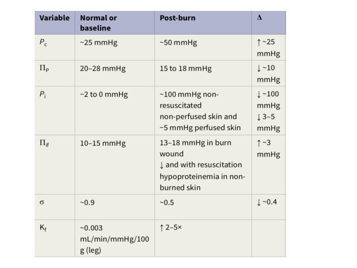
Inflamation and Odema-Landis Starling Equation

Mediators involved in edema formation • Mast cells in the burned skin release histamine in large quantities immediately after injury, which elicits a characteristic response in venules by increasing intercellular junction space formation causing increased permeability • Serotonin released from aggregated platlets causes pulmonary vasoconstriction

• Mediators causing Increased permeability Prostaglandin E 2 and I 2 Free Oxygen Radicles Thromboxane A 2 & B 2 • Leukotrines B 4 and D 4 cause pulmonary hypertension • Angiotensin & Vasopressin responsible for systemic vasoconstriction and gut ischemia

• In the first 12 hours there is an abrupt increase in the fluid levels in the burn tissue • After 24 hour there is a more gradual increase in fluid content both in burned and non burned soft tissue • This gradual 3 rd space loss is eventually responsible for burn shock

• Prompt and adequate fluid resuscitation improves outcome of the burn patient • It is imperative to avoid Over –resuscitation as well • This trend of providing fluid in excess of the Parkland formula has been termed ‘fluid creep’

• Complications of fluid creep are Eye injuries due to elevated orbital pressures Pulmonary edema Prolonged mechanical ventilation Graft failure Need for fasciotomy of uninjured extremities Abdominal Compartment Syndrome
 >30 cm. H 2 O is defined as intra-abdominal")
• Intra-abdominal pressure (IAP) >30 cm. H 2 O is defined as intra-abdominal hypertension (IAH). • ACS is sustained IAH + clinically tense abdomen combined + ventilation aberrations due to elevated pulmonary inspiratory pressures OR oliguria despite aggressive fluid resuscitation

Myocardial Dysfunction • Myocardial contractility is depressed along with relaxation capacity leading to a stiff myocardium • Possible causes for this are Raised Intracellular calcium levels Circulating Myocardial depressant factor( not isolated) Raised TNF alpha levels

• Even though contractility is depressed , the cardiac Output may be increased upto 130150% for a period of 2 years • Adrenergic stimulation causes increased heart rate as well as raised Systemic and Pulmonary vascular resistance • Mortality occurs because of cardiac stress in a setting of myocardial dysfunction

GI System • Apoptosis of epithelium of Small intestine mucosa • Vesiculation of microvilli with breakdown of actin filaments in the microvilli of small intestine • Loss of brush border lipase activity – loss of fatty acids • Poor uptake of Glucose and amino acids from the lumen

• Increased gut permeability leading to fluid loss • Vasoconstriction leading to ischemia which causes bacterial and endotoxin translocation across the mucosa causing septicemia • Inverse relation between blood flow and gut permeability

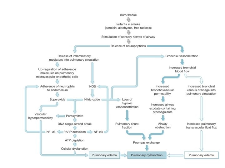
Lungs • In large burns there is a pronounced increase in pulmonary vascular resistance (PVR) • Both pre and post capilary vasoconstriction occurs which causes pulmonary odema • Hypo-protenemia still remains the dominant cause of pulmonary odema • In case of inhalational injury factors released due to injury to bronchial tree and lung parenchyma occurs

Renal Dysfunction • Local and Systemic cytokine release causes decreased renal blood flow which causes Acute Kidney Injury • Free Oxygen Radicles can cause direct tubular damage • Other factor maybe myoglobinurea following rhabdomyolysis ( Myoglobin> 1500 -3000 ng/ml) • AKI may occur despite adequate fluid resuscitation by Parkland Formula

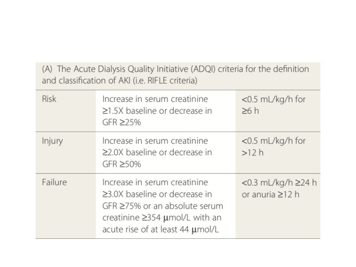
• Imperative to identify and diagnose Acute Kidney Injury so that patient can be shifteds for renal replacement therapy( Dialysis) • RIFLE and AKIN criteria developed to aid diagnosis and plan therapy • AKIN is modification of RIFLE with only change that it should be applied within 48 hours of burn injury
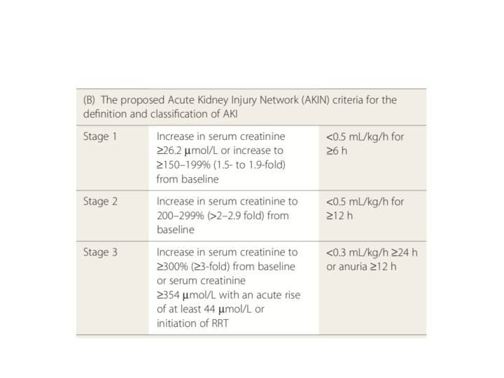

• Even though creatinine is not the ideal biochemical marker of kidney dysfunction it still remains the gold standard • New markers of AKI, such as cystatin-C, have shown promise as earlier detectors of changes in GFR • In order to differentiate pre renal from renal failure it is important to analyze the following indices
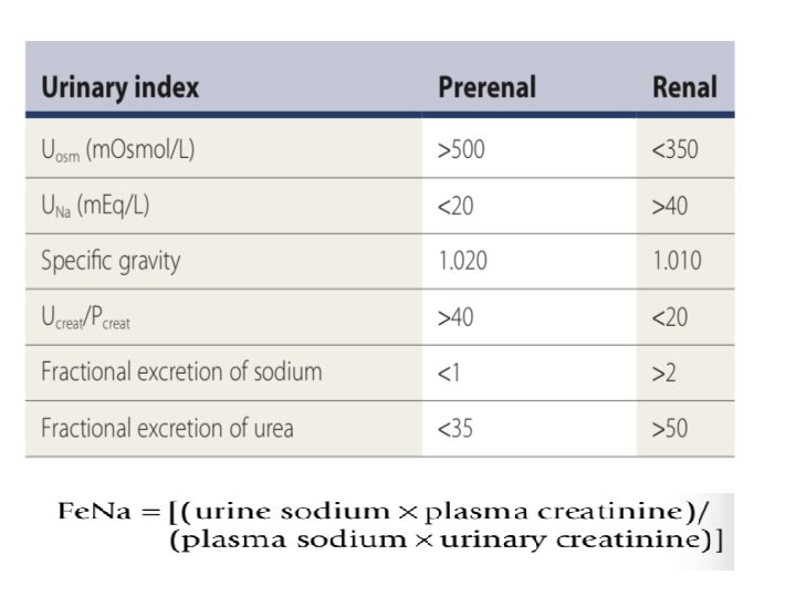

• Fractional excretion of urea is a more reliable indicator as it negates the effect of diuretic use

Change in Membrane Potentials • Membrane potential in non burned distant tissues such as skeletal muscles , nerves, Gi tissues partially depolarize ( from -90 to -70) • Cell death can occur at resting potentials of -60 • This change causes action potential dampening which may be responsible for tissue dysfunction • This change is brought about increased sodium conductance

• The factor which leads to this has not yet been identified but it has a complex and probably dynamic structure
- Slides: 50