Department of pathology Li shuhua nontoxic goiter Toxic


 Pathogenesis: autoimmune disorder")


- Slides: 33
Department of pathology Li shuhua
nontoxic goiter Toxic goiter adenom a adenocarcino ma
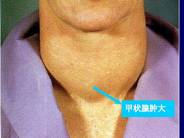
Diffuse nontoxic goiter Pathogenesis: Dietary iodine deficiency endemic Not apparent sporadic Impaired synthesis of thyroid hormone Rise TSH level in serum Hypertrophy and hyperplasia of thyroid follicular cells Gross enlargement of the thyroid gland
morphology Diffuse hyperplastic goiter Diffuse colloid goiter Nodular goiter
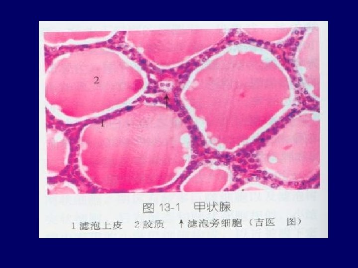
Diffuse hyperplastic goiter 1. Diffuse,symmetric enlargement of the gland; <150 g 2. follicle are lined by crowded columnar cells, which may pile up and form projections. There is only little colloid in
Diffuse colloid goiter 1. Cut surface: brown, glassy, translucent; 200 -300 g 2. Colloid is abundant in the follicles; 3. follicular epithelium are flattened or cuboidal or hyperplasia.
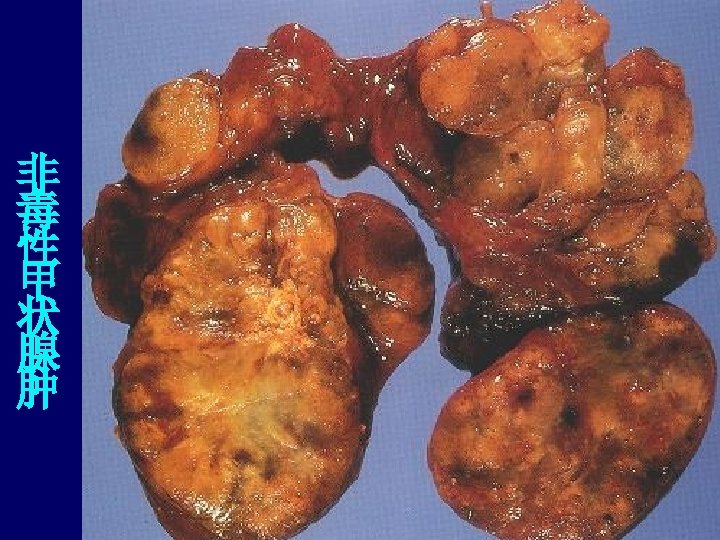
Nodular goiter macroscopically: • Multilobulated, asymmetrically enlarged glands • Cut surface: irregular nodules containing variable amount of brown, gelatinous colloid;
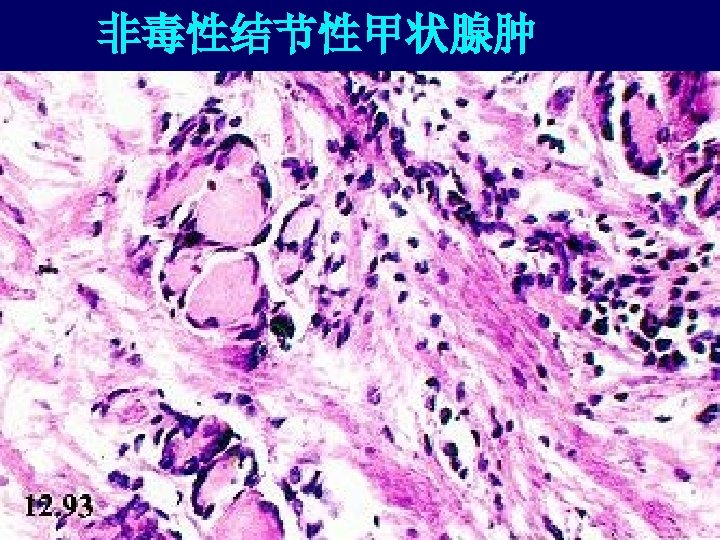
microscopically 1. Regressive changes: fibrosis, hemorrhage, calcification, cystic 2. Colloid-rich follicles lined by flattened epithelium and areas of follicular epithelial hypertrophy and hyperplasia;
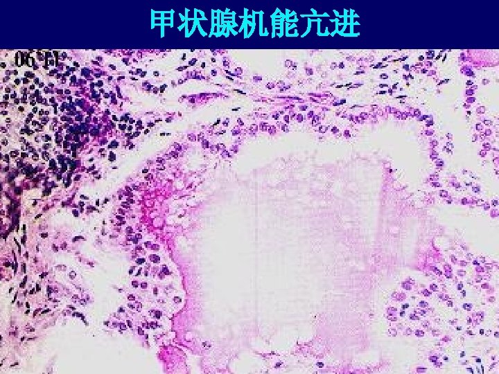
Diffuse toxic goiter (grave’s disease) Pathogenesis: autoimmune disorder
Morphology: 1. diffusely enlarged, gland is smooth and soft, capsule is intact 2. microscopically: follicular epithelial cells are tall, columnar , crowed, formation of small papillae; colloid is pale with scalloped margins. 3. vessel and lymphoid aggregates
Clinical features diffuse hyperplasia of the thyroid, ophthalmopathy, dermopathy
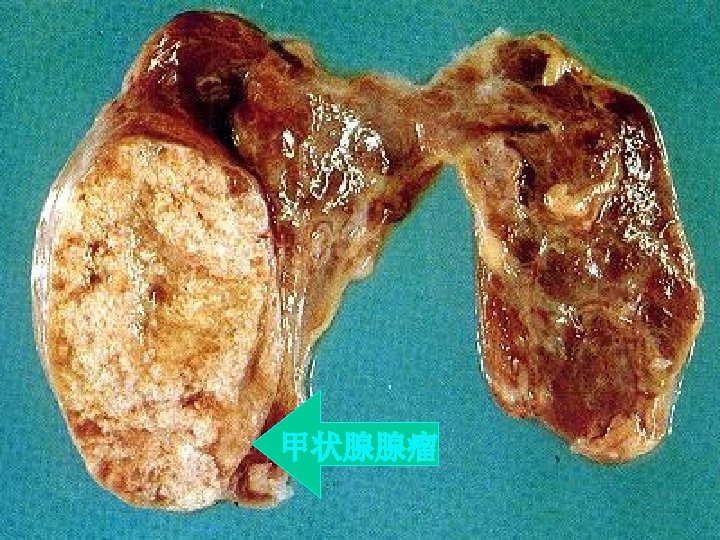
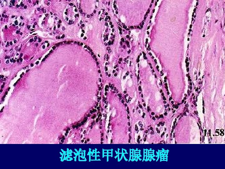
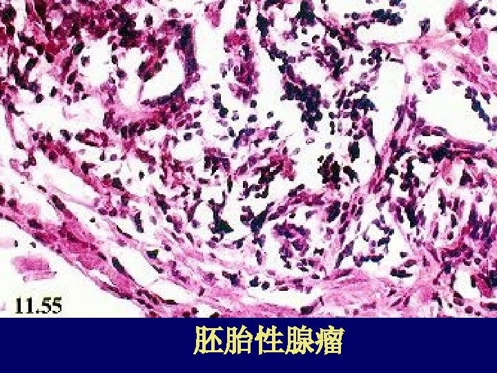
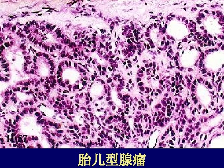
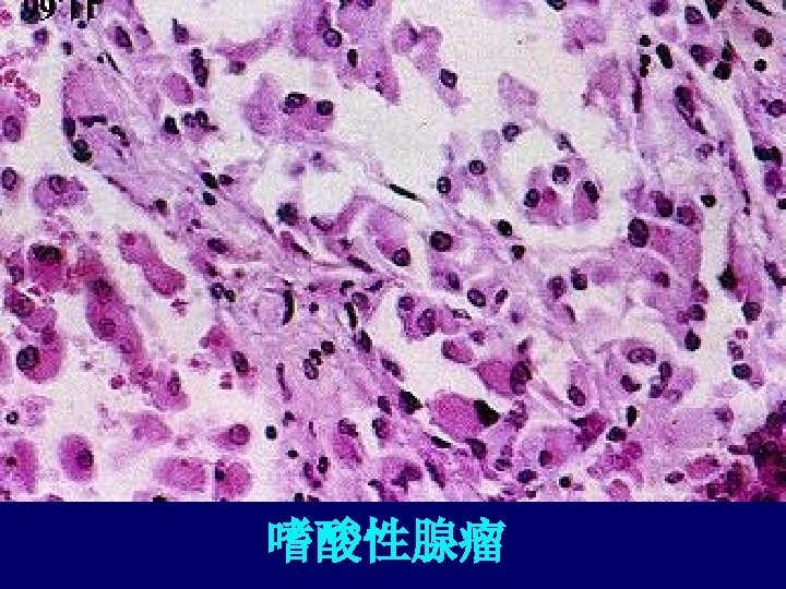
adenomas 1. solitary, spherical, encapsulated lesion 2. cut surface: gray white to red brown; regressive change 3. microscopically : form relatively uniform, normal-appearing follicles that contain colloid 4. Well-defined, intact capsule 5. histologic subtypes
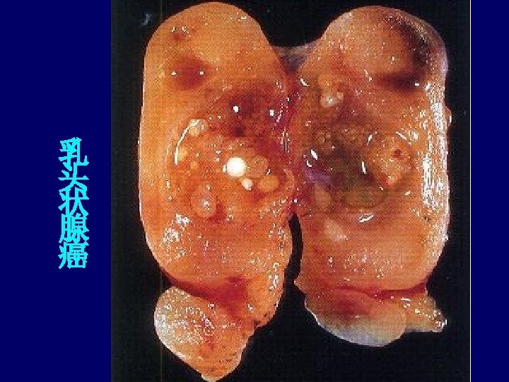
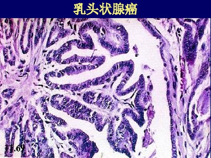
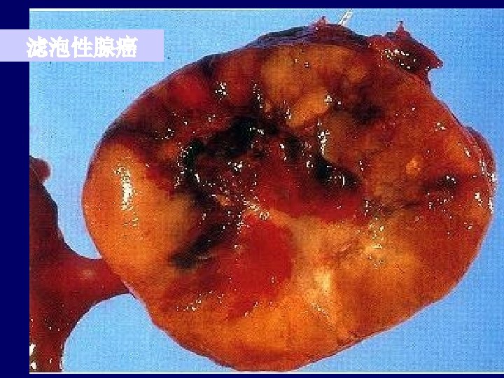
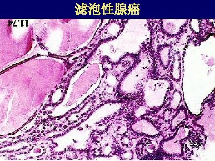
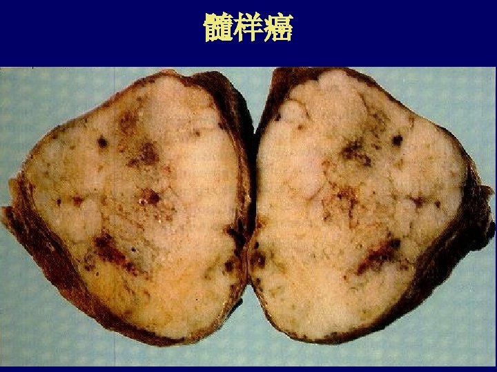
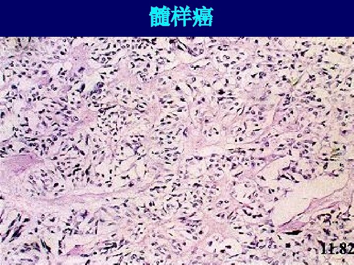
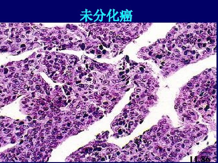
carcinoma 1. papillary carcinoma 2. follicular carcinoma 3. medullary carcinoma 4. anaplastic carcinoma