Department of Human Anatomy KNMU MUSCLES AND TOPOGRAPHY


 can be")


- Slides: 64
Department of Human Anatomy KNMU MUSCLES AND TOPOGRAPHY OF THE UPPER AND LOWER LIMBS Slide-lecture for students of the 6 Faculty of Medicine Lector – associate professor Zharova Nataliya 2015
• PLAN 1. 2. 3. 4. 5. 6. 7. 8. 9. 10. 11. 12. 13. Muscles of the upper limb. Muscles and topography of the shoulder girdle. Muscles and topography of the upper arm. Muscles and topography of the forearm. Muscles and topography of the hand. Fasciae of the upper limb. The synovial bursae of the upper limb. The muscles of the lower limb. Muscles and topography of the pelvic girdle. Muscles and topography of the thigh. Muscles and topography of the leg. Muscles and topography of the foot. The synovial bursae of the lower limb.
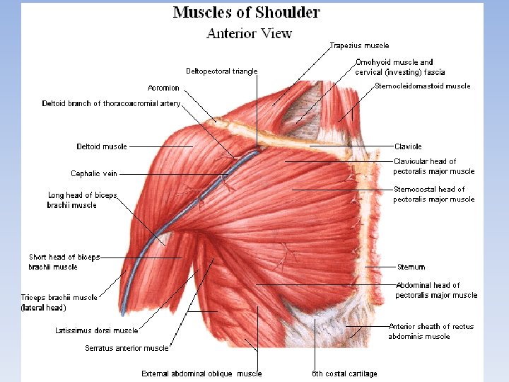
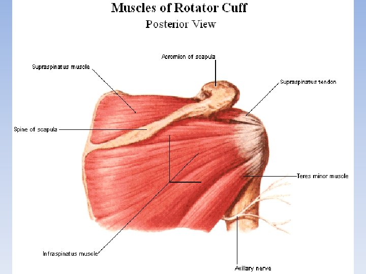
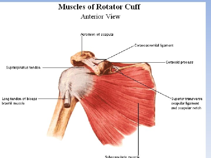
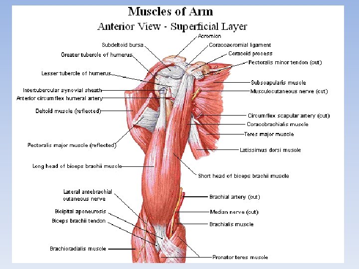
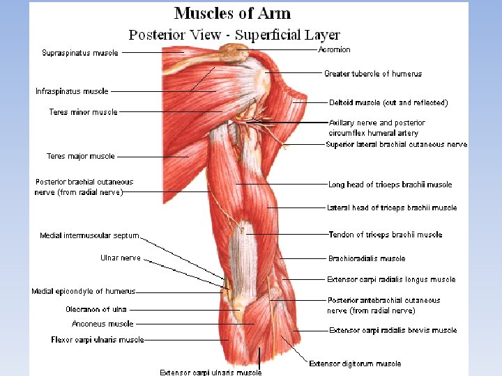
MUSCLES OF THE UPPER LIMB The muscles of the upper limb can be classified into the muscles of the shoulder joint, the muscles of the arm, the muscles of the forearm and the muscles of the hand. Muscles of the Shoulder joint The Dorsal group 1. The deltoid muscle 2. The supraspinatus muscle 3. The infraspinatus muscle 4. The teres minor muscle 5. The teres major muscle The Ventral group 1. The subscapular muscle
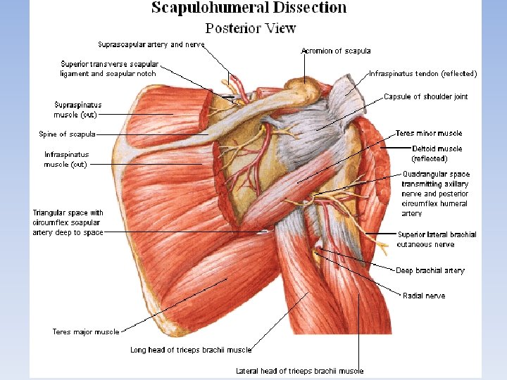
The axillary cavity Walls. The axillary cavity has four walls formed by the following muscles: • anterior — pectoralis major and minor; • posterior — subscapularis, teres major, and latissimus dorsi; • medial — serratus anterior; • lateral — short head of the biceps brachii, coracobrachialis, and humerus. On the posterior wall are 2 openings: • quadrangular opening bounded by the teres major (below), subscapularis (above), long head of the triceps brachii (medially), and humerus (laterally); • triangular opening bounded by the teres major (below), subscapularis (above), and long head of the triceps brachii (laterally).
Triangles of the anterior wall of the axillary cavity. For better orientation, three triangles are distinguished on the anterior wall of the axillary cavity: • clavipectoral triangle resides between the clavicle and the upper border of the pectoralis minor; • pectoral triangle corresponds to the projection of the pectoralis minor; • subpectoral triangle located between the inferior borders of the pectoralis minor (above) and pectoralis major (below); laterally it is bounded by the deltoid. Between the pectoralis major and deltoid there is a deep deltoidopectoral groove.
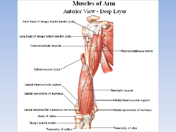
Muscles of the Upper Arm The muscles of the upper arm are concerned with the movements at the shoulder and elbow joints on the frontal axis and, therefore, are situated on the anterior (two flexors) and posterior (two extensors) surfaces of the upper arm and attach to the bones of the forearm. Anterior muscles Posterior muscles of the Upper Arm (extensors) (flexors) 1. The biceps brachii muscle 1. The triceps brachii muscle 2. The coracobrachial muscle 2. Elbow’s muscle
The topography of the upper arm • Radial canal is located behind the humerus. It is bounded by the radial groove of the humerus in the front and by the triceps brachii in the back. The canal has a spiral path. The radial canal begins on the medial surface of the arm between the medial and lateral heads of the triceps brachii and exits on the lateral surface of the arm between the brachialis and brachio-radialis. The canal gives passage to the radial nerve and a. profunda brachii. • Medial bicipital groove lies medially between the biceps brachii and brachialis; it transmits the main neurovascular bundle of the upper arm. • Lateral bicipital groove resides laterally between the biceps brachii and brachialis; it gives passage to the cephalic vein.
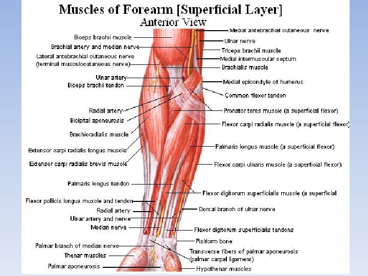
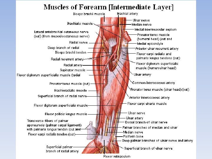
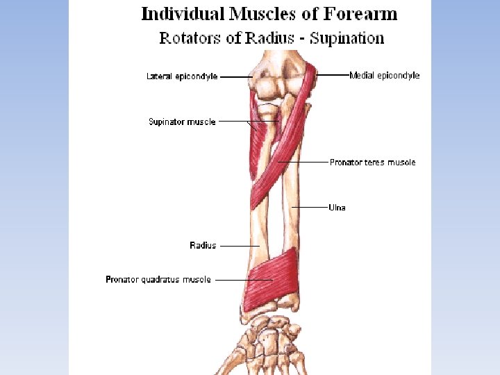
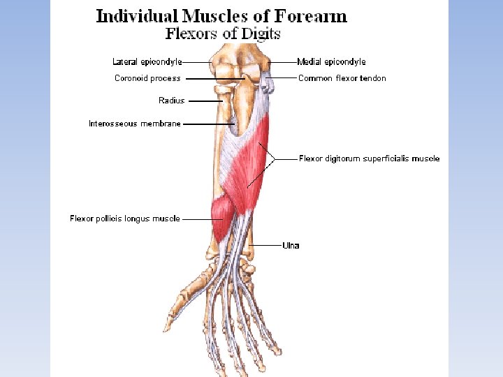
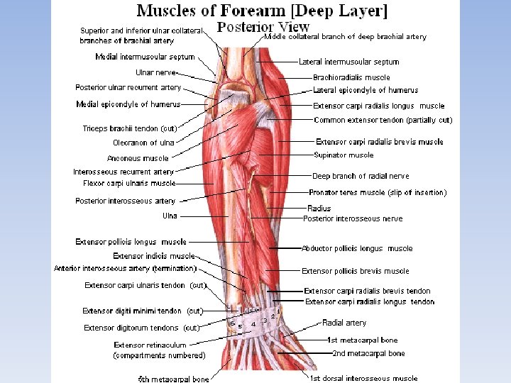
Muscles of the Forearm The muscles of the forearm are separated into two groups according to position and function: the anterior group is composed of flexors and pronators, the posterior group is composed of extensors and supinator. Each group consists of a superficial and deep layers. The Anterior group, Superficial layer The Anterior Group, (flexors) Deep layer (flexors) 1. The round pronator muscle 6. The long flexors of the thumb 2. The radial flexor of the wrist 7. The deep flexor of the fingers 3. The long palmar muscle 8. The square pronator muscle 4. The ulnar flexor of the wrist 5. The superficial flexor of the fingers
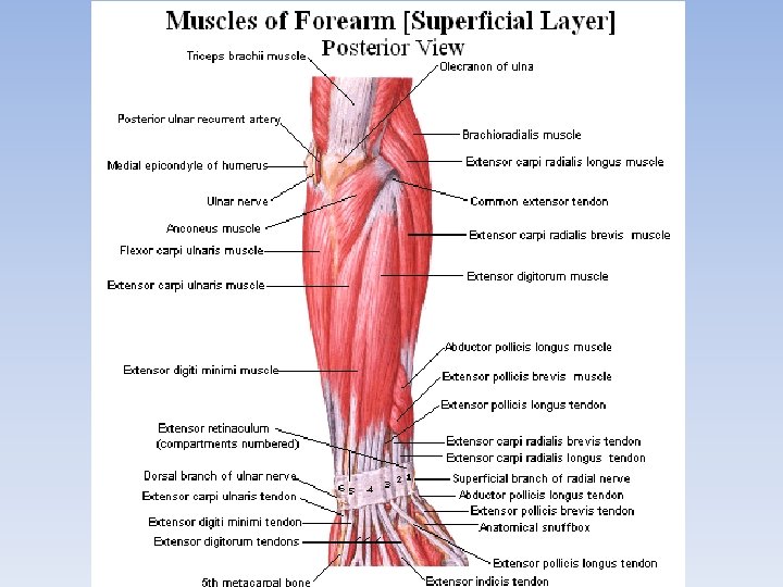
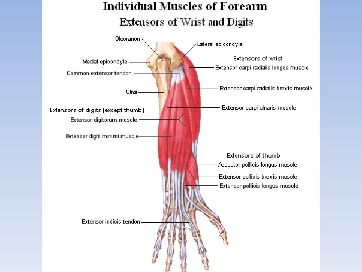
The Posterior group The muscles of the posterior group of the forearm are divided into two subgroups: radial and ulnar. The first occupies the anterolateral surface of the forearm, while the second occupies the posterior surface and is formed by the superficial and deep layers. The Posterior Radial Group 1. The brachioradial muscle The Posterior Ulnar group Superficial layer 2. The long radial extensor of the wrist 4. The common extensor of the fingers 3. The short radial extensor of the wrist 5. The extensor of the little finger 6. The ulnar extensor of the wrist Deep layer 7. The supinator muscle 8. The long abductor of the thumb 9. The short extensor of the thumb 10. The long extensor of the thumb 11. The extensor of the index
The topography of the forearm • The cubital fossa is bounded by the brachialis above; below it is bounded by the brachioradialis (laterally) and pronator teres (medially). Within the borders of the cubital fossa there are two grooves located on each side from the brachialis — medial cubital groove and lateral cubital groove.
The topography of the forearm The ulnar groove of the forearm lies between the flexor carpi ulnaris and flexor digitorum superficialis. It transmits the ulnar nerve, artery, and vein. • The median groove of the forearm resides in the lower part of the forearm between the flexor carpi radialis and flexor digitorum superficialis. It contains the median nerve. • • The radial groove runs between the flexor carpi radialis and brachioradialis. It transmits the radial artery, vein, and nerve.
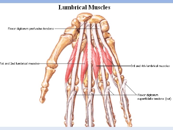
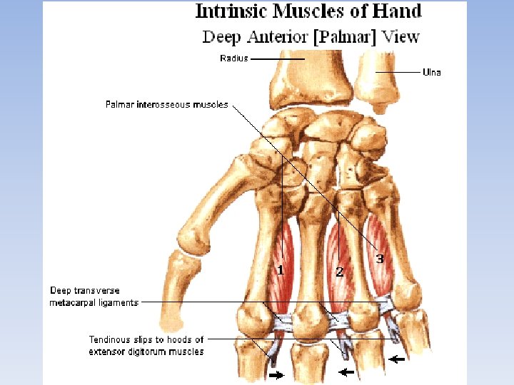
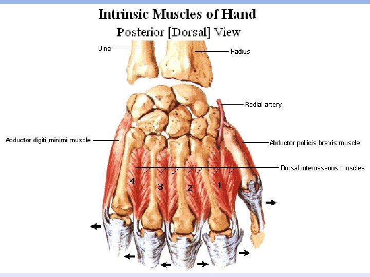
Muscles of the Hand Thenar muscles of The Hypothenar muscles Muscles of the Hollow of the. Hand of the Hand 1. The short abductor of the 1. The short palmar muscle 1. The lumbrical (worm-like) thumb 2. The abductor of the little muscles 2. The short flexor of the finger 2. The interossei muscles thumb 3. The short flexor of the -The palmar interossei 3. The opponens muscle of little finger muscles the thumb 4. The opponens muscle of -- The dorsal interossei 4. The adductor muscle of the little finger muscles the thumb
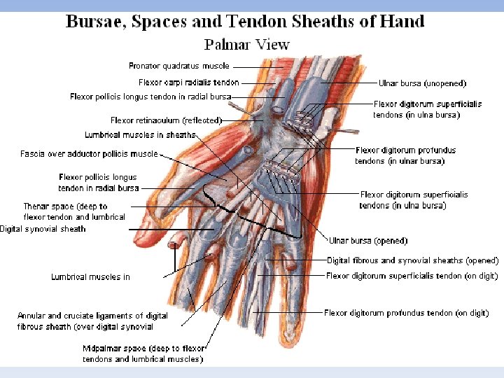
The topography of the hand The carpal canal is located in the region of the carpus underneath the flexor retinaculum. It gives passage to the tendons of the flexor digitorum superficialis, flexor digitorum profundus, flexor pollicis longus, and the median nerve. • On both sides from the carpal canal, the flexor retinaculum splits and forms another two canals — the radial carpal canal and ulnar carpal. • The radial carpal canal gives passage to the tendon of the flexor carpi radialis, while the ulnar carpal canal transmits the ulnar artery, vein, and nerve. The fibrous sheaths of the digits of the hand are formed by the dense fibrous lamina, which fuse with the bones. As a result, osteofibrous canals are formed, which contain tendons of the flexors covered by the synovial sheath. Each fibrous sheath consists of the anular part and cruciform part, the less dense cruciform part is situated in the joint region.
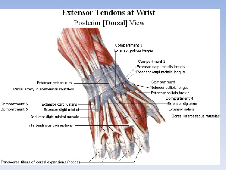
Six canals on the dorsal surface of the wrist joint are counted from the radial to the ulnar border and transmit following tendons: 1. the first canal transmits the tendons of the m. abductor pollicis longus and m. extensor pollicis brevis; 2. the second canal transmits the tendons of the m. m. extensor carpi radialis longus and brevis; 3. the third canal transmits the tendon of the m. extensor pollicis longus, and crosses the preceding canal obliquely; 4. the fourth canal transmits the tendons of the m. extensor digitorum and m. extensor indicis; 5. the fifth canal transmits the tendon of the m. extensor digiti minimi; 6. the sixth canal transmits the tendon of the m. extensor carpi ulnaris.
FASCIAE OF THE UPPER LIMB • • • 1. The deltoid fascia 2. The supraspinous fascia 3. The infraspinous fascia 4. The brachial fascia 5. The antebrachial fascia 6. The palmar aponeurosis 7. The deep palmar fascia 8. The dorsal fascia of the hand 9. flexor retinaculum 10. extensor retinaculum
THE SYNOVIAL BURSAE OF THE UPPER LIMB 1. 2. 3. 4. 5. 6. 7. subdeltoid bursa resides between the deltoid and greater tubercle of the humerus; subacromial bursa lies under the acromion of the scapula; subcutaneous acromial bursa lies superficially under the skin in the region of the acromion of the scapula; subtendinous bursa of subscapularis is located near the attachment point of the subscapularis; the bursa communicates with the cavity of the shoulder joint; subtendinous bursa of latissimus dorsi resides under the tendon near the point of attachment of the muscle on the humerus; there is also another small bursa under the tendon of the teres major. In the region of the elbow joint the following bursae are found: bicipitoradial bursa resides between the tendon of the biceps brachii and the tuberosity of the radius; subcutaneous olecranon bursa wide, lies under the skin of the olecranon.
• Clinical applications. The inflammatory diseases of the synovial bursae (bursitis) can be acute or chronic. In acute bursitis, pus can accumulate in the bursa, which requires surgical intervention. Chronic bursitis (gygroma) is often an occupational disease. In the shoulder girdle area, inflammatory processes are most common in the subcutaneous acromial bursa and in the subdeltoid bursa (among porters). In the region of the elbow joint, the subcutaneous olecranon bursa may become inflamed (among tanners and etchers).
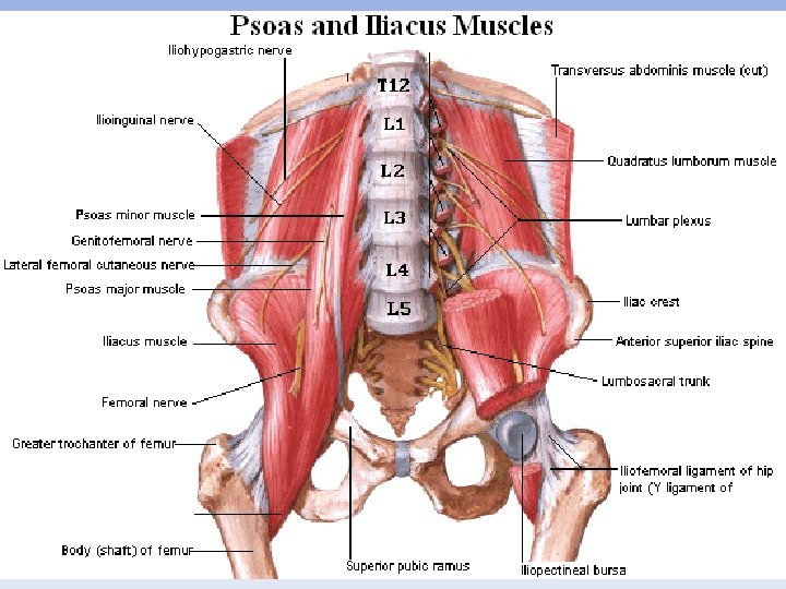
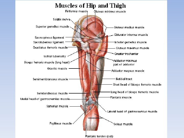
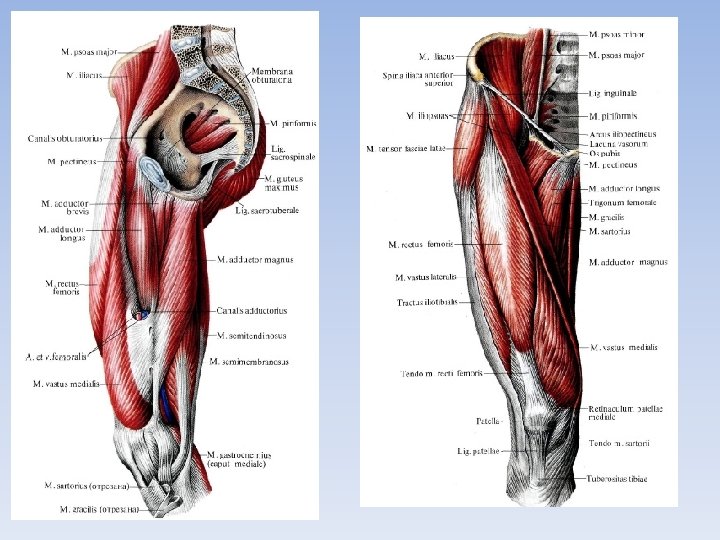
THE MUSCLES OF THE LOWER LIMB All other muscles of the lower limb are autochthonous. They are grouped into the muscles of the pelvic girdle, thigh, leg, and foot. MUSCLES OF THE PELVIC GIRDLE The muscules of the pelvic girdle are divided into anterior group (flexors) and posterior group (extensors, rotators and abductors), which pass from the pelvic girdle to the femur and allow movements at the hip joint. The Anterior Group 1. The iliopsoas muscle 1) The greater psoas muscle 2) The iliacus muscle 2. The lesser psoas muscle The Posterior group 1. The gluteus maximus muscle 2. The gluteus medius muscle 3. The gluteus minimus muscle 4. The tensor fascial latae muscle 5. The piriformis muscle 6. The obturator internus muscle 7 -8. The gemellus superior and inferior muscles 9. The quadratus femoris muscle 10. The obturator externus muscle
THE TOPOGRAPHY OF THE LOWER LIMB On the lower limb there are several topographo-anatomical structures, which have a clinical significance. THE REGION OF THE PELVIC GIRDLE – The suprapiriform foramen resides in the region of the ischium within the greater sciatic foramen above the piriformis. It gives passage to the superior sciatic vessels and nerve. – The infrapiriformf oramen is located below the piriformis in the same area. This foramen transmits the inferior sciatic vessels and nerves. – The obturator canal is bounded by the obturator groove of the pubis, obturator membrane, and by the obturator internus. The canal transmits the obturator vessels and nerve. • Clinical applications. The supra- and infra- piriform foramina as well as the obturator canal may serve as the exit location for the hernias (sciatic and obturator hernias). The purulent masses from the pelvis may spread through these openings into the gluteal region.
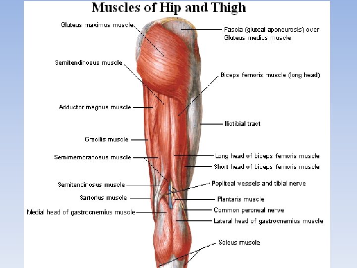
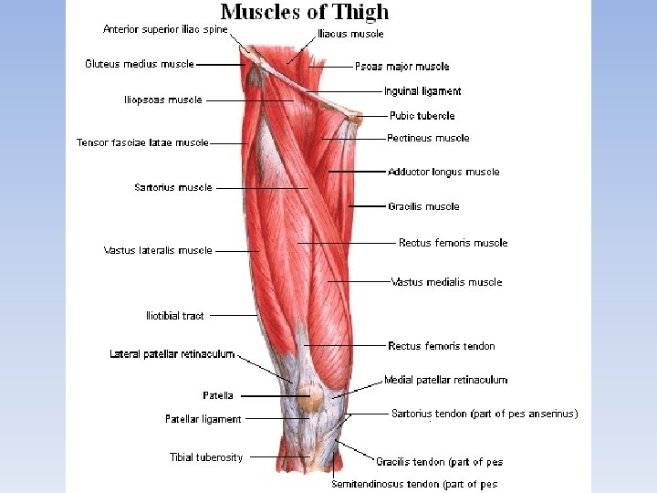
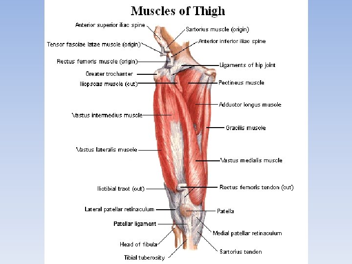
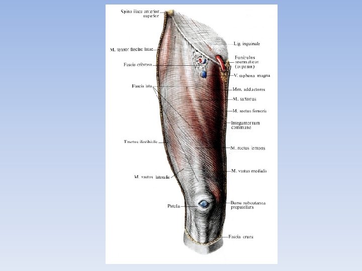
MUSCLES OF THE THIGH The muscles of the thigh are divided into three groups: anterior (extensors of the leg and flexors of the thigh), posterior (flexors of the leg and extensors of the thigh) and medial (adductors). The last group acts only on the hip joint, where as the first two groups act on the knee joint also. The Anterior Group The Posterior Group (extensors) (flexors) 1. The quadriceps femoris muscle 1. The semitendinosus muscle 1) The rectus femoris muscle 2. The semimembranosus muscle 2) The vastus lateralis muscle 3. The biceps femoris muscle 3) The vastus medialis muscle 4. The popliteus muscle 4) The vastus intermedius muscle 2. The sartorius or tailor’s muscle 3. The articular muscle of the knee
THE REGION OF THE THIGH The muscular space resides below the inguinal ligament on the lateral side. It is bounded: • medially — by the thickened region of the fascial layer — iliopectineal arch, in the front and above — by the inguinal ligament, • laterally and in the back — by the ilium. The muscular space gives passage to the iliopsoas and femoral nerve. The vascular space resides behind the inguinal ligament, medially from the muscular space, from which it is separated by the iliopectineal arch. • In the front and above the vascular space is bounded by the inguinal ligament, • posteriorly — by the thick periosteum of the pubis, • laterally — by the iliopectineal arch, • medially — by the lacunar ligament. The vascular space transmits the femoral artery (resides laterally) and femoral vein (resides medially).
THE REGION OF THE THIGH The femoral triangle resides on the anterior surface of the thigh. • It is bounded by the inguinal ligament above, sartorius laterally, and adductor longus medially. • The triangle contains the main neurovascular bundle of the thigh and lymph nodes.
THE REGION OF THE THIGH The femoral ring resides behind the inguinal ligament, medially from the femoral vein; the ring is a part of the vascular space. The femoral vein forms the lateral wall of the femoral ring. From the medial side it is bounded by the arcuate fibers, which descend from the inguinal ligament and called the lacunar ligament. The anterior wall of the femoral ring is formed by the inguinal ligament, and posterior — by the thickened periosteum of the pubis. • The femoral canal. The femoral ring is a weak spot under the inguinal ligament, which can serve as an exit location for the femoral hernias. When the hernia passes through the femoral canal is formed. The internal aperture of the femoral canal is the femoral ring. Passing through the femoral ring, the hernia lies between the superficial and deep layers of the fascia lata, which form the anterior and posterior walls of the femoral canal. The lateral wall of the canal is formed by the femoral vein. The hernias emerge onto the skin through the saphenous opening in the superficial layer of the fascia lata, which becomes the external aperture of the femoral canal.
The iliopectineal groove lies between the pectineus and iliopsoas within the borders of the femoral triangle. The anterior femoral groove passes between the vastus medialis and adductor longus. It is bounded by the vastus medialis laterally and adductor longus and magnus medially. It is anterior wall is represented by the tendineous plate, which bridges over these muscles, lamina vastoadductoria. At the downward-facing apex of the femoral triangle the anterior femoral groove transforms into an adductor canal. • The canal leads into the popliteal fossa, where it opens. This opening is formed by the cleft in the tendon of the adductor magnus — adductor hiatus. The canal gives passage to the femoral vessels and saphenous nerve.
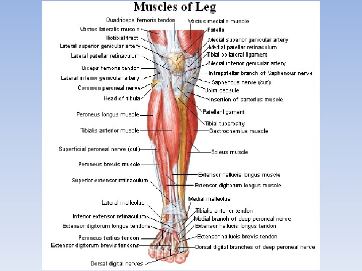
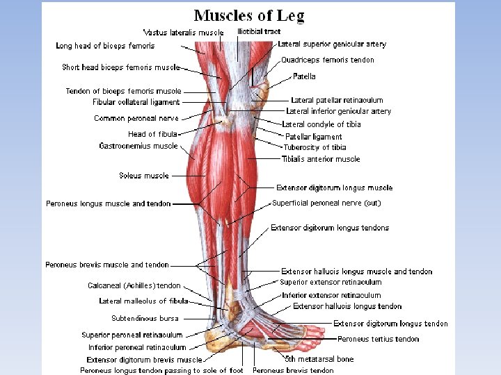
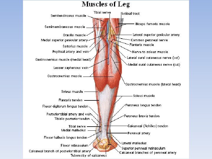
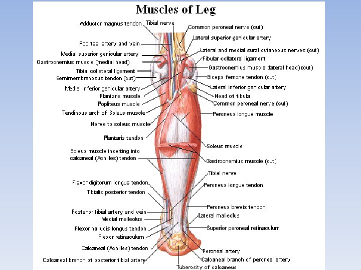
MUSCLES OF THE LEG The muscles of the leg are divided into three groups: the anterior (extensors), the posterior and the lateral (flexors). The Anterior Group (extensors) 1. The anterior tibial muscle 2. The long extensor of the toes 3. The peroneus tertius muscle 4. The long extensor of the big toe The Lateral Group (flexors) 1. The long peroneal muscle 2. The short peroneal muscle The Posterior Group (flexors) Superficial layer 1. The triceps surae muscle 2. The gastrocnemius muscle 3. The soleus muscle 4. The plantaris muscle Deep layer 1. The long flexor of the toes 2. The posterior tibial muscle 3. The long flexor of the big toe
THE REGION OF THE LEG The popliteal fossa resides in the back on • • the border between the thigh and the leg. It is rhomboid in shape. Above, it is bounded by the biceps femoris (laterally), semitendinosus and semimembranosus (medially). Below, the fossa is bounded by the two heads of the gastrocnemius. The bottom of the popliteal fossa (anterior wall) is formed by the popliteal surface, facies poplitea, of the femur and the capsule of the knee joint. The main neurovascular bundle passes through the popliteal fossa. The lymph nodes and vessels occupy the fossa along with the adipose tissue.
THE REGION OF THE LEG The cruropopliteal canal leads from the popliteal fossa into the leg. It resides in the back between the deep muscles of the leg and the soleus. • Therefore, its anterior wall is formed by the tibialis posterior, while the anterior wall — by the soleus. • The canal has three openings — superior, inferior, and anterior. The superior opening of the canal is bounded by the popliteus in the front and by the tendineous arch of the soleus in the back. • The inferior opening resides between the tibialis posterior and soleus, where the latter becomes the Achilles tendon. The anterior opening is located in the upper part of the interosseous membrane of the leg. The inferior musculoperoneal canal is the branch of the cruropopliteal canal in the lateral direction. Its anterior wall is formed by the posterior surface of the fibula, while its posterior wall — by the flexor hallucis longus. It transmits the fibular vessels. The superior musculoperoneal canal is an independent canal, which resides in the upper third of the leg between the lateral surface of the fibula and peroneus longus. It gives passage to the superficial peroneal nerve.
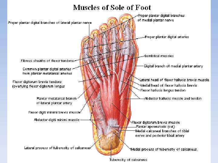
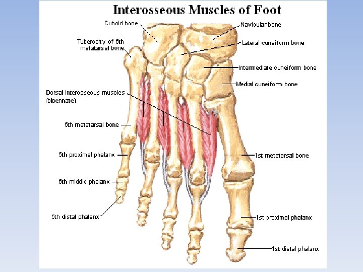
MUSCLES OF THE FOOT • The foot, like the hand, in addition to tendons of the long muscles of the leg descending on it, has its own short muscles among which are dorsal and plantar muscles. The Dorsal muscles of the Foot • The short extensor of the toes (m. extensor digitorum brevis) lies on the dorsal surface of the foot under the tendons of the extensor digitorum longus. The Plantar muscles of the Foot • The muscle of the sole of the foot form three groups: medial (muscles of the big toe), lateral (muscles of the little toe) and middle group situated in the middle of the sole.
Synovial bursae on the lower limb: In the region of the hip joint: 1. subcutaneous trochanteric bursa spacious, resides under the skin in the region of the greater trochanter of the femur; 2. trochanteric bursae of gluteal muscles located between the greater trochanter and tendons of each of the gluteal muscles — gluteus maximus, medius, and minimus; 3. bursa of piriformis resides between its tendon and the greater trochanter; 4. sciatic bursa of obturator intemus lies where the muscle passes around the margin of the lesser sciatic notch; 5. subtendinous bursa of iliacus resides betweenthe tendon of the iliopsoas and lesser trochanter of the femur. Clinical applications. In the region of the hip joint, inflammations (bursitis) of the subtendineous bursa of the iliac occur, which cause painful sensations. Frequently, the subcutaneous trochanteric bursa becomes inflamed. In this case, pain and swelling are located in the region of the greater trochanter.
In the region of the knee joint: 1. suprapatellar bursa resides under the tendon of the quadriceps femoris; it is extensively connected with the knee joint (see the knee joint); 2. subcutaneous prepatellar bursa spacious, resides under the skin in front of the patella; 3. subcutaneous infrapatellar bursa resides under the skin below the patella; 4. deep infrapatellar bursa resides between the tendon of the quadriceps femoris (patellar ligament, Iig. patellae) and tibia; 5. anserine bursa lies under the tendineous extension of the sartorius, gracilis, and semitendinosus near their attachments on the tibia; 6. subtendinous bursae reside near the points of attachments of the tendons of the biceps femoris and lateral head of the gastrocnemius. • Clinical applications. In the region of the knee joint, inflammations of the subcutaneous prepatellar bursa are fairly common (prepatellar bursitis), which are characterized by the appearance of a round fluctuating swelling in the front of the patella. In the popliteal fossa, inflammation of the bursae located under the muscular tendons also occurs.
In the region of the talocrural joint: • • • subcutaneous calcaneal bursa resides under the skin in the region of the calcaneal tuberosity; bursa of tendo calcaneus (Achilles) lies between the calcaneal tendon and the calcaneus; subcutaneous bursae of malleoli reside under the skin in the regions of the malleoli.
THE END