Department of Anatomy DEFINITION The term connective tissue


 Specialised for synthesis & storage of fat. Each cell shows")


- Slides: 44
Department of Anatomy
DEFINITION The term connective tissue is applied to a tissue which fills the spaces between more specialized elements and serves to hold them together and support them.
TYPES 1. Ordinary Connective tissue 2. Mucoid tissue 3. Special Connective tissue
Ordinary connective tissue Irregular Connevtive Tissue Loose connective tissue –seen where fat is absent-eyelid penis scrotum, labia minora Dense connective tissue –dermis, muscles vessels, nerves Adipose tissue Dense Regular Connective Tissue: Fascia , Tendons, aponeurosis
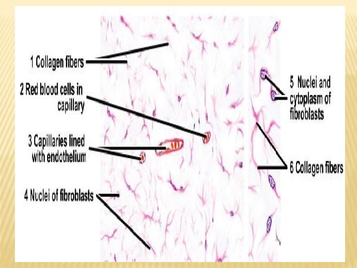
1. ORDINARY CONNECTIVE TISSUE Made up of Cells - fixed - wandering Intercellular matrix - ground substance - fibres
Fibres of Connective Tissue 1. Collagen fibres 2. Elastic fibres 3. Reticular fibres
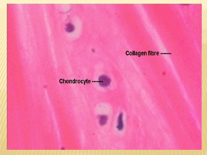
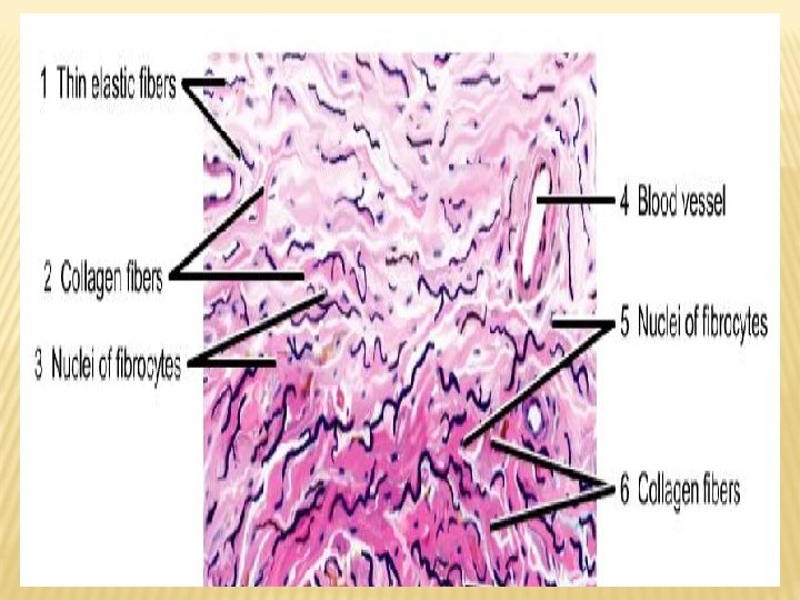
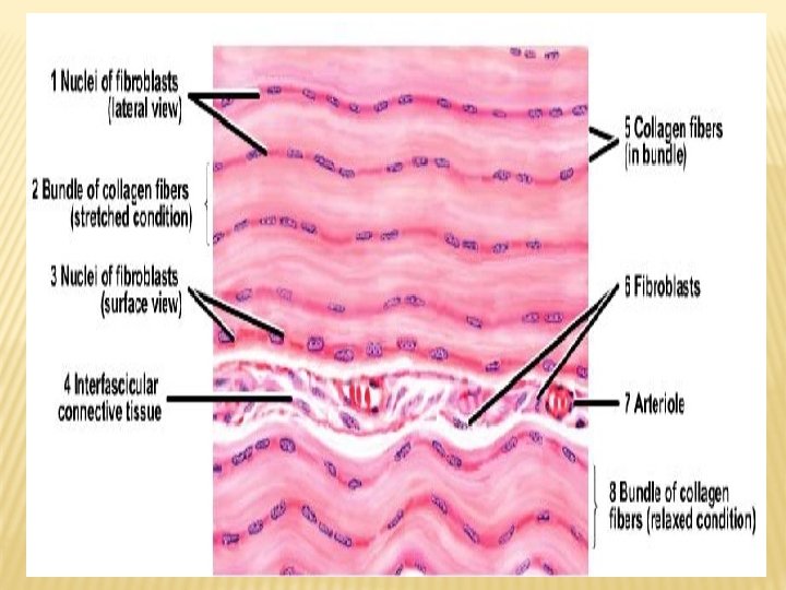
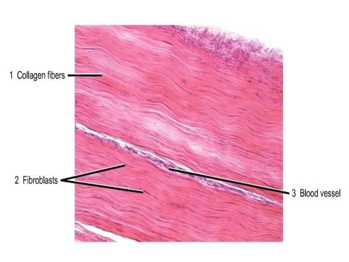
1. COLLAGEN FIBRES Made up of wavy bundles of collagen fibrils. Bundles split into branches, individual fibres do not branch. Synthesized by fibroblasts. Stains pink with H & E Staining. In Unstained preparations looks white – white fibres. Can resist considerable tensile forces.
Chemically, protein is COLLAGEN, made up of molecules of TROPOCOLLAGEN
Types Of Collagen Fibres Type - in tendons, ligaments I - larger, prominent Type II-striations in hyaline cartilage - smaller, less prominent striations Type III - reticular fibres Type IV - lens capsule More than 20 types of fibres have been identified
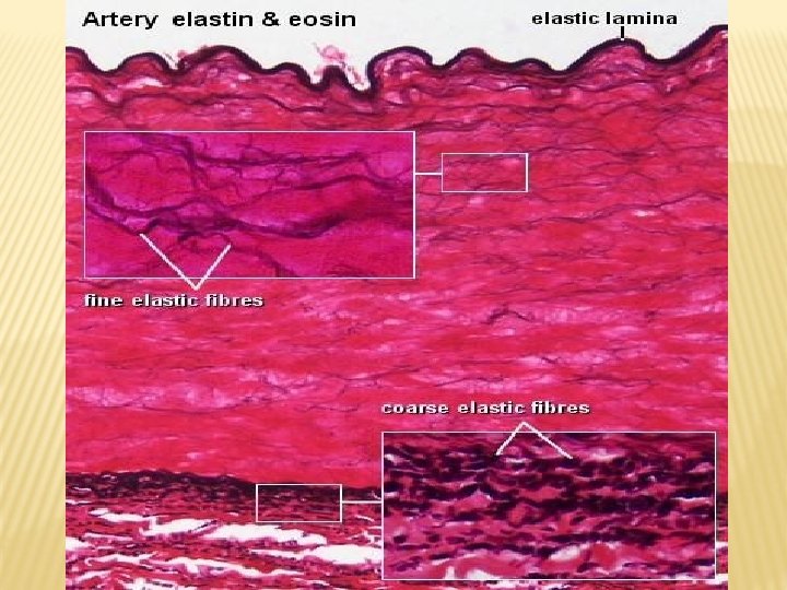
2. ELASTIC FIBRES Run singly not in bundles The individual fibres branch & anastomose with one another. When fibres are cut , the ends retract & recoil. Synthesized by fibroblasts Take a weak pink stain with H & E. Unstained fibres appear Thinner than collagen fibres (0. 1 - 0. 2 nm) Can be stretched like a rubber band & return to original yellow in color.
Chemically, protein is ELASTIN, made up of molecules of TROPOELASTIN. Also contains a glycoprotein FIBRILLIN.
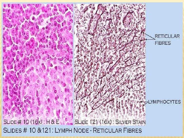
3. RETICULAR FIBRES Very fine branched fibres that are characteristically arranged in delicate networks. The networks hold the cells in place. Seen in lymphoid organs, liver & bone marrow. Do not take up H & E stain, can be stained by silver salts, -ARGENTOPHIL FIBRES. Synthesized by reticular cells or probably by fibroblasts.
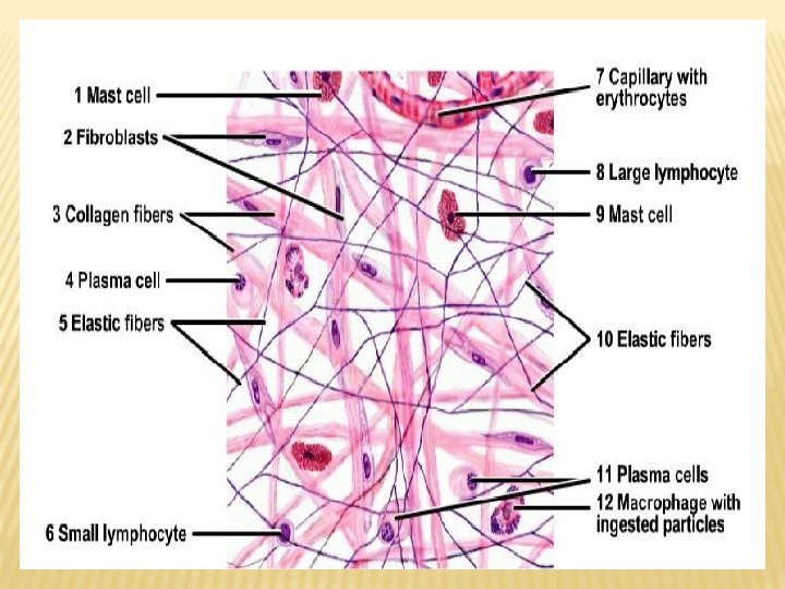
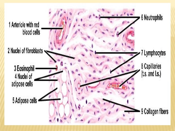
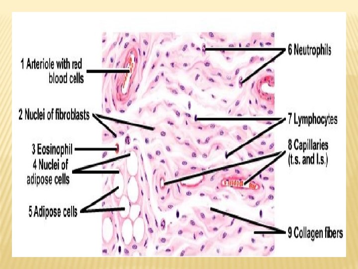
CELLS OF CONNECTIVE TISSUE FIXED CELLS - fibroblasts, fat cells. WANDERING CELLS – macrophages, plasma cells, mast cells & lymphocytes
1. FIXED CELLS 1. Fibroblasts: Stem cells with multiple processes. Spindle shaped Inactive –FIBROCYTES Found in all types of connective tissue. Produce & maintain fibres & ground substance.
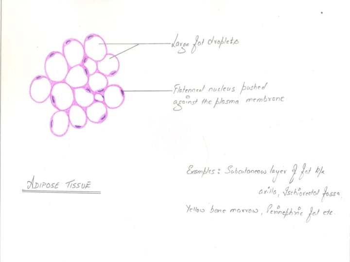
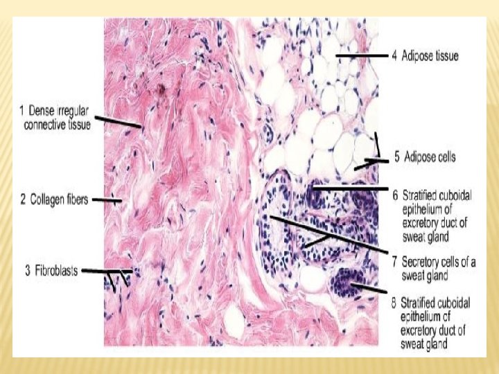
2. Fat cells (Adipocytes) Specialised for synthesis & storage of fat. Each cell shows a large globule of fat. The nucleus is flattened pushed to the periphery. The cytoplasm appears as a thin rim around the fat globule. Signet ring appearance.
2. WANDERING CELLS 1. Macrophages : Histiocytes Big eater, oval in shape. Found in the connective tissues of almost all organs of the body. Phagocytosis - worn out RBCs , Carbon particles.
2. Plasma Cells Components of loose areolar tissue as well as lymphatic tissue. Cell is rounded but eccentrically placed nucleus. Chromatin in the nucleus is often arranged like spokes of a wheel (cart wheel appearance). Synthesizes and secretes Antibodies. They develop from B lymphocytes.
3. Mast Cells Round or Ovoid in shape. Nucleusis small and centrally placed. Cytoplasm is packed with coarse granules. These granules contain Histamine & Heparin.
4. WBCs like Lymphocytes & Neutrophils Lymphocytes – small or large rounded cells with a large darkly stained nucleus. . Neutrophils – 3 to 5 lobes of nuclei with small or fine eosinophilic
GROUND SUBSTANCE The interfibrillar substance is called ground substance or matrix. It is made of Glycosaminoglycans, Proteoglycans & water.
Hyaluronic acid Chondrat in Dermata n Kerata n Hepara n Vitreous body Cartilag e Ski n Corne a Live r
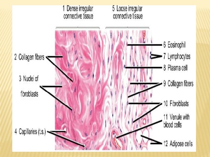
TYPES OF ORDINARY CONNECTIVE TISSUE 1. LOOSE CONNECTIVE TISSUE – found almost everywhere in the body as a thin filling & made of fibres, cells & ground substance.
2. DENSE CONNECTIVE TISSUE Here the fibres are closely packed with very little matrix and cells. 2 types Irregular & regular
DENSE IRREGULAR CONNECTIVE TISSUE Found in regions which experience considerable mechanical stress & where protection is needed. Fibre bundles interweave in 3 dimensions giving it considerable strength. Eg : Adventitia , Periosteum
DENSE REGULAR CONNECTIVE TISSUE Forms sheets or bundles wherein the direction of arrangement of fibres is related to the stresses which they undergo. Eg : Tendons , ligaments
2. MUCOID TISSUE An embryonic type of connective tissue typically seen in the Whartons jelly. Consists of copious matrix, fine mesh of collagen & stellate cells with long processes. In adults it is seen in the Vitreous body of the eye & nucleus pulposus of intervertebral disc.
3. SPECIAL CONNECTIVE TISSUE Bone Cartilag e
SUMMARY Connective tissue is one of the basic tissues of the body which connects various structures. It is mainly made up of fibres, cells & ground substance. The composition may vary – dense, loose or mucoid. 3 types of fibres – collagen, elastic & reticular fibres. The connective tissue cells may be fixed or they may wander in the surrounding tissues. Connective tissue helps in regenerations and repair of tissues.
Applied Anatomy 1. Disease of the collagen fibre. Fibrinoid Necrosis: Rhematoid Fever Rhematoid arthritis 2. Marfans Syndrome : Caused by defective Fibrillin gene Results abnormal development of the elastic fiberes