DentinDentinogenesis Dentin Forms the bulk of the tooth

Dentin-Dentinogenesis

Dentin • • • Forms the bulk of the tooth, elasticity Protected tissue (by enamel and cementum) Predentin (dentin) ~ Osteoid (bone) Dentinal tubules Odontoblasts (present in the pulp) Mature dentin: 70% inorganic, 20% organic, 10% water • Primarily type I (~56%), small amounts of III & V • Other dentin proteins – DPP, DSP, DGP, DMP 1, osteocalcin, osteopontin etc, amelogenin – Promoters and inhibitors of mineralization

Non-collagenous proteins • • DPP dentin phosphoprotein/phosphophorin DSP dentin sialoprotein DGP dentin glycoprotein DMP 1 Osteonectin (SPARC) Osteocalcin Bone sialoprotein Osteoprotein

Dentin Sialophosphoprotein • Composed of – DPP binds calcium; collagen, initiates hydroxyapatite formation – DSP peritubular dentin; prevent occlusion of tubules – DGP
 dentin: Most of the dentin – Mantle dentin (FIRST DENTIN):")
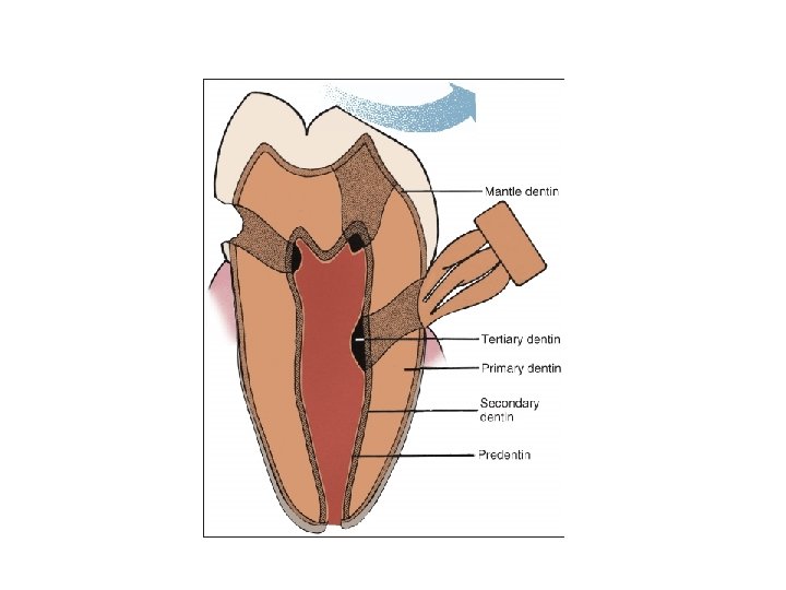
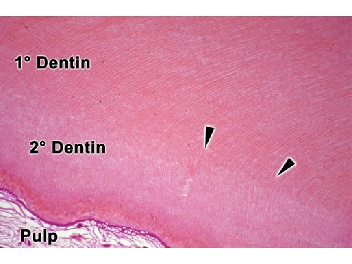
Dentin • Primary (circumpulpal) dentin: Most of the dentin – Mantle dentin (FIRST DENTIN): outer layer of coronal dentin. • Different properties (collagen distribution and orientation) in the crown compared to the root • Secondary – – – After root formation By odontoblasts that form primary Demarcation line Continuous but slower odontoblastic activity Reduction of pulpal chamber size (recession) • Tertiary – Reactionary (preexisting odontoblasts) or reparative (newly differentiated cells) dentin – Reaction to attrition, caries or restorative procedure – Site-specific. Produced only by those cells directly stimulated – Tubular or atubular / osteodentin

Essentials of Oral Histology and Embryology, Ed: James Avery, 3 rd edition.

Essentials of Oral Histology and Embryology, Ed: James Avery, 3 rd edition.
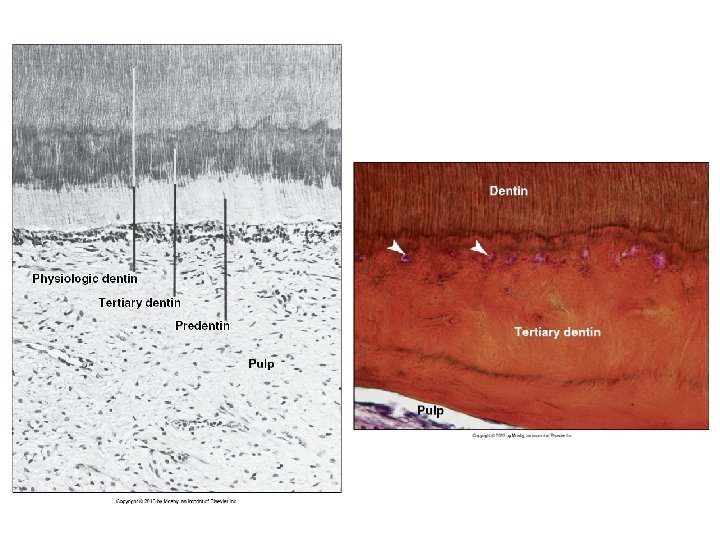

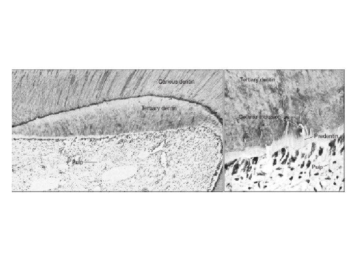
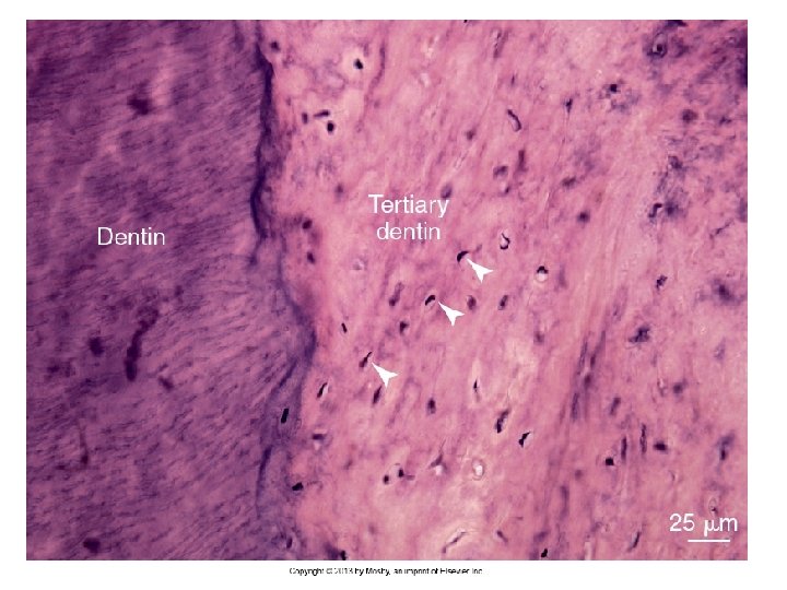
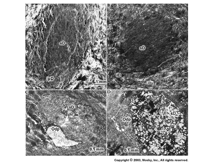
Tertiary Dentin Rate of deposition depends on the degree of injury More severe injury: more rapid rate of dentin deposition Due to rapid rate of deposition cells get entrapped in the newly formed matrix and tubular pattern becomes grossly distorted Essentials of Oral Histology and Embryology, Ed: James Avery, 3 rd edition.

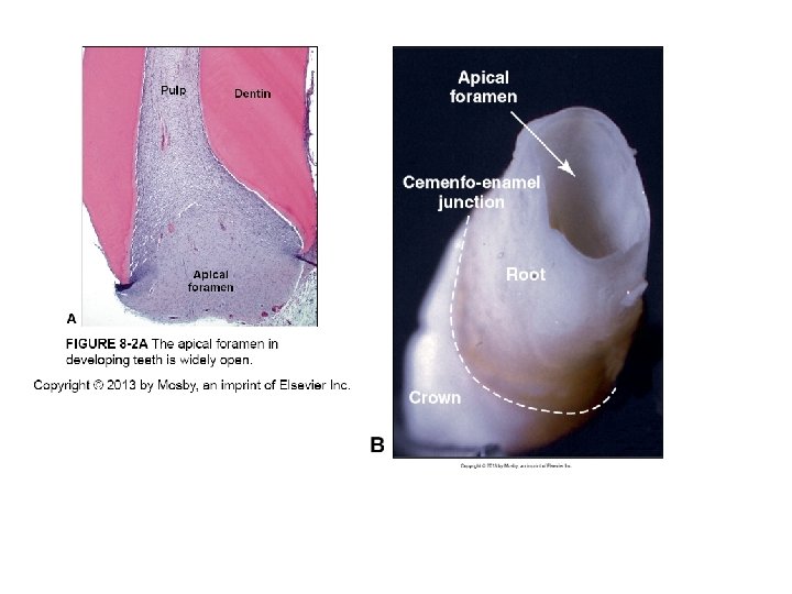
Dentin formation • • Begins at bell stage From cusp tips and down the slope Coronal and radicular dentin Completion of root dentin occurs after tooth eruption (open apex) • Completion of root dentin formation does not occur in the primary tooth until 18 months after eruption; and 2 -3 years for permanent teeth after eruption (during this period the tooth is said to have an open apex)

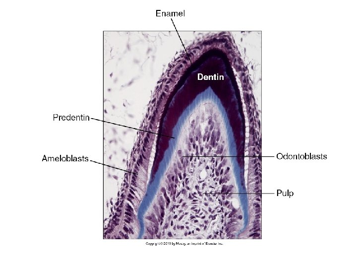
Ameloblasts First layer of enamel Dentin Odontoblasts

Rates of dentin deposition varies both within various regions of a tooth and also among different teeth Dentin formation continues throughout the life of the tooth resulting in a gradual and progressive reduction in the size of the pulp cavity
 Role of inner enamel epithelium Acellular zone (collagen fibrils)")
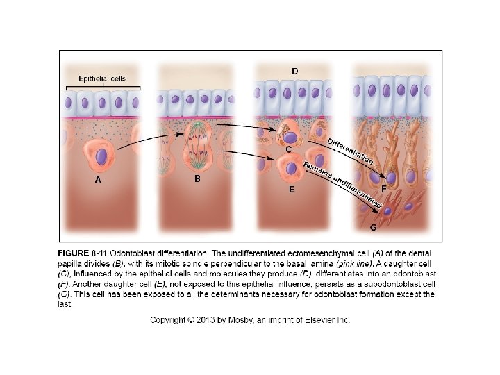
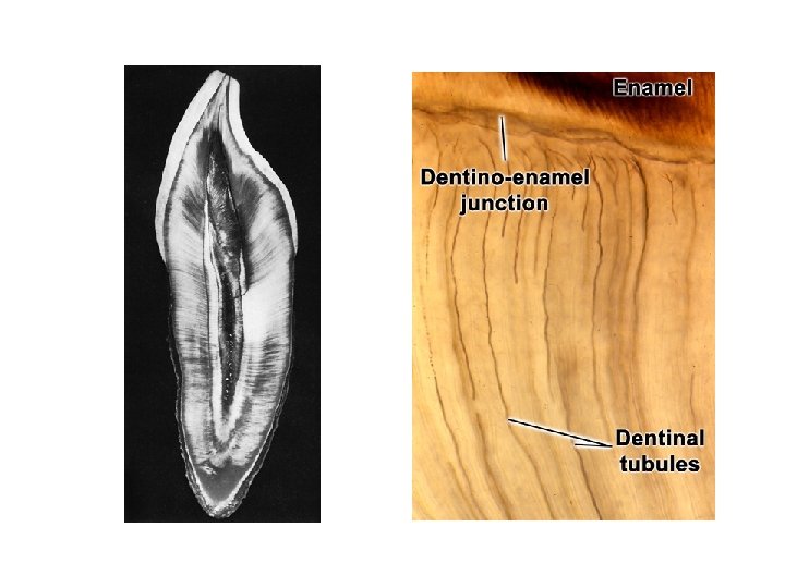
Dentinogenesis • • Odontoblasts (preodontoblasts) Role of inner enamel epithelium Acellular zone (collagen fibrils) First product: type III collagen fibrils with fibronectin (von Korff’s fibers) • Then type I collagen parallel to DE junction • Enamel spindles • Odontoblastic processes

Von Korff’s fibers
: Observed deep in between odontoblasts")
Von Korff’s fibers (arrowheads): Observed deep in between odontoblasts

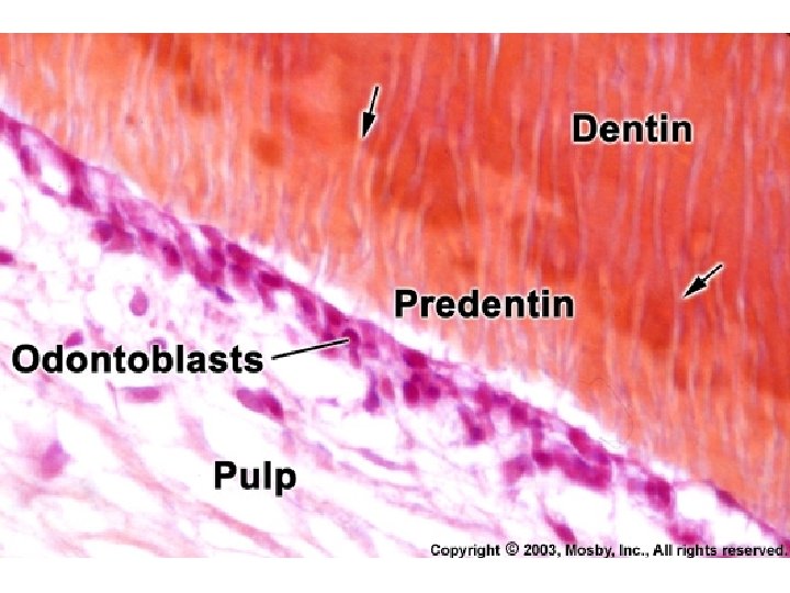
Mineralization of Dentin Occurs by formation of matrix vesicles Mineralization first appears as single crystals seeded by phospholipids in the vesicle membrane These crystals grow rapidly and rupture from the confines of the vesicle to spread as a cluster of crystallites that fuse with adjacent clusters to form a continuous layer of mineralized matrix Deposition of mineral lags behind the formation of the organic matrix so that a layer of predentin (organic layer) is always present between odontoblasts and the mineralization front Following this, noncollagenous protein secreted by odontoblasts will now regulate mineral deposition

Two recognized patterns of dentin mineralization: linear and globular depending on the rate of dentin formation Mantle dentin: predominantly globular Circumpulpal dentin: both linear and globular Globular pattern occurs when mineralization is the fastest Linear pattern occurs when rate of formation is slow Linear Globular

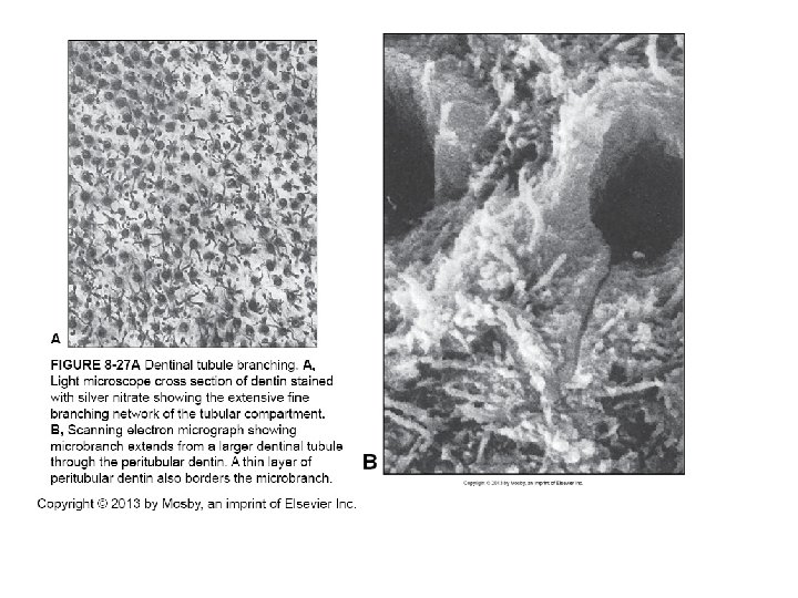
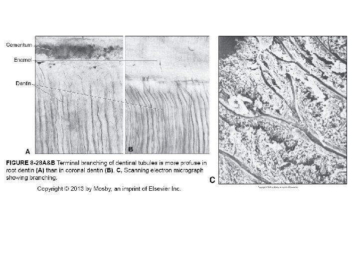
Dentinal tubules • Contain cytoplasmic processes • Extend through the entire thickness • S-shaped path (curvatures) due to crowding in the crown – Secondary curvatures • • Almost straight in the incisal area Straight in root dentin Tapered (largest diameter near the pulp) Branches (more frequent in the root) that derive from branching of the odontoblastic process
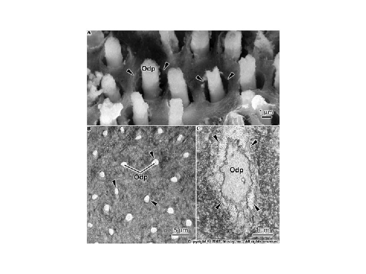

In predentin, odontoblastic processes are surrounded by meshwork of collagen. In dentin, the odontoblastic process is within dentinal tubule surrounded by peritubular dentin that is poor in collagen and more mineralized

Essentials of Oral Histology and Embryology, Ed: James Avery, 3 rd edition.
 • Collar of hypermineralized dentin •")
Peritubular dentin • Intratubular dentin (old wrong term) • Collar of hypermineralized dentin • Little collagen but DSP, DMP 1

Intertubular dentin • Between tubules • Primary secretory product of odontoblasts

Essentials of Oral Histology and Embryology, Ed: James Avery, 3 rd edition.

Sclerotic dentin • Occluded dentinal tubule – In ground sections dentin look translucent – Starts in the teens – Can be just minerals without well formed dentin • Increases with age • Root and middle of crown • Reduces dentin permeability prolonged pulp vitality Essentials of Oral Histology and Embryology, Ed: James Avery, 3 rd edition.

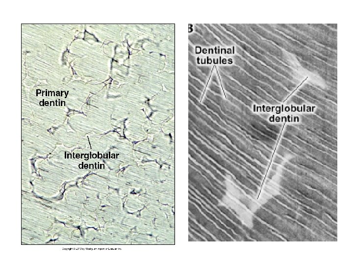
Interglobular dentin • Areas of undermineralized or hypomineralized dentin – defect in mineralization • Primary teeth • Just below the mantle dentin in the circumpulpal area • Especially prevalent in patients with Vitamin D deficiency or exposed to high levels of fluoride at the time of dentin formation • No defect in matrix formation. So the architecture of tubules is normal as they run uninterrupted through the interglobular areas. • However, there is lack of peritubular dentin

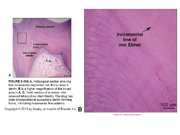
Incremental growth lines • Organic matrix deposited as a rate of 4μm per day • Changes in the orientation from day to day • At the 5 th day there is more exaggerated change in orientation (Lines of von Ebner) - ~ 20 μm apart. • At right angle to tubules and generally mark the rhythmic, linear pattern of dentin deposition in an inward and rootward direction • Rate of mineralization is 2μm every 12 hours. So the organic matrix of dentin is deposited rhythmically at a daily rate of about 4 μm/day and is mineralized in a 12 -hour cycle
 • They characterize areas of")
Contour lines of Owen (Another type of incremental pattern) • They characterize areas of deficient mineralization due to trauma • Original description by Owen: Lines occurring from the secondary curvature of dentinal tubules • Neonatal line: defines the disturbance of mineralization during birth Essentials of Oral Histology and Embryology, Ed: James Avery, 3 rd edition.

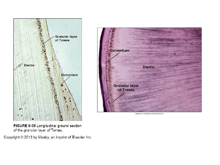
Granular layer of Tomes • Root • Ground sections • Progressive increase from the CE junction to the apex of the tooth • ? Hypomineralization of interglobular dentin – Initial theory • Sections made through loops of dentinal tubules and an optical phenomenon • Special arrangement of collagen and noncollagen matrix proteins at the interface between dentin and cementum

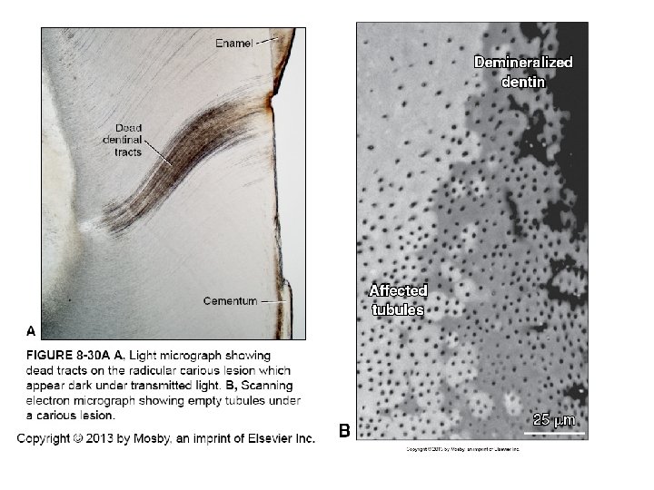
Age changes • Dentinal tubule complete closure Sclerotic dentin • Dead tracts of dentin – Retracted processes from tubules e. g. caries – Empty tubules with entrapped air in ground sections – Coronal dentin – Filled with reparative dentin

Essentials of Oral Histology and Embryology, Ed: James Avery, 3 rd edition.

Hereditary Abnormalities of Dentin • Dentinogenesis imperfecta • Dentin dysplasia • Vitamin D resistant rickets

Osteogenenesis/Dentinogenesis Imperfecta

Hereditary Opalescent Dentin

Dentin Dysplasia Type II

Dentin Dysplasia Type II

Vitamin D-resistant Rickets

Essentials of Oral Histology and Embryology, Ed: James Avery, 3 rd edition.
- Slides: 54