DENTIN STRUCTURE The dentin provides the bulk and

DENTIN STRUCTURE

• The dentin provides the bulk and general form of the tooth and is characterized as a hard tissue with tubules throughout its thickness. Since it begins to form slightly before the enamel, it determines the shape of the crown, including the cusps and ridges, and the number and size of the roots.

• As a living tissue it contains within its tubules the processes of the specialized cells, the odontoblasts. The cell bodies of the odontoblast are arranged along the pulpal surface of the dentin. • Physically and chemically the dentin closely resembles bone. • The main morphologic difference between bone and dentin is that some of the osteoblasts exist on the surface of bone, and when one of these cells becomes enclosed within its matrix, it is called an osteocyte. • The odontoblasts’ cell bodies remain external to dentin, but their processes exist within tubules in dentin. Both are considered vital tissues because they contain living protoplasm.

PHYSICAL AND CHEMICAL PROPERTIES • 1 -In the teeth of young individuals the dentin usually is light yellowish in color, becoming darker with age. • 2 -Unlike enamel, which is very hard and brittle, dentin is viscoelastic and subject to slight deformation. It is somewhat harder than bone but considerably softer than enamel. Dentin hardness varies slightly between tooth types and between crown and root dentin. The dentin of primary teeth is slightly less hard than that of permanent teeth.

• 3 -The lower content of mineral salts in dentin renders it more radiolucent than enamel Dentin consists of 35% organic matter and water and 65% inorganic material. The organic substance consists of collagenous fibrils embedded in the ground substance of mucopolysaccharides (proteogly- cans and glycosaminoglycans).

• 4 -Type I collagen is the principal type of collagen found in the dentin. the matrix contains growth factors like transforming growth factor (TGF), fibroblast growth factor (FGF), insulin-like growth factors (IGFs), bone morphogenic proteins (BMPs), epidermal growth factor (EGF), platelet derived growth factor (PDGF), placenta growth factor (PLGF), vascular endothelial growth factor (VEGF), and angiogenic growth factor (AGF).

• 5 -The inorganic component consist of hydroxyapatite, as in bone, cementum, and enamel. Each hydroxyapatite crystal is composed of several thousand unit cells. The unit cells have a formula of 3 Ca 3(PO 4)2 • Ca(OH)2. The crystals are plate shaped and much smaller than the hydroxyapatite crystals in enamel. Dentin also contains small amounts of phosphates, carbonates, and sulfates. Organic and inorganic substances can be separated by either decalcification or incineration.

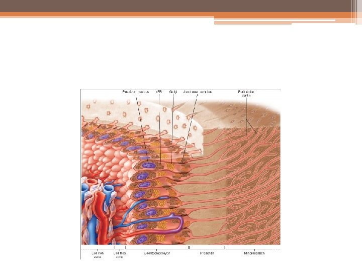
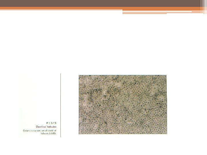
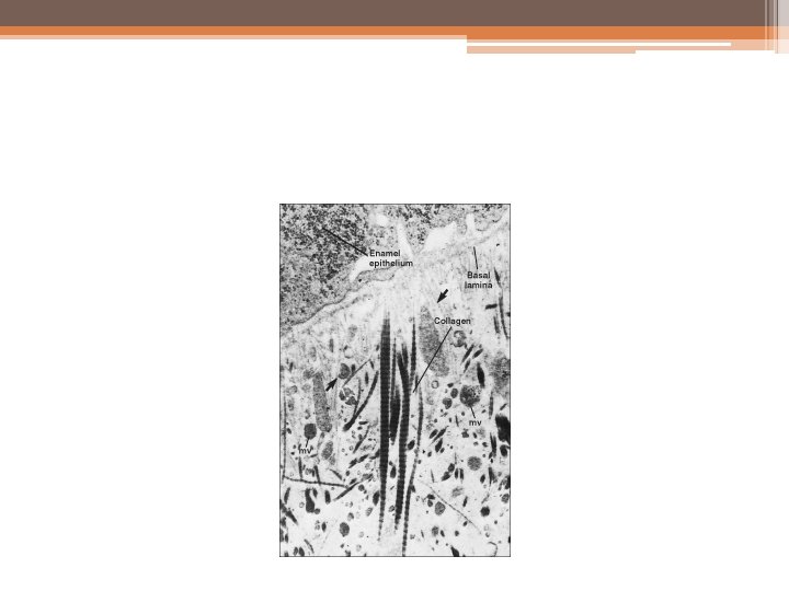
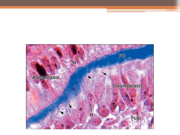
STRUCTURE: • The dentinal matrix of collagen fibers is arranged in a network. As dentin calcifies, the hydroxyapatite crystals mask the individual collagen fibers. • The bodies of the odontoblasts are arranged in a layer on the pulpal surface of the dentin, and only their cytoplasmic processes are included in the tubules in the mineralized matrix. • Each cell gives rise to one process, which traverses the predentin and calcified dentin within one tubule and terminates in a branching network at the junction with enamel or cementum. Tubules are found throughout normal dentin and are therefore characteristic of it.
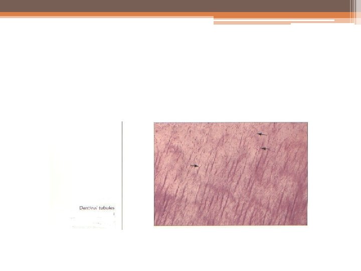
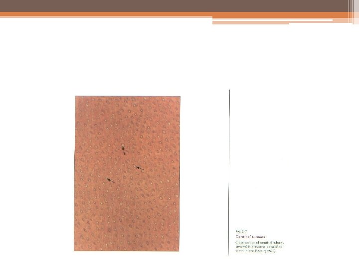
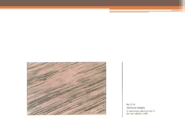

Dentinal tubules: • The course of the dentinal tubules follows a gentle curve in the crown, less so in the root, where it resembles a gentle S (sigmoid course) in shape. These curvatures are called primary curvatures. Starting at right angles from the pulpal surface, the first convexity of this doubly curved course is directed toward the apex of the tooth. These tubules end perpendicular to the DEJ and DCJ.

• Branches of the dentinal tubules near the terminals are referred to as terminal branches. The terminal branching is more profuse in the root dentin than in the coronal dentin. Near the root tip and along the incisal edges and cusps the tubules are almost straight. Over their entire lengths the tubules exhibit minute, relatively regular secondary curvatures that are sinusoidal in shape.

• The tubules are longer than the dentin, and are thick because they curve through dentin. Dentin thickness varied not only from tooth to tooth but also in different surfaces of the same tooth. The buccal surfaces showed maximum thickness, followed by lingual, and there was no difference in thickness between mesial and distal surfaces.

• There are more tubules per unit area in the crown than in the root. The dentinal tubules have lateral branches throughout dentin, which are termed canaliculi or microtubules and originate more or less at right angles to the main tubule every 1 to 2 μm along its length.

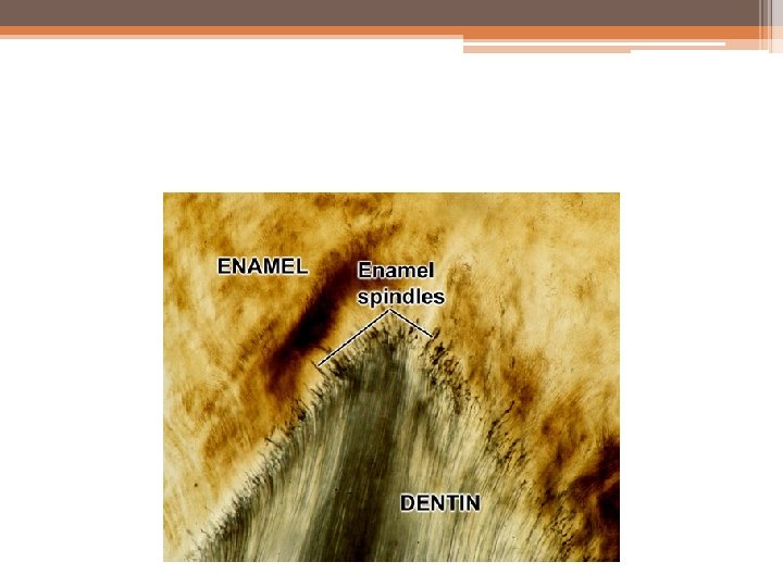
• Some of them enter adjacent or distant tubules while others end in the intertubular dentin. A few odontoblastic processes extend through the DEJ into the enamel for several millimeters. These are termed enamel spindles

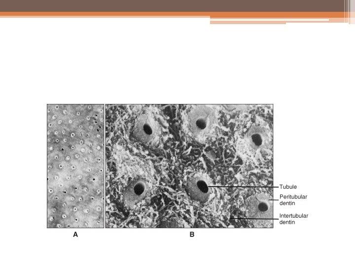
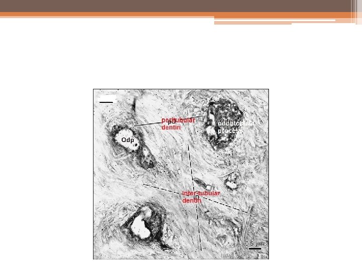
Peritubular dentin • The dentin that immediately surrounds the dentinal tubules is termed peritubular dentin. This dentin forms the walls of the tubules in all but the dentin near the pulp. It is more highly mineralized (about 9%) than the dentin present between the tubules (the intertubular dentin). Peritubular dentin differs from intertubular dentin by its matrix composition. The crystal arrangement appears to be similar.

• Since the deposition of the minerals occurs in the inner wall of the tubule rather on the outer wall, the term ‘Intratubular dentin’ is considered to be more appropriate than the term peritubular dentin. • the calcified tubule wall has an inner organic lining termed the lamina limitans. This is described as a thin organic membrane, high in glycosaminoglycan (GAG) and similar to the lining of lacunae in cartilage and bone.

Intertubular dentin : • The main body of dentin is composed of intertubular dentin. It is located between the dentinal tubules or, more specifically, between the zones of peritubular dentin. About one half of its volume is organic matrix, specifically collagen fibers, which are randomly oriented around the dentinal tubules

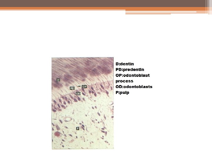
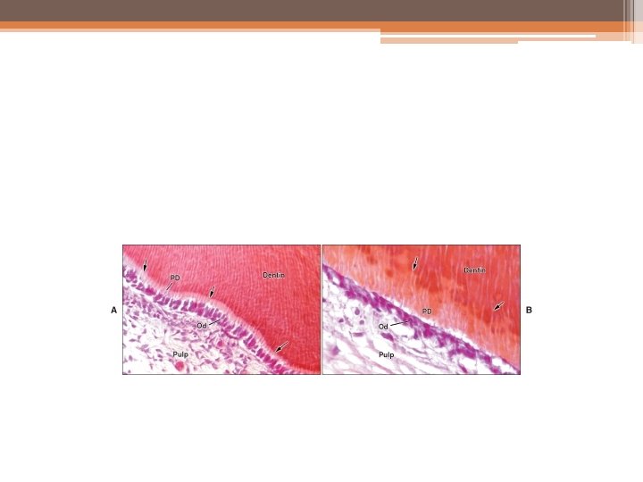
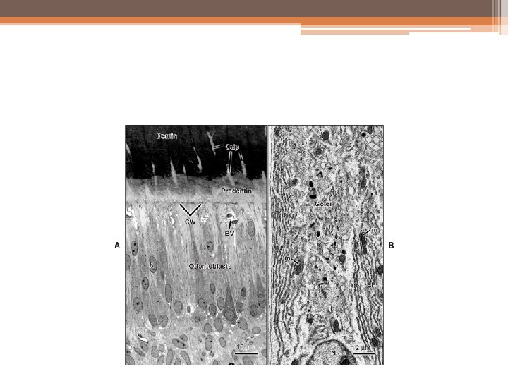
Predentin: • The predentin is located always adjacent to the pulp tissue and is 2 to 6 m wide, depending on the extent of activity of the odontoblast. It is not mineralized. The predentin appears to be pale staining than the mineral- ized dentin owing to differences in composition of the matrix. As the collagen fibers undergo mineralization at the predentin- dentin junction, the predentin becomes dentin and a new layer of predentin forms circumpulpally.

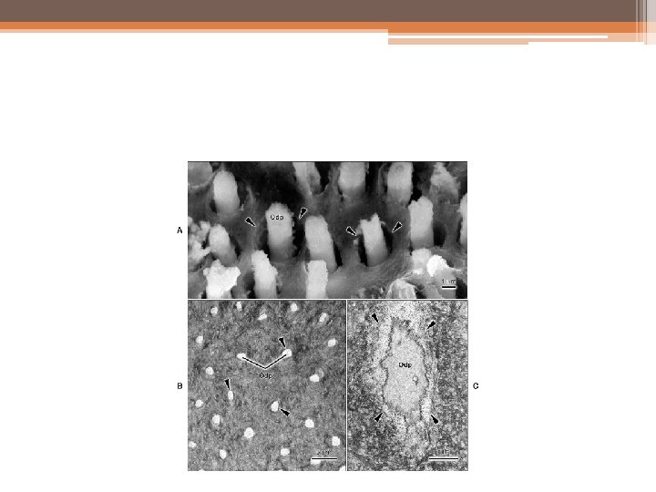
Odontoblast process • The odontoblast processes are the cytoplasmic extensions of the odontoblasts. The odontoblast cells reside in the peripheral pulp at the pulppredentin border and their processes extend into the dentinal tubules. The processes are largest in diameter near the pulp and taper to approximately into the dentin.

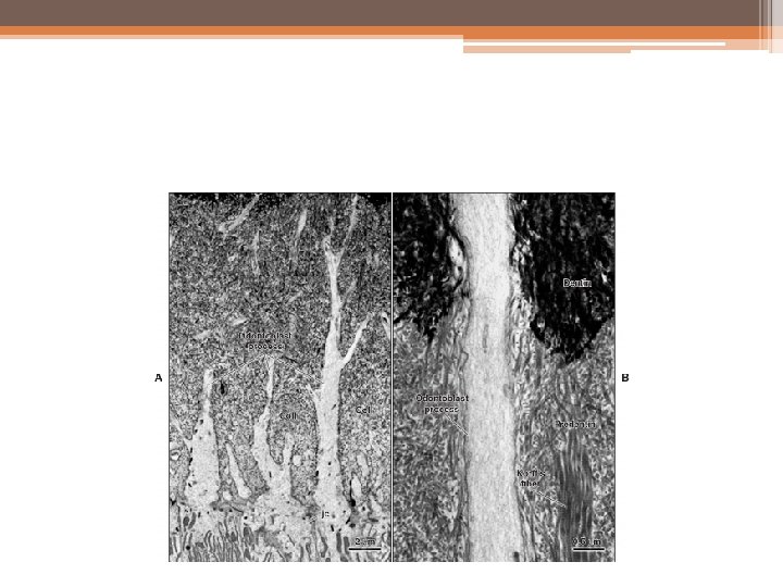

• The life span of the odontoblasts is equal to the age of the tooth as once differentiated, they cannot undergo further division. The odontoblastic processes narrow to about half the size of the cell as they enter the tubules. • The odontoblast processes divide near the DEJ and may indeed extend into enamel in the enamel spindles. Periodically along the course of the processes side branches (lateral branches) appear that extend laterally into adjacent tubules.

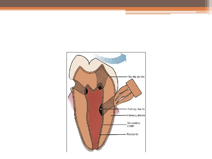
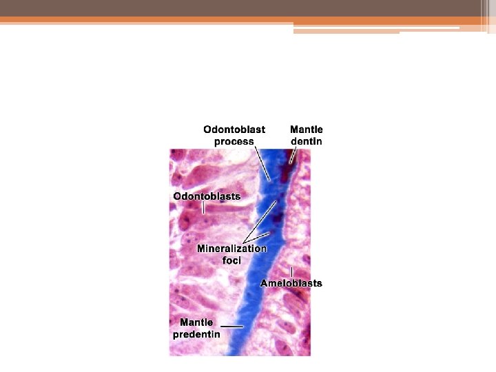
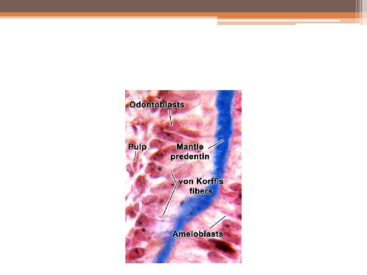
PRIMARY DENTIN: • Dentin which are formed before root completion are known as primary dentin. The primary dentin are of two types—mantle dentin and the circumpulpal dentin. • Mantle dentin is the name of the first-formed dentin in the crown underlying the DEJ. This zone below the DE junction is soft and thus provides cushioning effect to the tooth. The fibrils formed in this zone are perpendicular to the DEJ and the organic matrix is composed of larger collagen fibrils than are present in the rest of the primary dentin (circumpulpal dentin).

• The larger diameter collagen fibers are known as von Korff’s fibers. They contain mainly type III collagen. Compared to circumpulpal dentin, mantle dentin is less mineralized. Mantle dentin also has fewer defects than circum- pulpal dentin. Unlike, rest of the dentin, matrix vesicles are involved in the mineralization of mantle dentin. Mantle dentin undergoes globular mineralization whereas the circumpulpal dentin mineralizes either by globular or linear pattern.

• When the rate of formation progresses slowly, the mineralization front appears more uniform and the process is said to be linear. • Circumpulpal dentin forms the remaining primary dentin or bulk of the tooth. The collagen fibrils in circumpulpal dentin are much smaller in diameter and are more closely packed together compared to the mantle dentin. The circumpulpal dentin may contain slightly more mineral than mantle dentin.
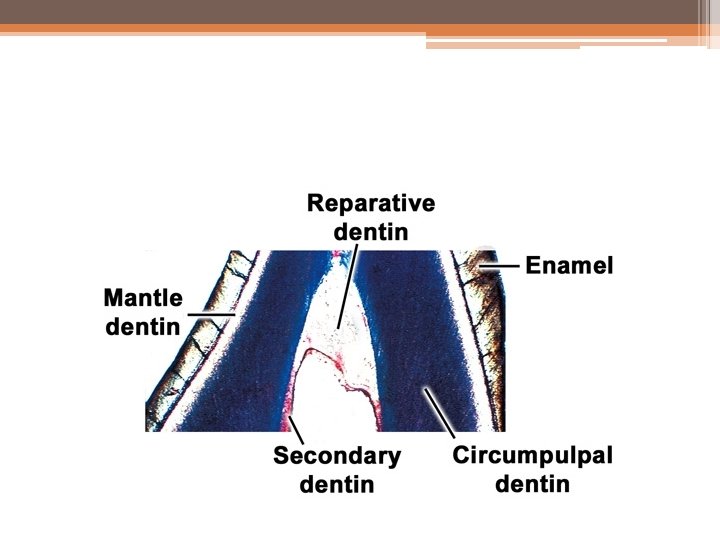

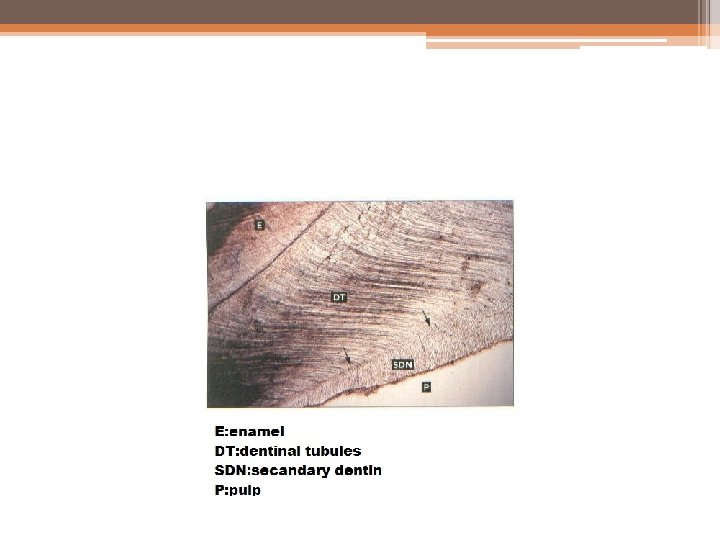
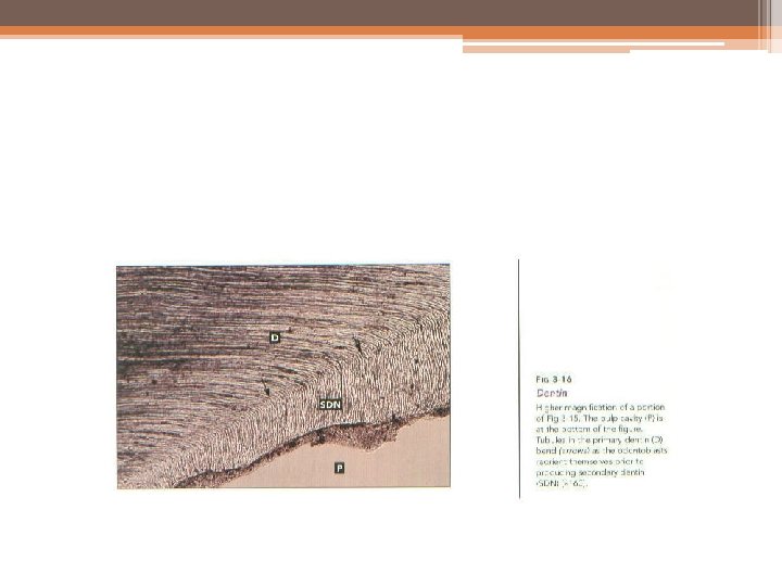
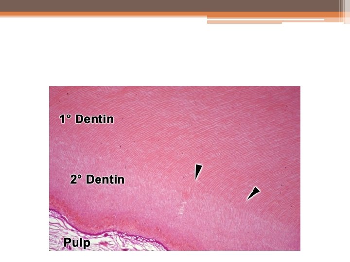
• SECONDARY DENTIN • Secondary dentin is a narrow band of dentin bordering the pulp and representing that dentin formed after root completion. This dentin contains fewer tubules than primary dentin. secondary dentin is formed more slowly than primary dentin and that it looks similar to primary dentin but contains fewer tubules.
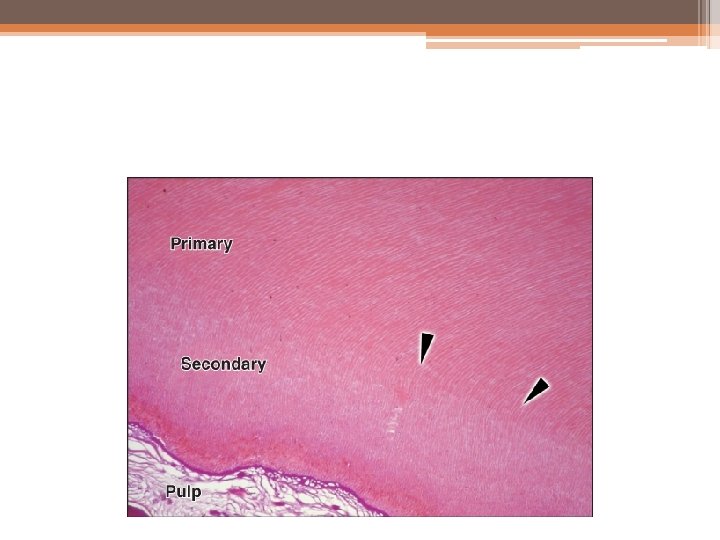

• Secondary dentin is not formed uniformly and appears in greater amounts on the roof and floor of the coronal pulp chamber, where it protects the pulp from exposure in older teeth. The secondary dentin formed is not in response to any external stimuli and it appears very much like primary dentin. Due to the regular arrangement of dentinal tubules, it is known as regular secondary dentin. The apical dentin shows irregularity in the dentinal tubules of both primary and secondary dentin.

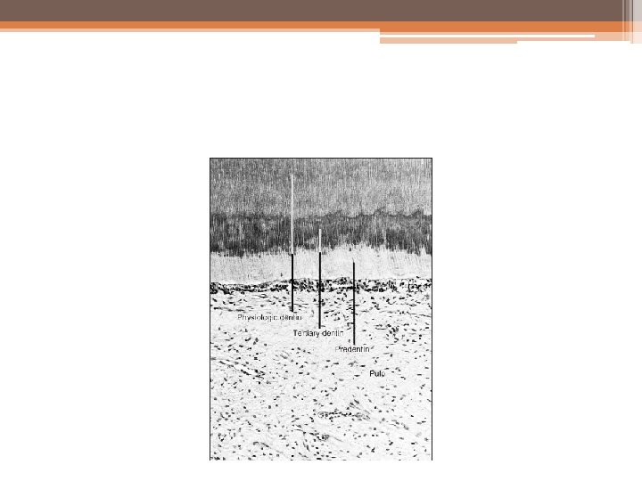
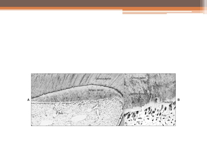
TERTIARY DENTIN • Tertiary dentin is reparative, response, or reactive dentin. This is localized formation of dentin on the pulp-dentin border, formed in reaction to trauma such as caries or restorative procedures. This type of dentin is described reparative dentin.

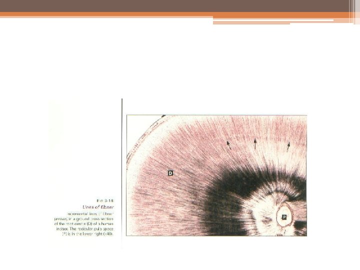
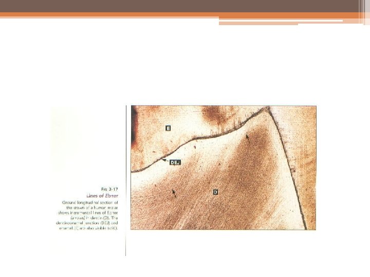
INCREMENTAL LINES • The incremental lines of von Ebner, appear as fine lines or striations in dentin. They run at right angles to the dentinal tubules and correspond to the incremental lines in enamel or bone. These lines reflect the daily rhythmic, recurrent deposition of dentin matrix as well as a hesitation in the daily formative process. The daily increment decreases after a tooth reaches functional occlusion.


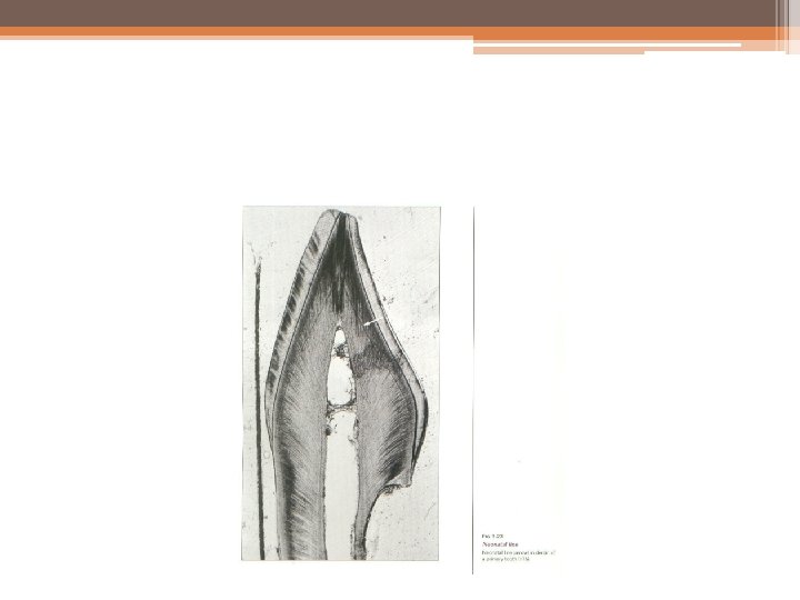
• Such lines are readily demonstrated in ground sections and are known as contour lines of Owen these lines to represent hypocalcified bands. In the deciduous teeth and in the first permanent molars, where dentin is formed partly before and partly after birth, the prenatal and postnatal dentin are separated by an accentuated contour line.

• This is termed the neonatal line and is seen in enamel as well as dentin This line reflects the abrupt change in environment that occurs at birth. The dentin matrix formed prior to birth is usually of better quality than that formed after birth, and the neonatal line may be a zone of hypocalcification.

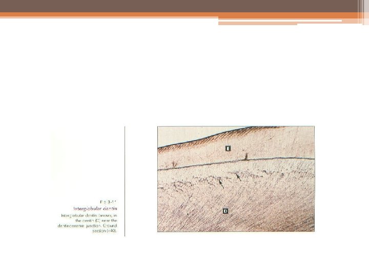
INTERGLOBULAR DENTIN • Sometimes mineralization of dentin begins in small globular areas that fail to coalesce into a homogenous mass. This results in zones of hypomineralization between the globules. These zones are known as globular dentin or interglobular spaces.
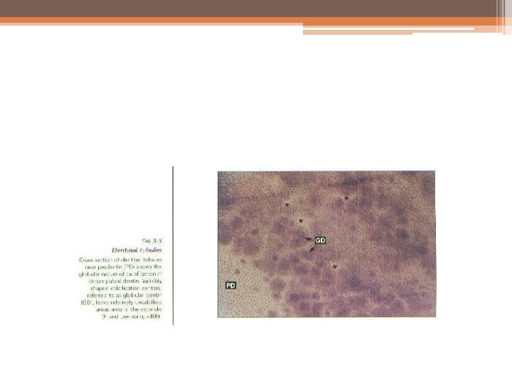
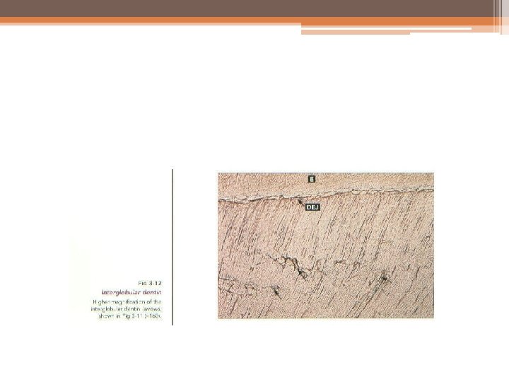
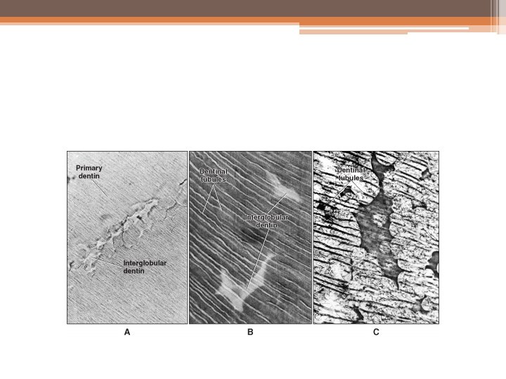

• This dentin forms in the crowns of teeth in the circumpulpal dentin just below the mantle dentin, and it follows the incremental pattern. The dentinal tubules pass uninterruptedly through interglobular dentin, thus demonstrating defect of mineralization and not of matrix formation

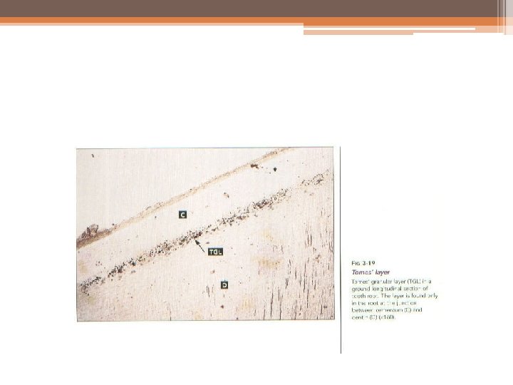
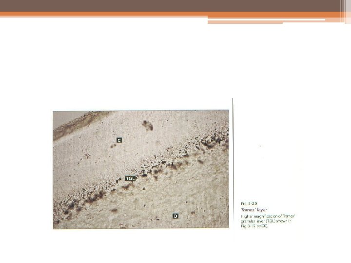
GRANULAR LAYER • When dry ground sections of the root dentin are visualized in transmitted light, a zone adjacent to the cementum appears granular. This is known as (Tomes’) granular layer. • This zone increases slightly in amount from the cementoenamel junction to the root apex and is believed to be caused by a coalescing and looping of the terminal portions of the dentinal tubules. Such a process is considered possible as a result of the odontoblasts turning on themselves during early dentin formation. These areas remain unmineralized, like interglobular dentin.
- Slides: 68