Degas Bony and membranous labyrinth Vestibular system Dr

Degas Bony and membranous labyrinth. Vestibular system. Dr. Emese Pálfi/Dr. Gábor Baksa Semmelweis University Department of Anatomy, Histology and Embryology

Vestibulocochlear organs Organon statoacusticum Balance Organon staticum Hearing Organon acusticum

Bony labyrinth cochlea vestibule: recessi + maculae cribrosae three semi-circular canals: ant. , post. , lat. perilymph aqueductus vestibuli + canaliculcus cochleae 7 Anterior semicircular canal 9 Lateral semicircular canal 11 Posterior semicircular canal (posterior canal) 14 Fenestra vestibuli 15 Cupula of cochlea 18 Tympanic cavity and fenestra cochleae (probe) 19 External acoustic meatus 20 Facial canal 21 Base of cochlea and musculotubal canal
")
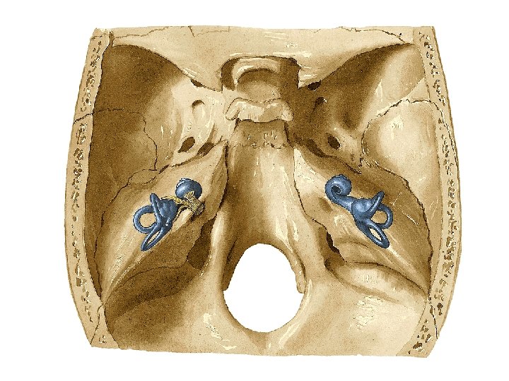
Membranous labyrinth cochlear duct semi-circular ducts utricle saccule endolymph 1 Ampulla (anterior semicircular canal) 2 Elliptical recess 4 Spherical recess 6 Base of cochlea 7 Anterior semicircular canal 8 Crus commune or common Limb 9 Lateral semicircular canal 10 Posterior bony ampulla 11 Posterior semicircular canal (posterior canal) 12 Fenestra cochleae 13 Bony ampulla 14 Fenestra vestibuli 15 Cupula of cochlea

Balance - function Upright posture: Vestibular organ: 2 x 3 semicircular canals, saccule and utricle + nuclei and pathways Information: receptors of the tendons, muscles, joints, skin autochtone back muscles, antigravity muscles of the extremities, neck muscles visual system Expected or unexpected change in body or head position, fixation of the field of view: 2 x 3 semicircular canals, saccule and utricle + nuclei and pathways Information: receptors of tendons, muscles, joints, skin appropriate muscles visual system

Receptor fields Macula of the utricle Macula of the saccule Crista ampullaris Otholitic organs linear acceleration rotations
 a large kinocillium is")
Vestibular receptors - - secondary sensory epithelial cells (neuroepithelial cells) a large kinocillium is accompanied by a large number of stereocillia the mechanoelectric transduction is based on mechanosensitive ion channels innervated by the distal processes of the bipolar cells and efferents from the center

Otholit organs: forward and backward movements, gravitational forces - otoconia - otholit membrane Crista ampullaris: spatial orientation - cupula
")
Ganglion and nerves Vestibular ganglion of Scarpa in the internal auditory canal (bipolar neurons) Superior and inferior part facial area sup. vestibular area inf. vestibular area cochlear area foramen singulare Vestibulocochlear nerve (N. VIII. ): • cochlear division for hearing • vestibular division for balance: utriculoampullary nerve (ant. and lat. ampullary nerve, utricular nerve) saccular nerve post. ampullary nerve

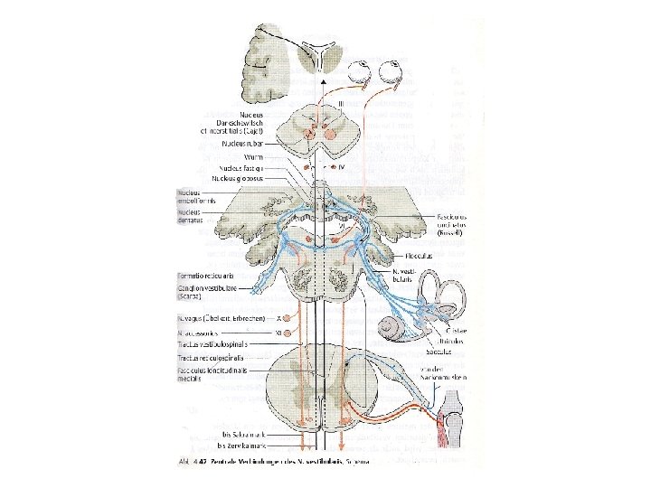
Central connections peripheral processes innervate the receptors central processes vestibular component of the vestibulocochlear nerve all give afferents to the four vestibular nuclei: • • superior vestibular nucleus (Bechterew) medial vestibular nucleus (Schwalbe) lateral vestibularis nucleus (Deiters) inferior vestibular nucleus (Roller) There are fibers which are ending directly in the cerebellum.
 • to the")
All vestibular nuclei give fibers through the medial longitudinal fasciculus (MLF) • to the cranial nerve nuclei of N. III. , IV. and VI. (ascending fibers) • to the cerebellum Superior vestibular nucleus (Bechterew) Lateral vestibularis nucleus (Deiters): function: fixation of the gaze in the original direction Medial vestibular nucleus (Schwalbe): function: coordination of the neck and eye movements Inferior vestibular nucleus (Roller): function: comparison and integration of the information
: lateral vestibulospinal tract α and δ motoneurons of")
Spinal cord Lateral vestibularis nucleus (Deiters): lateral vestibulospinal tract α and δ motoneurons of the anterior horn Medial vestibularis nucleus (Schwalbe): medial vestibulospinal tract α and δ motoneurons of the anterior horn function: control the tone of the neck, thoracal and limb muscles its dorsal part gets inhibitory fibers from the cerebellum and spinal cord
 Inferior vestibular nucleus (Roller) vestibulocerebellar tract vestibulocerebellum = archicerebellum")
Cerebellum Medial vestibular nucleus (Schwalbe) Inferior vestibular nucleus (Roller) vestibulocerebellar tract vestibulocerebellum = archicerebellum = flocculonodular lobe Purkinje cells of the vestibulocerebellum project on the fastigial nucleus through FASTIGIOBULBAR TRACT the fibers reaching the vestibular nuclei and the reticular formation Reticular formation of the bridge (medial reticulospinal tract) and nucl. of Deiters increase muscle tone in the antigravity muscles of the extremities Cerebellar regulation of balance have a control loop: vestibulocerebellar control loop

Nystagmus Via MLF all vestibular nuclei are reaching the nucl. III. , IV. and VI. nerves. vestibulo-ocular reflex (VOR): during movements of the object / head / body, the position of the light changes on the retina and the image become blurred the eyes are adjusting the image is always projected on the fovea centralis This creates the physilogical nystagmus (= involuntary, rapid and repetitive movement of the eyes)

physiological or pathological Has a slow and a fast component Is characterized by direction, frequency and amplitude. Peripheral nystagmus: damage in the vestibular system Central nystagmus: damage elsewhere (e. g. , cerebellum, eye, etc. )
: change in field of")
Vestibular system Information from: • the visual system (visual cortex): change in field of view • the skin receptors and proprioceptors: e. g. stretching in the muscle spindle • the vestibular receptors (maculae and cristae): lienar / angular acceleration

Vestibular disorders Symptoms are also dependent on the disease. The most common symptoms are dizziness, headache, nausea, vomiting. "Sensory conflict" between vestibular, sensory and visual stimuli "Dizziness": vertigo feeling of insecurity Buoyancy Hypothesis

Caloric nystagmus Cold water ~ 33 ° C endolymph falls within the semicircular canal mimics a head turn to the contralateral side both eyes will turn toward the ipsilateral ear Warm water ~ 44 ° C endolymph in the ipsilateral horizontal canal rises mimics a head turn to the ipsilateral side both eyes will turn toward the contralateral ear Róbert Bárány Nobel prize: 1915

Thank you for your attention! Literature: Dr. Gábor Baksa: Vestibuläres System Dr. Márk Kozsurek: A vestibularis apparatus Rohen, Yokochi: Color atlas of anatomy (2011) Sobotta: Atlas and Text-book of Human Anatomy (1911) Grey: Anatomy of the human body (1918)
- Slides: 21