
Definations Stroke TIA HIE Pathophysiology Classical Symtoms Weakness


- Slides: 112
Definations Stroke TIA HIE
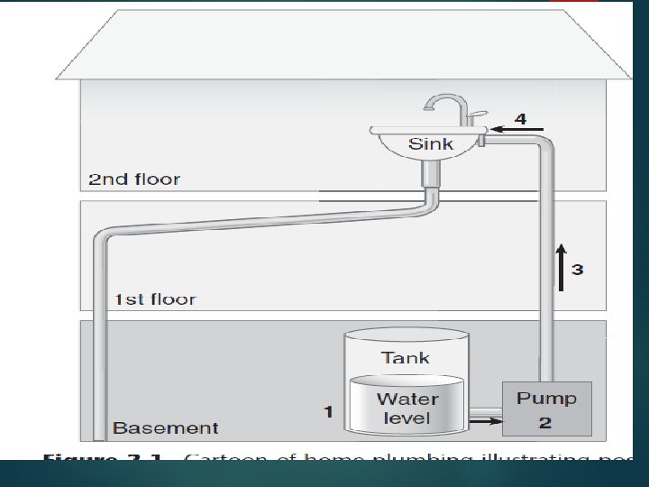
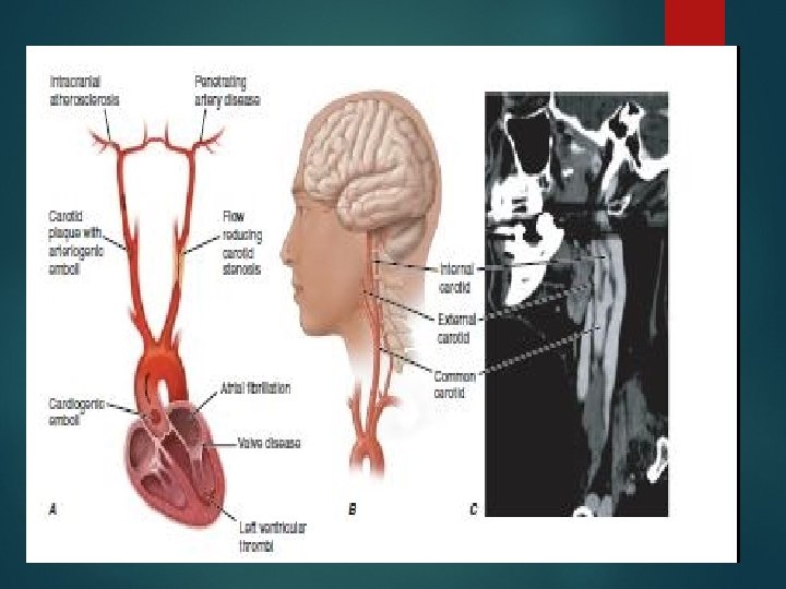
Pathophysiology
Classical Symtoms Weakness Vision loss Gait Ability to speak or understand Severe Headache
TOAST classification Small vessel Large vessel Cardiogenic Iatrogenic Cryotogenic
Oxford Classification Anterior Partial Ant Posterior
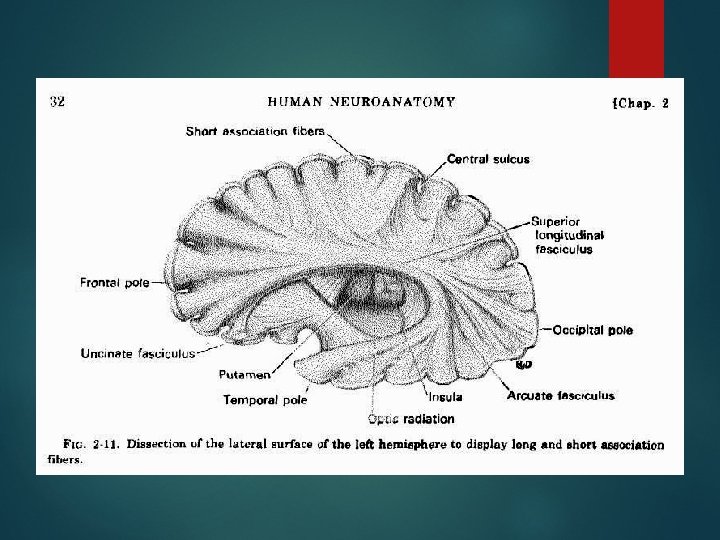
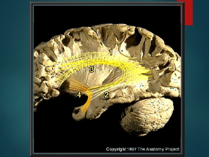
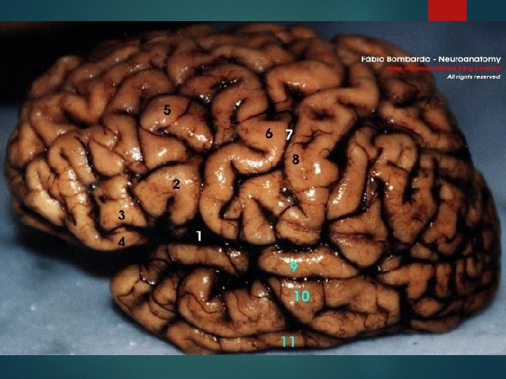
Localization Cortical signs Subcorical Brainstem Spinal cord Others
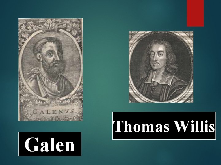
Galen Thomas Willis
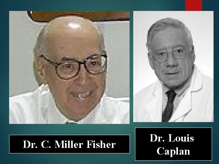
Dr. C. Miller Fisher Dr. Louis Caplan
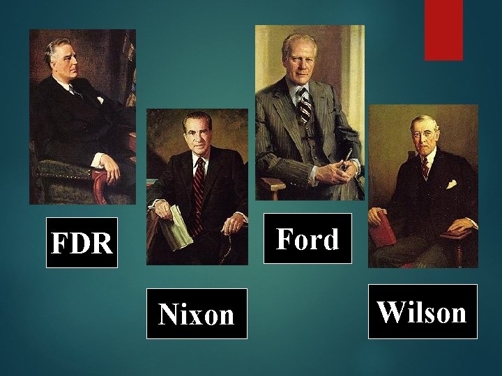
Ford FDR Nixon Wilson
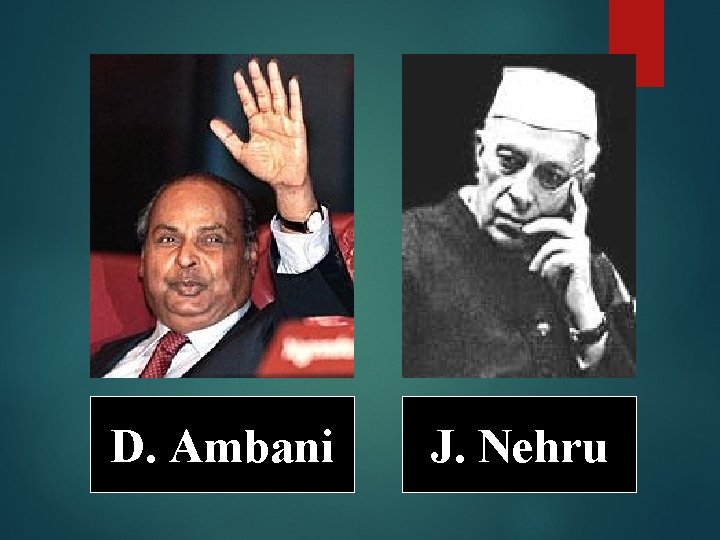
D. Ambani J. Nehru
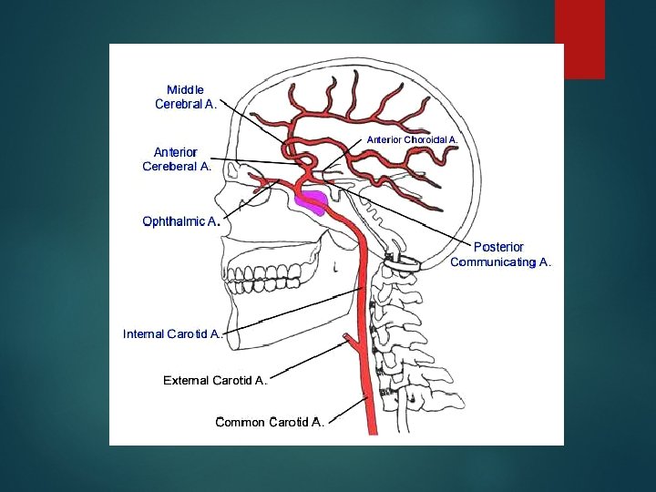
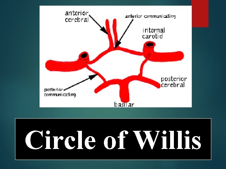
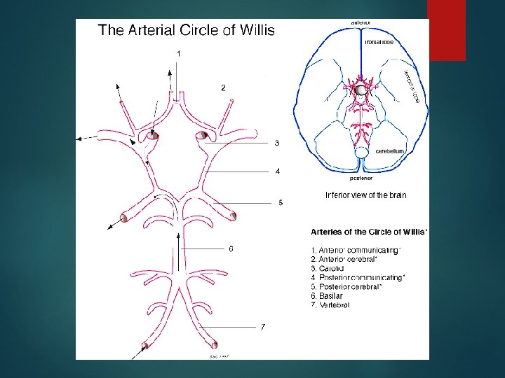
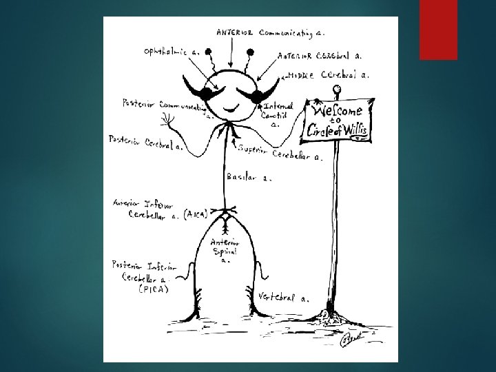
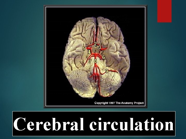
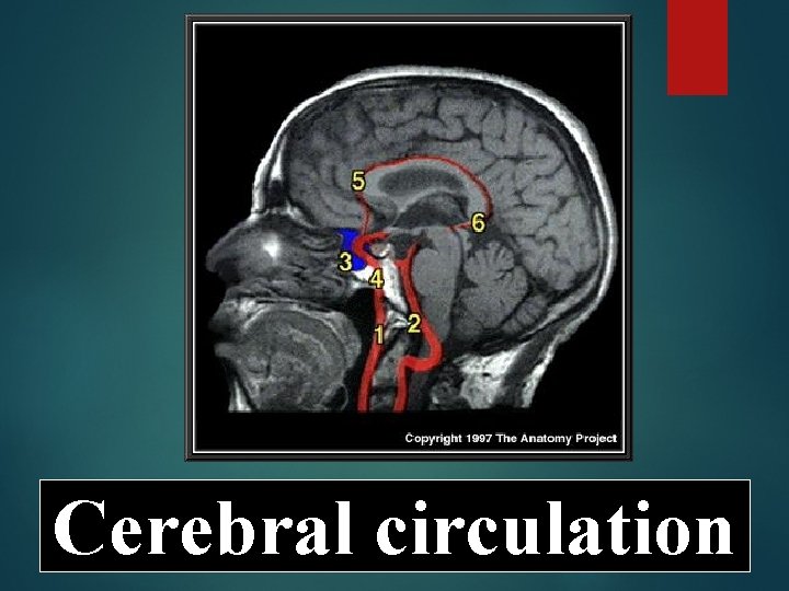
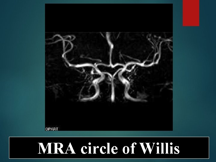
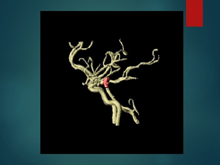
Circle of Willis
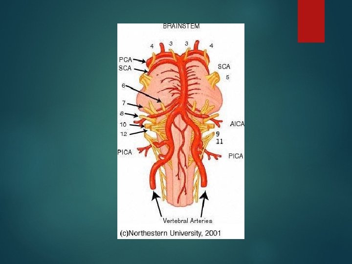
Cerebral circulation
Cerebral circulation
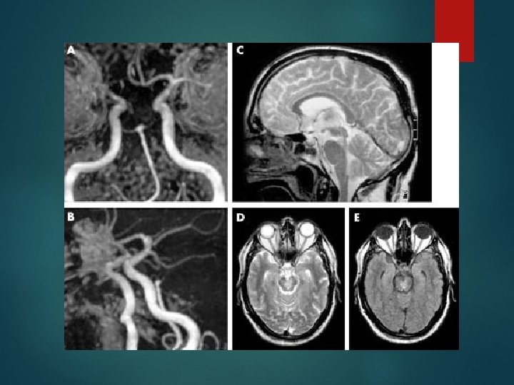
MRA circle of Willis
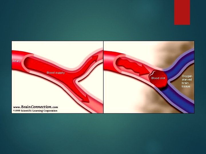
Normal blood flow Thrombosis Artery Blood supply Blood clot Oxygen starved brain tissue
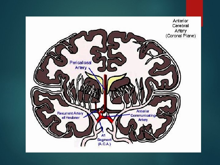
ACA Syndrome Supply: Frontal pole Superomedial border and medial frontal and parietal lobes including paracentral lobule and cingulate Anterior 4/5 of corpus callosum Heubner’s: Anterior limb of the internal capsule, head of caudate
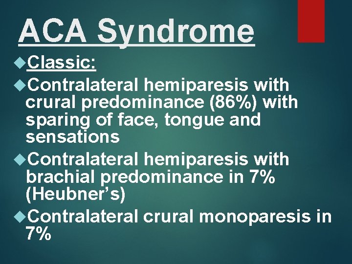
ACA Syndrome Classic: Contralateral hemiparesis with crural predominance (86%) with sparing of face, tongue and sensations Contralateral hemiparesis with brachial predominance in 7% (Heubner’s) Contralateral crural monoparesis in 7%
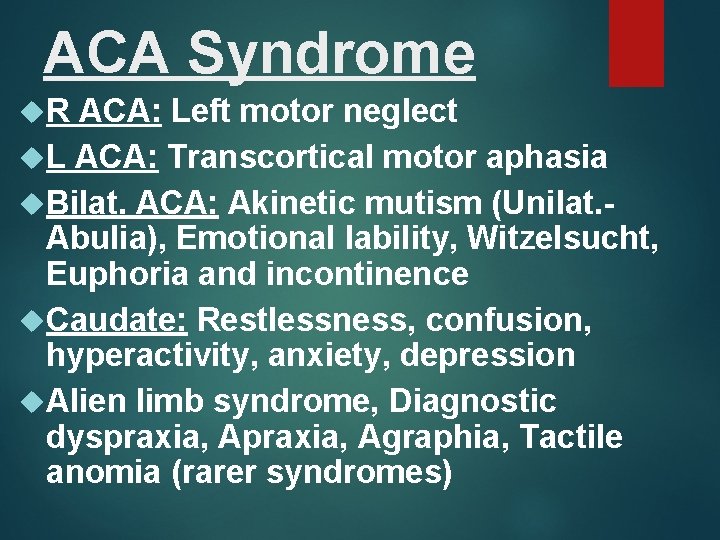
ACA Syndrome R ACA: Left motor neglect L ACA: Transcortical motor aphasia Bilat. ACA: Akinetic mutism (Unilat. Abulia), Emotional lability, Witzelsucht, Euphoria and incontinence Caudate: Restlessness, confusion, hyperactivity, anxiety, depression Alien limb syndrome, Diagnostic dyspraxia, Agraphia, Tactile anomia (rarer syndromes)
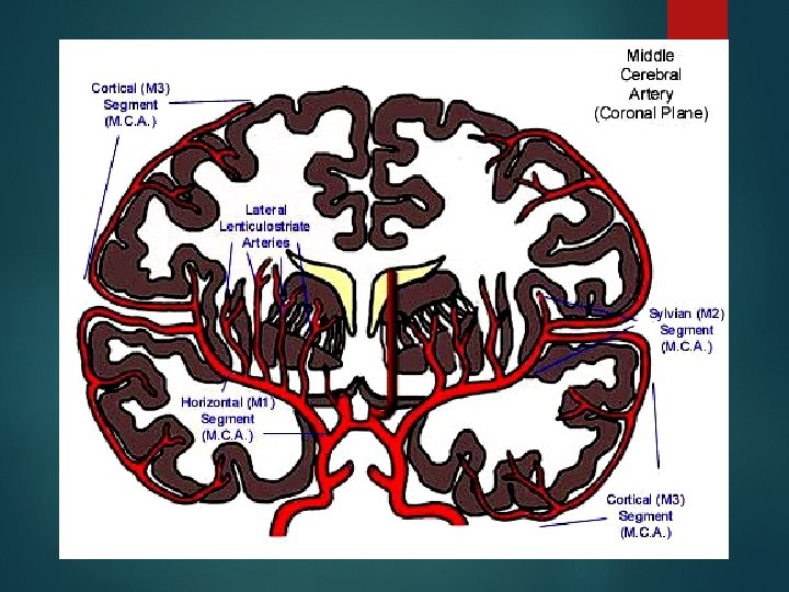
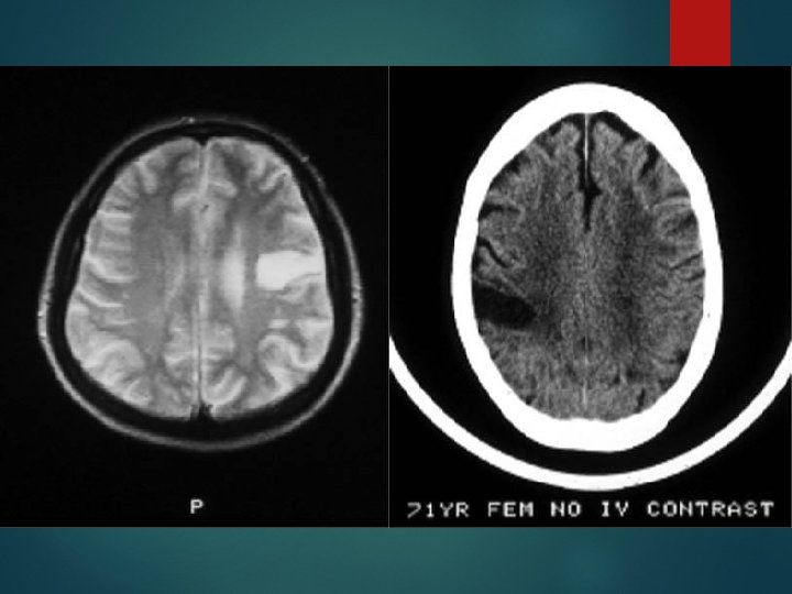
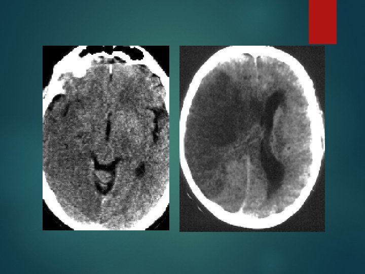
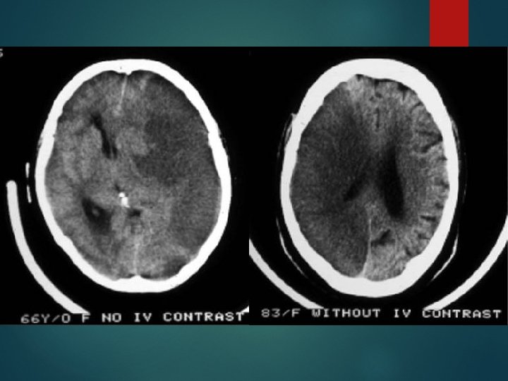
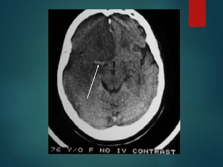
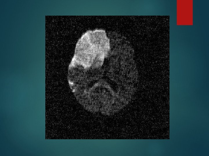
MCA Syndrome Supply: Entire lateral hemisphere including sensorimotor cortex, language areas Lenticulostriate: Caudate, putamen and posterior limb of internal capsule, corona radiata
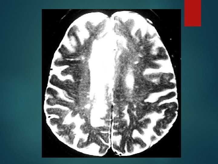
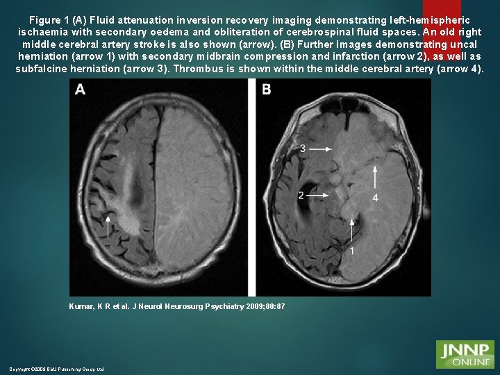
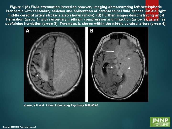
Figure 1 (A) Fluid attenuation inversion recovery imaging demonstrating left-hemispheric ischaemia with secondary oedema and obliteration of cerebrospinal fluid spaces. An old right middle cerebral artery stroke is also shown (arrow). (B) Further images demonstrating uncal herniation (arrow 1) with secondary midbrain compression and infarction (arrow 2), as well as subfalcine herniation (arrow 3). Thrombus is shown within the middle cerebral artery (arrow 4). Kumar, K R et al. J Neurol Neurosurg Psychiatry 2009; 80: 87 Copyright © 2009 BMJ Publishing Group Ltd.
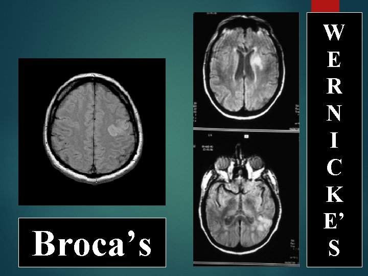
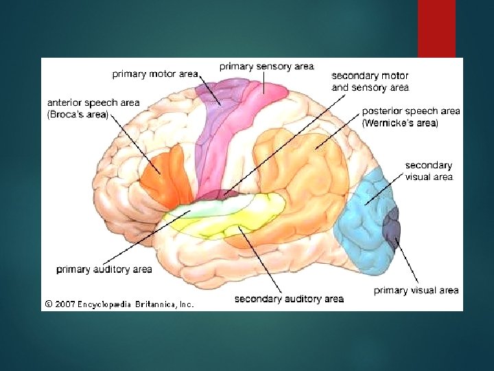
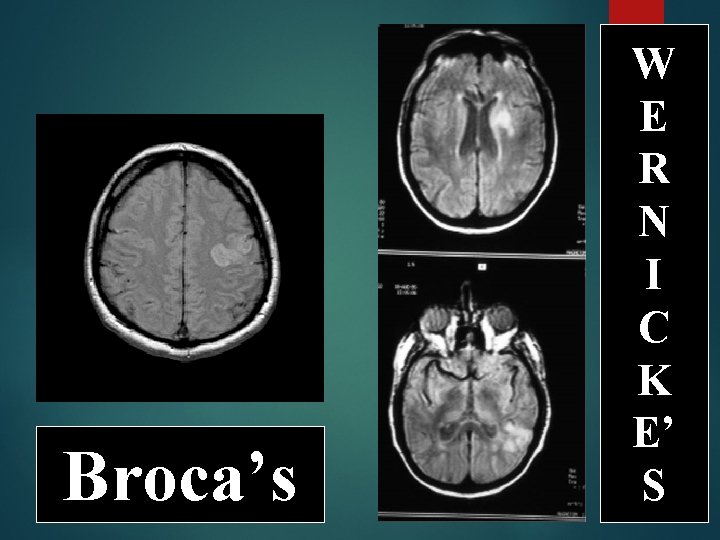
Broca’s W E R N I C K E’ S
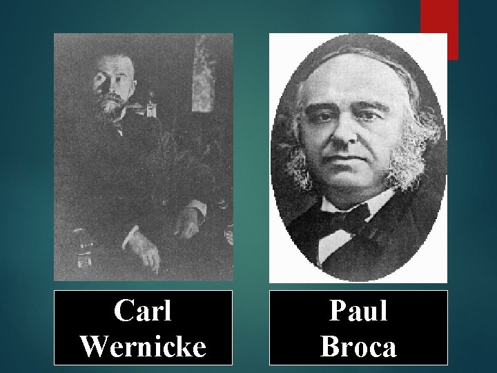
Carl Wernicke Paul Broca
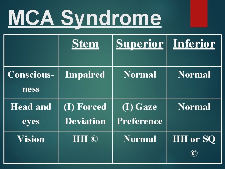
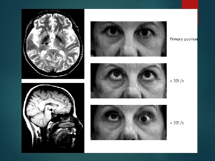
MCA Syndrome Stem Superior Inferior Consciousness Impaired Head and eyes (I) Forced (I) Gaze Deviation Preference Vision HH © Normal HH or SQ ©
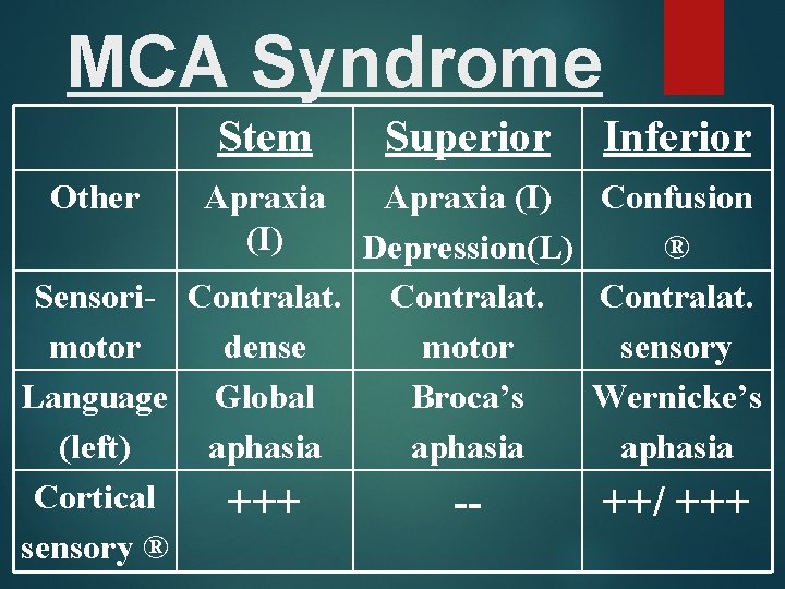
MCA Syndrome Stem Other Apraxia (I) Superior Inferior Apraxia (I) Confusion Depression(L) ® Sensori- Contralat. motor dense motor sensory Language Global Broca’s Wernicke’s (left) aphasia Cortical +++ -++/ +++ sensory ®
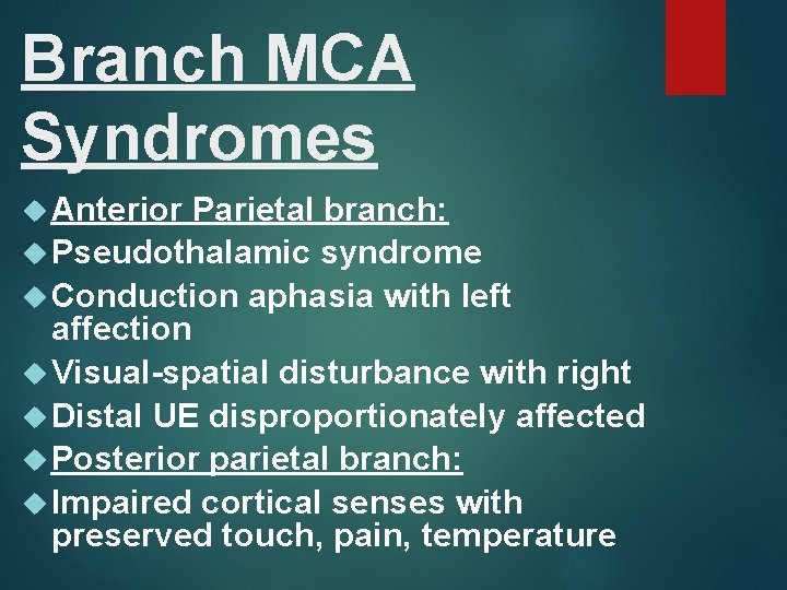
Branch MCA Syndromes Anterior Parietal branch: Pseudothalamic syndrome Conduction aphasia with left affection Visual-spatial disturbance with right Distal UE disproportionately affected Posterior parietal branch: Impaired cortical senses with preserved touch, pain, temperature
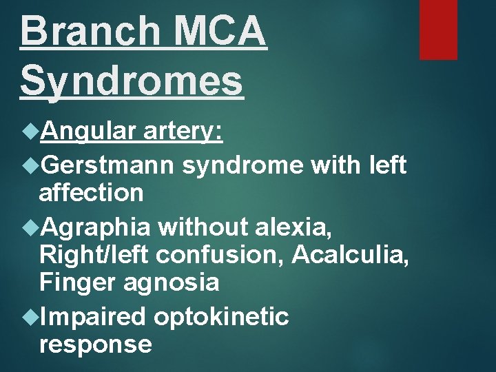
Branch MCA Syndromes Angular artery: Gerstmann syndrome with left affection Agraphia without alexia, Right/left confusion, Acalculia, Finger agnosia Impaired optokinetic response
Lacunar MCA Syndromes Pure motor stroke Sensori-motor stroke Ataxic hemiparesis
ACh. A Syndrome Supply: Posterior limb of internal capsule, posterior corona radiata, Globus pallidus, LGB, Optic tract
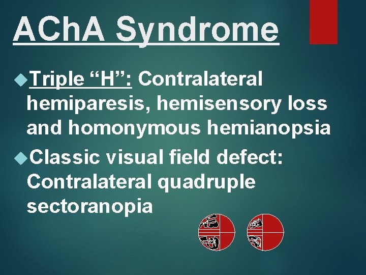
ACh. A Syndrome Triple “H”: Contralateral hemiparesis, hemisensory loss and homonymous hemianopsia Classic visual field defect: Contralateral quadruple sectoranopia
Adolf Wallenberg
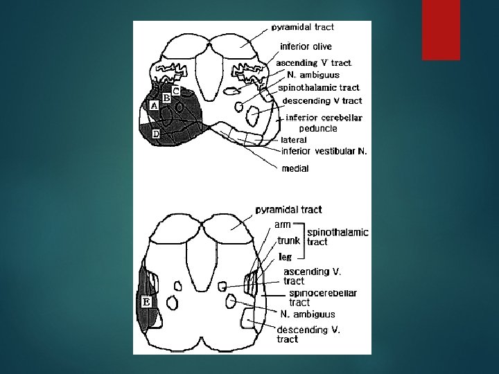
PICA Syndrome Supply: Posterolateral medulla and posterior and inferior cerebellum
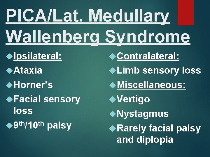
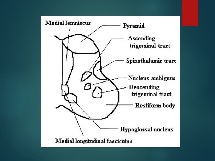
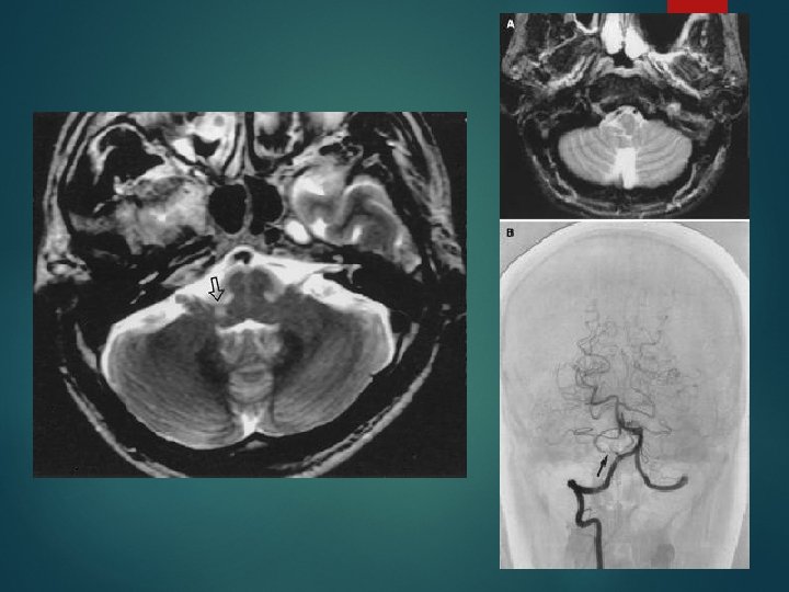
PICA/Lat. Medullary Wallenberg Syndrome Ipsilateral: Contralateral: Ataxia Limb Horner’s Miscellaneous: Facial Vertigo sensory loss 9 th/10 th sensory loss Nystagmus palsy Rarely facial palsy and diplopia
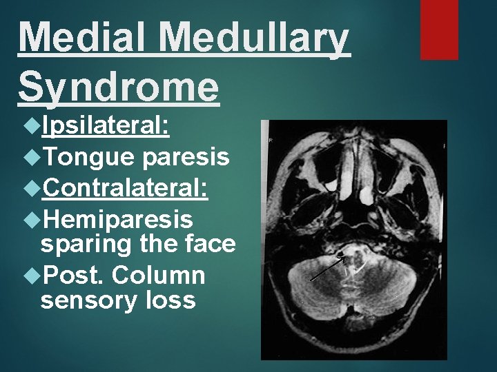
Medial Medullary Syndrome Ipsilateral: Tongue paresis Contralateral: Hemiparesis sparing the face Post. Column sensory loss
Weber
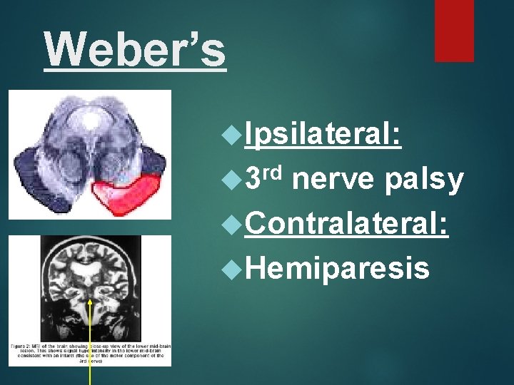
Weber’s Ipsilateral: rd 3 nerve palsy Contralateral: Hemiparesis
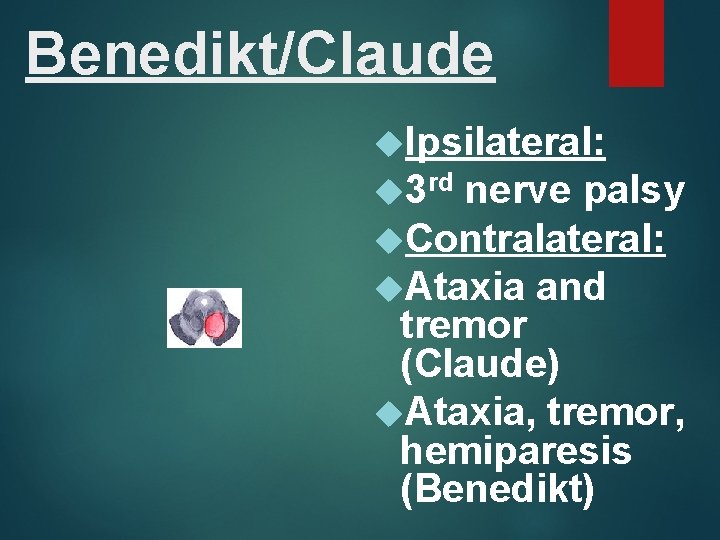
Benedikt/Claude Ipsilateral: 3 rd nerve palsy Contralateral: Ataxia and tremor (Claude) Ataxia, tremor, hemiparesis (Benedikt)
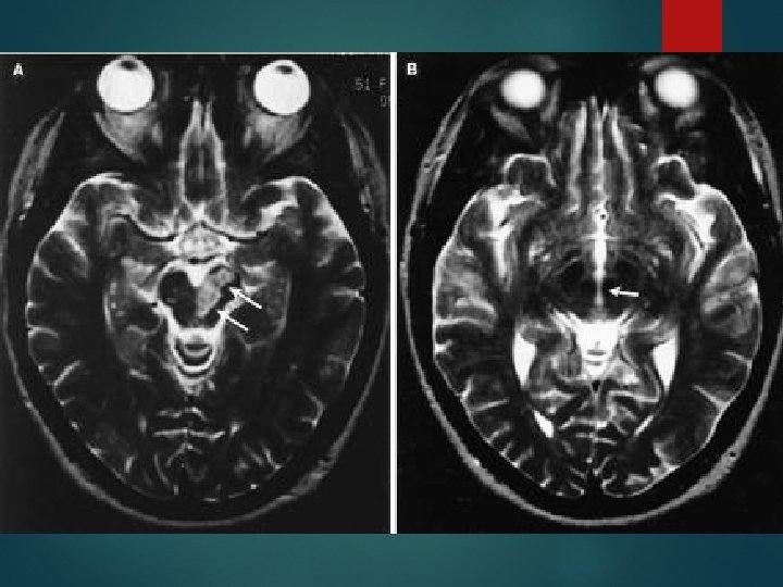
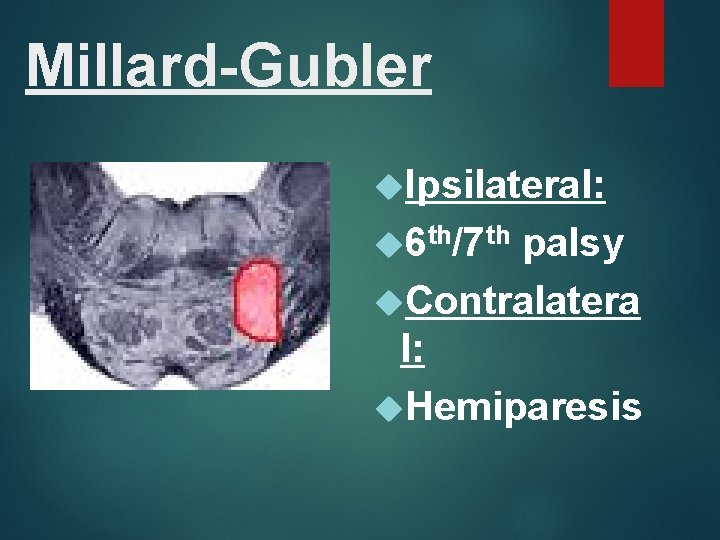
Millard-Gubler Ipsilateral: 6 th/7 th palsy Contralatera l: Hemiparesis
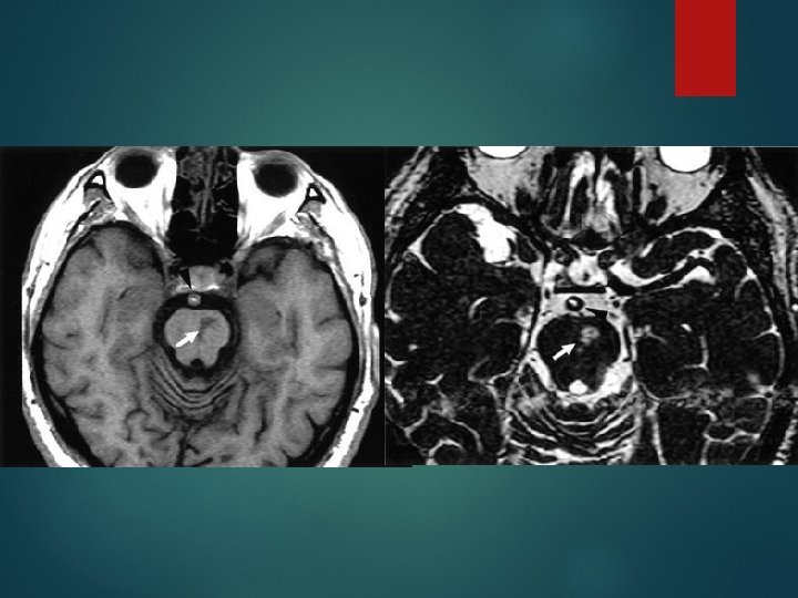
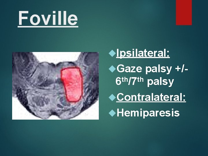
Foville Ipsilateral: Gaze palsy +/6 th/7 th palsy Contralateral: Hemiparesis
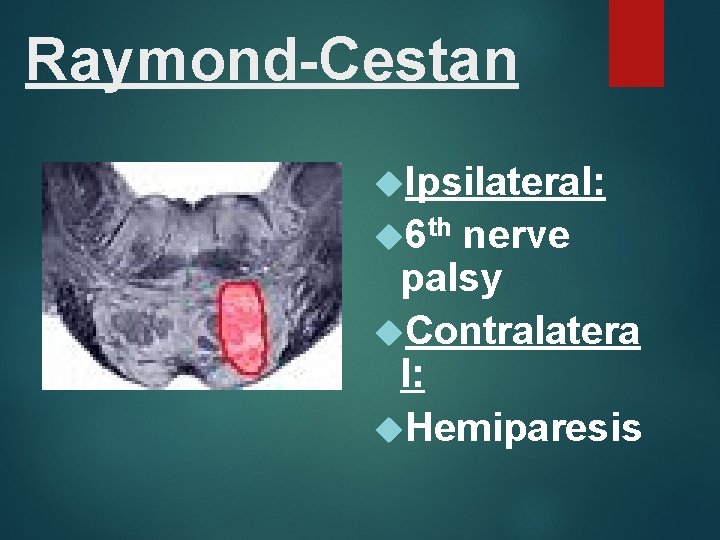
Raymond-Cestan Ipsilateral: 6 th nerve palsy Contralatera l: Hemiparesis
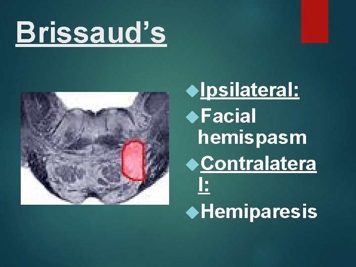
Brissaud’s Ipsilateral: Facial hemispasm Contralatera l: Hemiparesis
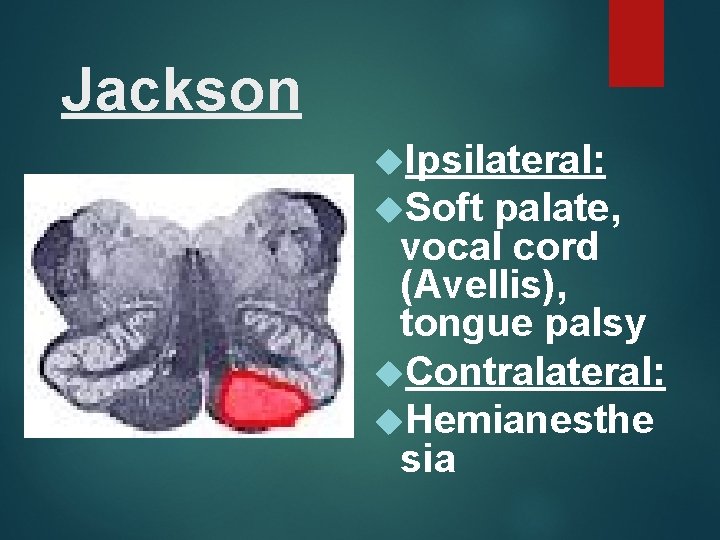
Jackson Ipsilateral: Soft palate, vocal cord (Avellis), tongue palsy Contralateral: Hemianesthe sia
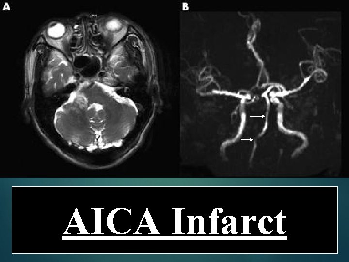
AICA Syndrome Supply: Anterior and inferior cerebellum and posterolateral pons
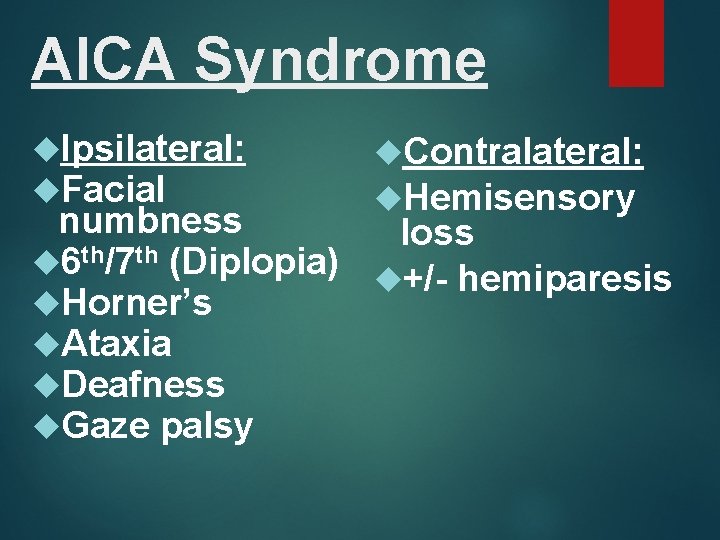
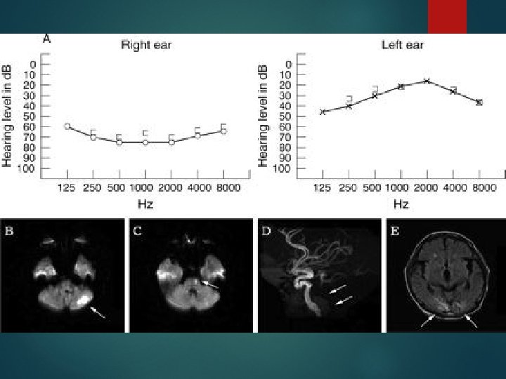
AICA Syndrome Ipsilateral: Facial Contralateral: Hemisensory numbness loss 6 th/7 th (Diplopia) +/- hemiparesis Horner’s Ataxia Deafness Gaze palsy
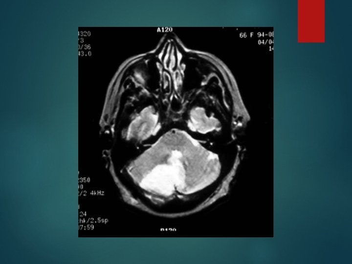
AICA Infarct
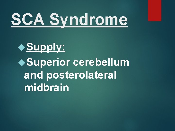
SCA Syndrome Supply: Superior cerebellum and posterolateral midbrain
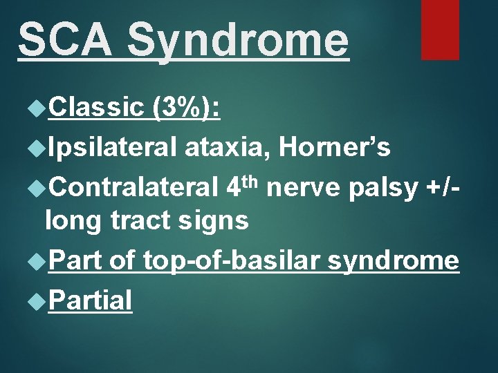
SCA Syndrome Classic (3%): Ipsilateral ataxia, Horner’s Contralateral 4 th nerve palsy +/long tract signs Part of top-of-basilar syndrome Partial
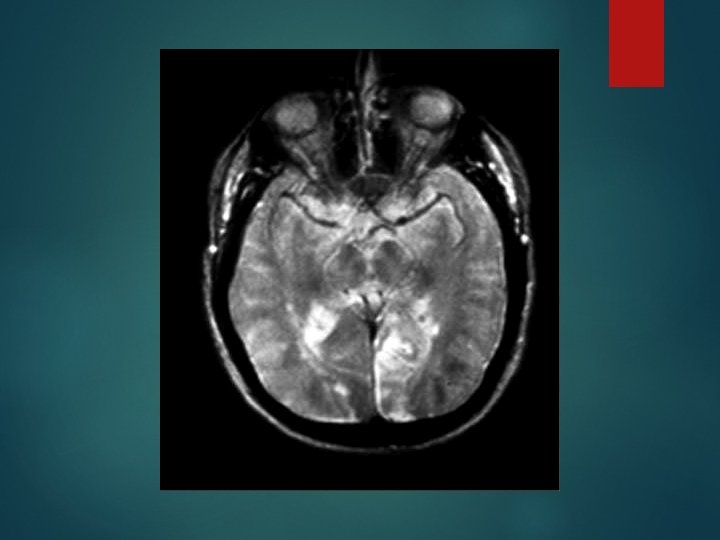
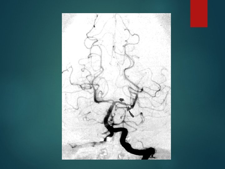
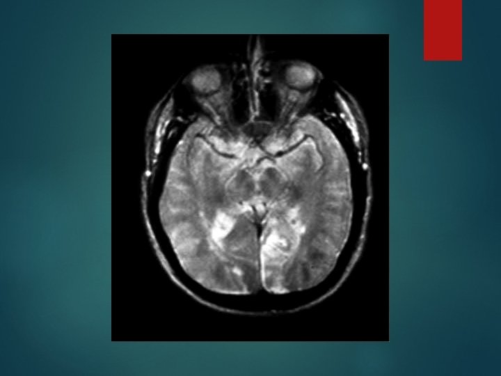
PCA Syndrome Supply: Thalamus Midbrain crus cerebri Hemispheric: Medial temporal and occipital cortex including visual cortex, posterior 3/5 of corpus callosum including splenium
PCA Syndrome Pre-communal: Contralateral hemiparesis, hemisensory loss +/- HH Post-communal ambient cistern segment: Contralateral hemisensory loss +/- HH
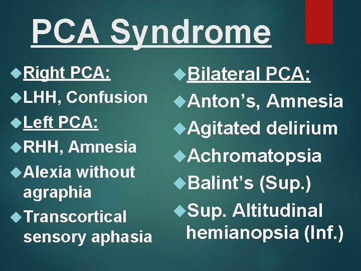
PCA Syndrome Right PCA: LHH, Confusion Left PCA: RHH, Amnesia Alexia without agraphia Transcortical sensory aphasia Bilateral PCA: Anton’s, Amnesia Agitated delirium Achromatopsia Balint’s (Sup. ) Sup. Altitudinal hemianopsia (Inf. )
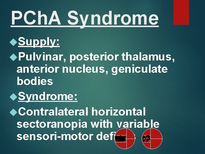
PCh. A Syndrome Supply: Pulvinar, posterior thalamus, anterior nucleus, geniculate bodies Syndrome: Contralateral horizontal sectoranopia with variable sensori-motor deficits
Lacunar Syndromes Pure motor stroke Pure sensory stroke Ataxic hemiparesis Dysarthria-Clumsy hand syndrome Sensori-motor stroke
Aphasias Disorder of language, acquired secondary to brain damage. Dysphasia: Developmental language disorder Differentiate from dysarthria, dysphonia, stuttering, speech apraxia, thought disorders
Aphasias Language components: Spontaneous speech Naming Auditory comprehension Repetition Reading Writing
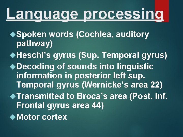
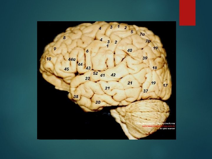
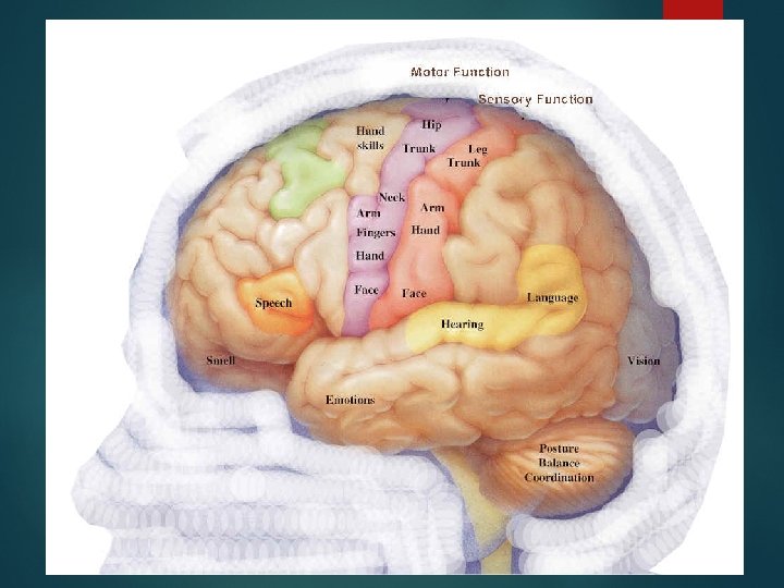
Language processing Spoken words (Cochlea, auditory pathway) Heschl’s gyrus (Sup. Temporal gyrus) Decoding of sounds into linguistic information in posterior left sup. Temporal gyrus (Wernicke’s area 22) Transmitted to Broca’s area (Post. Inf. Frontal gyrus area 44) Motor cortex
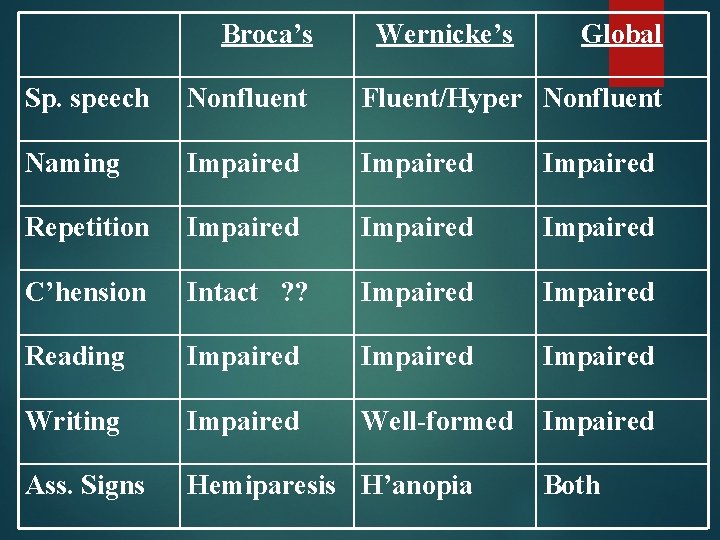
Broca’s Wernicke’s Global Sp. speech Nonfluent Fluent/Hyper Nonfluent Naming Impaired Repetition Impaired C’hension Intact ? ? Impaired Reading Impaired Writing Impaired Well-formed Impaired Ass. Signs Hemiparesis H’anopia Both
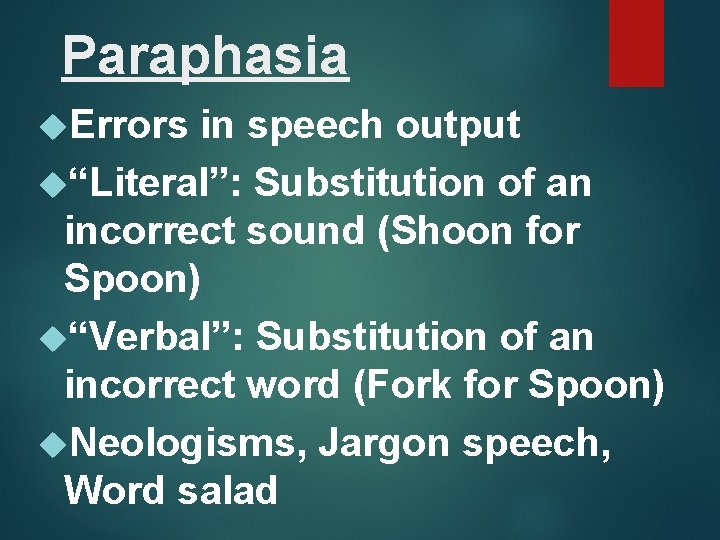
Paraphasia Errors in speech output “Literal”: Substitution of an incorrect sound (Shoon for Spoon) “Verbal”: Substitution of an incorrect word (Fork for Spoon) Neologisms, Jargon speech, Word salad
Figure 1 (A) Fluid attenuation inversion recovery imaging demonstrating left-hemispheric ischaemia with secondary oedema and obliteration of cerebrospinal fluid spaces. An old right middle cerebral artery stroke is also shown (arrow). (B) Further images demonstrating uncal herniation (arrow 1) with secondary midbrain compression and infarction (arrow 2), as well as subfalcine herniation (arrow 3). Thrombus is shown within the middle cerebral artery (arrow 4). Kumar, K R et al. J Neurol Neurosurg Psychiatry 2009; 80: 87 Copyright © 2009 BMJ Publishing Group Ltd.
Broca’s W E R N I C K E’ S
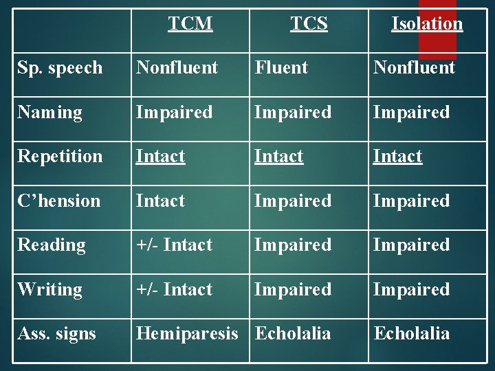
TCM TCS Isolation Sp. speech Nonfluent Fluent Nonfluent Naming Impaired Repetition Intact C’hension Intact Impaired Reading +/- Intact Impaired Writing +/- Intact Impaired Ass. signs Hemiparesis Echolalia
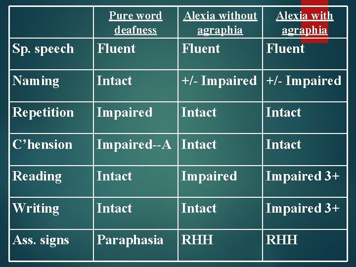
Pure word deafness Alexia without agraphia Alexia with agraphia Sp. speech Fluent Naming Intact +/- Impaired Repetition Impaired Intact C’hension Impaired--A Intact Reading Intact Impaired 3+ Writing Intact Impaired 3+ Ass. signs Paraphasia RHH
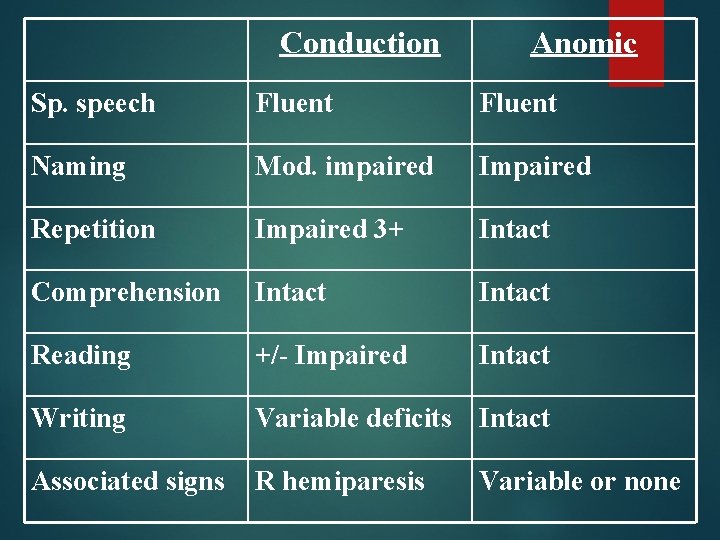
Conduction Anomic Sp. speech Fluent Naming Mod. impaired Impaired Repetition Impaired 3+ Intact Comprehension Intact Reading +/- Impaired Intact Writing Variable deficits Intact Associated signs R hemiparesis Variable or none
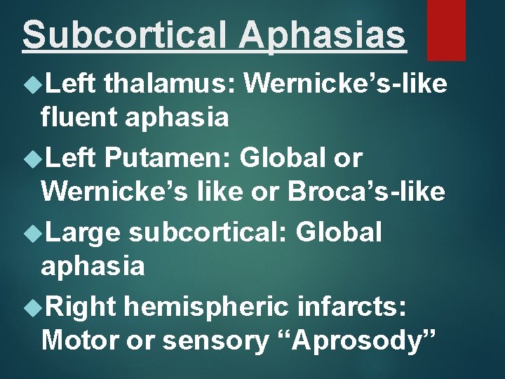
Subcortical Aphasias Left thalamus: Wernicke’s-like fluent aphasia Left Putamen: Global or Wernicke’s like or Broca’s-like Large subcortical: Global aphasia Right hemispheric infarcts: Motor or sensory “Aprosody”
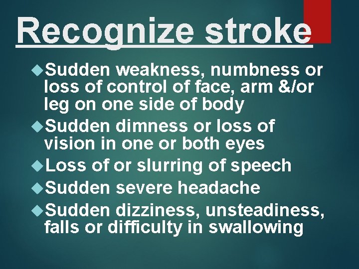
Recognize stroke Sudden weakness, numbness or loss of control of face, arm &/or leg on one side of body Sudden dimness or loss of vision in one or both eyes Loss of or slurring of speech Sudden severe headache Sudden dizziness, unsteadiness, falls or difficulty in swallowing
Acute Stroke: Rapid clinical assessment Management Urgent plain head CT CBC, Electrolytes, Sugar, PT/a. PTT Consider thrombolysis if <3 hours Other drug options, medical management CXR, EKG, Carotid doppler, 2 D echo MRA or DSA Other tests as indicated
Acute Stroke: General Rx Management of HTN: Do Not lower unless > 220/120 Do Not use sublingual preparations IV Labetelol or Enalaprilet, Oral Captopril, Candesartan Avoid direct arterial vasodilators Lower BP if specific indications
Acute Stroke: General RX DM: Euglycemia (Avoid hypo. ) Temperature regulation Fluid management Prevention and Rx of aspiration pneumonia Prevention and Rx of DVT Gastric prophylaxis Management of seizures
Acute Stroke: General RX Cardio-respiratory care Bladder and bowel care Prevention and Rx of spasticity Prevention and Rx of contractures Prevention and Rx of bed sores Always aim for early mobilization and ambulation
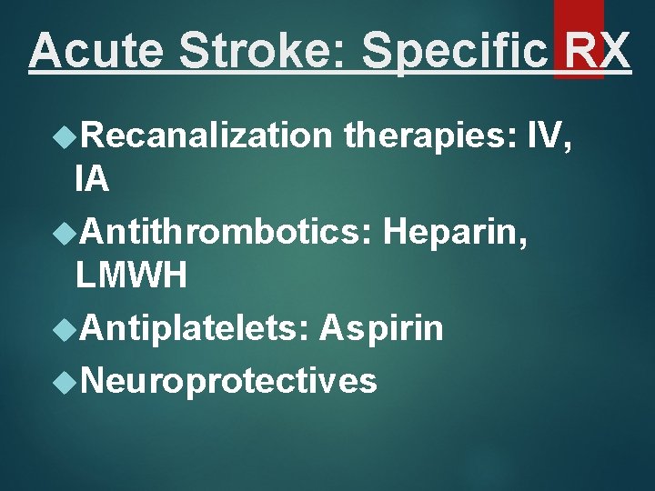
Acute Stroke: Specific RX Recanalization therapies: IV, IA Antithrombotics: Heparin, LMWH Antiplatelets: Aspirin Neuroprotectives
Quote Law of encounters: The probability of meeting someone you know increases when you are with someone you don’t want to be seen with!
Thank You…. .