Deep Stabilizing Core Muscle Activation and Training Aim
 Muscle Activation and Training Aim : Proximal stability for distal mobility")
Deep Stabilizing (Core) Muscle Activation and Training Aim : Proximal stability for distal mobility The primary functions of the muscles of the trunk are to provide the stabilizing force so upright posture can be maintained against a variety of forces that disturb balance and to provide a stable base so the muscles of the extremities can execute their function efficiently and without undue stress to the spinal structures
 and longus colli and")
core musculature (Tr. A and Mf in the lumbar spine) and longus colli and other deep musculature (in the cervical spine)

Guidelines for Stabilization Training, Principles and Progression: 1. Begin training awareness of safe spinal motions and the neutral spine position ( functional ROM and position). 2. Have patient learn to activate the deep (core) stabilizing musculature while in the neutral position. 3. Add extremity motions to load the global musculature while maintaining a stable neutral spine position (dynamic stabilization). 4. Increase repetitions to improve holding capacity (endurance) in the stabilizing musculature; increase load (change lever arm or add resistance) to improve strength while maintaining a stable neutral spine position. 5. Use alternating isometric contractions and rhythmic stabilization techniques to enhance stabilization and balance with fluctuating loads. 6. Progress to movement from one position to another in conjunction with extremity motions while maintaining a stable neutral spine (transitional stabilization). 7. Use unstable surfaces to improve the stabilizing response and improve balance.
 or ( chin in). Lower Cervical and")
Cervical Musculature Deep Neck Flexors (“yes” motion) or ( chin in). Lower Cervical and Upper Thoracic Extensor to activate and control the muscles that control axial extension (cervical retraction) : capital flexion + slight flattening of the cervical lordosis, and flattening of the upper thoracic kyphosis
")
Deep Neck Flexors: Activation and Training (craniocervical flexion and gentle axial extension)

• Place the folded Stabilizer. TM pressure biofeedback unit under the upper cervical spine and inflate to 20 mm. Hg. • Instruct the patient to nod and increase pressure on the cuff to 22 mm. Hg and hold the pressure steady. • If the patient is successful (i. e. , can hold the position with minimal superficial muscle activity), have him or her relax and repeat the flexion, this time increasing pressure to 24 mm. Hg. Repeat this incremental activation up to 30 mm. Hg (total 10 mm. Hg increase). • The final pressure is the one at which the patient can hold steady. • Muscle endurance (holding or tonic capacity) of the deep neck flexors is measured by the number of 10 -second holds (up to 10) at the final pressure for 10 repetitions.

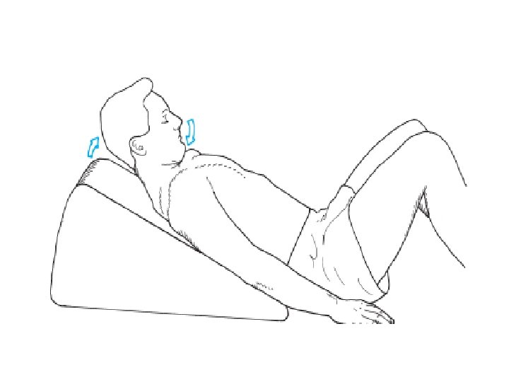
Lower Cervical and Upper Thoracic Extensor Activation and Training • Patient position and procedure: Prone with forehead on the treatment table and arms at the sides. Have the patient lift the forehead off the treatment table, keeping the chin tucked and eyes focused on the table to maintain the neutral spinal position hold 10 sec.
 Global Muscle Stabilization Exercises 1 - Stabilization Exercises for the Cervical Region")
Progression (A) Global Muscle Stabilization Exercises 1 - Stabilization Exercises for the Cervical Region (craniocervical flexion and gentle axial extension) (hold 10 seconds x 10 repetitions) 2 - Stabilization with Progressive Limb Loading Initially, the only resistance load comes from simple upper extremity movements. When the patient can perform multiple repetitions of the upper extremity motions, resistance is added with handheld weights or elastic resistance.
 External resistance : (free")
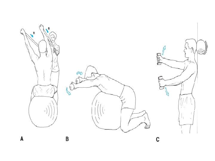
Stabilization with Progressive Limb Loading Extremity loading : (arms motions) External resistance : (free weights or elastic resistance) Unstable surfaces : ball while sitting, quadruped position, or standing supporting the ball between the head and the wall

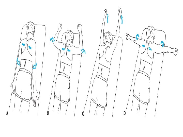
Cervical Stabilization with Progressive Limb Loading—Emphasis on Cervical Flexors Arm motioms: * Shoulder flexion to 90 * Shoulder abduction 90 * Shoulder external rotation with arms at sides * Shoulder flexion to end of range * Shoulder abduction combined with external rotation to end of range * Diagonal patterns * Reaching forward, outward, upward Patient position: Supine , Sitting (sitting on ball for less stability) , Standing with wall support (ball front head), and Standing with no support
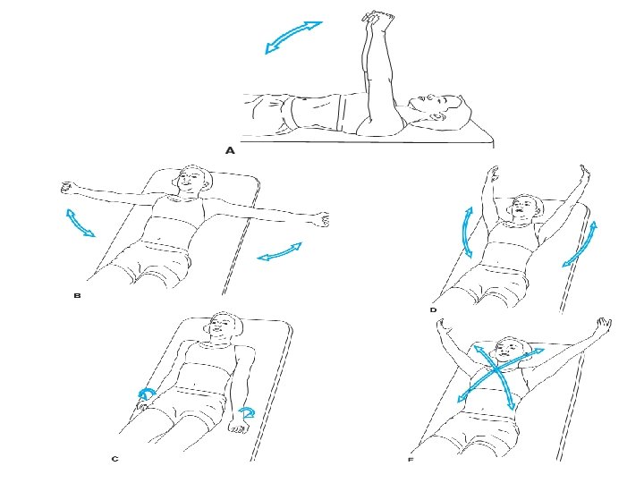

Cervical Stabilization with Progressive Limb Loading—Emphasis on Cervical and Thoracic Extensors Arm motion: * Arms at side: laterally rotate shoulders and adduct scapulae * Arms in 90/90 position (abducted and laterally rotated), horizontally abduct shoulders and adduct scapulae * Elevate shoulder in full flexion * Arms abducted to 90 and laterally rotated, elbows extended: horizontally abduct shoulders and adduct scapulae * Upper extremity diagonal patterns * Standing: reaching forward, outward, upward in functional patterns * Standing, no support: pushing/pulling and lifting activities Patient position: Prone forehead on treatment table, Quadruped over gym ball, Standing back supported by wall (ball behind head). and Standing no support
 ISOMETRIC AND DYNAMIC EXERCISES 1 - Isometric Resistance Activities by therapist or patient")
(B) ISOMETRIC AND DYNAMIC EXERCISES 1 - Isometric Resistance Activities by therapist or patient him self * Flexion. * Side bending. * Axial extension. * Rotation.
 cervical flexion and (B) axial extension.")
Self-resistance for isometric (A) cervical flexion and (B) axial extension.

2 - Dynamic Cervical Flexion Patient position and procedure: Supine. If the patient cannot tuck the chin and curl the neck to lift the head off the mat, begin with the patient on a slant board or large wedge-shaped bolster under the thorax and head to reduce the effects of gravity and Progress by decreasing the angle of the board or wedge and then adding manual resistance.

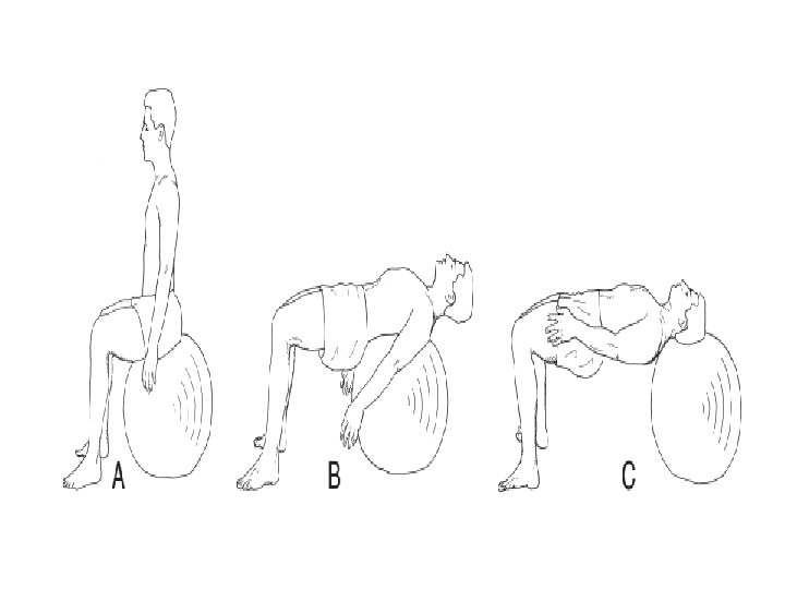
3 - Intermediate and Advanced Training For strengthening the cervical and upper thoracic flexors and extensors as stabilizers Begin by: (A) sitting on a large gym ball (B) walking forward while rolling the ball up the back. With the ball behind the mid-thoracic area, the cervical flexors must stabilize. (C) Continue walking forward until the ball is under the head; the cervical extensors now must stabilize. Walk back and forth between the two positions (B and C) to alternate control between the flexors and extensors. Progress by adding arm motions or arm motions with weights to increase resistance.

4 - Transitional Stabilization for the Cervical and Upper Thoracic Regions Standing with a basketball-size inflatable ball between the head and the wall. Have the patient roll the ball along the wall, using the head. This requires the patient to turn the body as he or she walks along. Sitting on a large gym ball. Have the patient walk the feet forward and backward , alternating stabilization between the flexors and extensors. Progress to advanced training by adding arm motions and then arm motions with weights in each of the positions.
 FUNCTIONAL TRAINING * Rolling. * Supine to sit/sit to lying down. * Sit")
(C) FUNCTIONAL TRAINING * Rolling. * Supine to sit/sit to lying down. * Sit to stand/stand to sit. * In and out of a car. * Walking.
")
Thoraco-lumbar Musculature Three techniques for abdominal muscle activation: 1 -Drawing-In Maneuver (Abdominal Hollowing Exercise) for Transverse Abdominis Activation Patient positions: Training may be easiest in the quadruped position in order to use the effects of gravity on the abdominal wall. Hook-lying (with knees 70 to 90 and feet resting on an exercise mat), prone-lying, or semireclined positions may be used if more comfortable for the patient. It is important to progress training to sitting and standing as soon as possible for functional activities. Procedure: Teach the patient using demonstration, verbal cues, and tactile facilitation. Explain that the muscle encircles the trunk; and when activated, the waistline draws inward. Palpation of the muscle is possible just distal to the anterior superior iliac spine (ASIS) and lateral to the rectus abdominis. When the internal oblique (IO) contracts, a bulge of the muscle is felt; when the Tr. A contracts, flat tension is felt. The goal is to activate the Tr. A with minimal or no contraction of the IO. This is a gentle contraction.

the drawingin maneuver Palpation of the transversus abdominis muscle

2 - Abdominal Bracing In contrast to the drawing-in maneuver, abdominal bracing occurs by setting the abdominals and actively flaring out laterally around the waist. There is no head or trunk flexion, no elevation of the lower ribs, no protrusion of the abdomen, and no pressure through the feet. The patient should be able to hold the braced position while breathing in a relaxed manner. This technique has been taught for a number of years as the method to stabilize the spine; and it has been shown to activate the oblique abdominal muscles consistent with their global stabilization function.

3 - Posterior Pelvic Tilt Pelvic tilt exercises principally activate the rectus abdominis muscle, which is used primarily for dynamic trunk flexion activity. It is not considered a core spinal stabilization muscle; therefore, it is not emphasized in the training for stabilization. 42 It is used mostly to teach awareness of movement of the pelvis and lumbar spine. It is activated when the patient explores his or her lumbar ROM with pelvic tilts to find the neutral position or functional spinal range.

Multifidus Activation and Training Patient position and procedure: Prone or side-lying. Place your palpating digits (thumbs or index fingers) immediately lateral to the spinous processes of the lumbar spine. Palpate each spinal level so comparisons in the activation of the multifidus (Mf) muscle can be made between each segment as well as from side to side. Instruct the patient to “swell the muscle” out against your digits. Palpate for consistency of muscle contraction at each level. Facilitation techniques include using the drawingin maneuver and gently contracting the pelvic floor muscles. In the side-lying position, facilitate by gently applying manual resistance to the thorax or pelvis to activate the rotation function of the Mf.
 Global Muscle Stabilization Exercises 1 -")
Stabilization Exercises for the thoraco- lumbar Region (A) Global Muscle Stabilization Exercises 1 - Stabilization with Progressive Limb Loading - Emphasis on Abdominals Level 1: core activation Draw in and hold 10 seconds Level 2: Opposite LE on mat; bent leg fall out in the supine position. This requires control to prevent pelvic rotation; stability is assisted by the opposite lower extremity while hook-lying.

Level 3: A, B, or C : Opposite LE is on table Level 4: A, B, or C: Hold opposite LE @ 90 of hip flexion with UE assistance Level 5: A, B, or C: Hold opposite LE @ 90 of hip flexion (no UE assistance) Level 6: A, B, or C: Bilateral LE movement
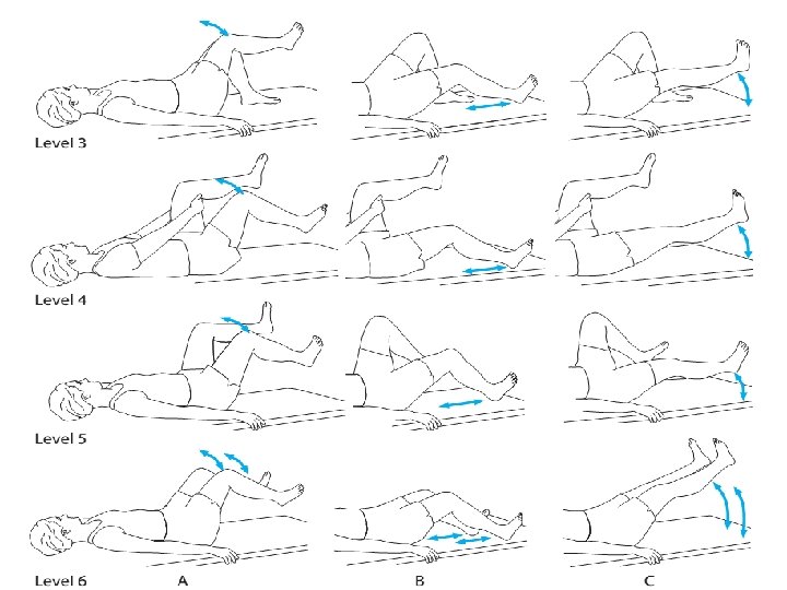

2 - Stabilization with Progressive Limb Loading - Emphasis on Trunk Extensors A- Flex one upper extremity (UE) B- Extend one lower extremity (LE) by sliding it along the exercise mat C- Extend one (LE) and lift 6– 8 inches off exercise mat D- Flex one UE and extend contralateral LE E- Extend one LE F- Extend both LE G- Lift head, arms and LE


3 -Quadratus Lumborum—Stabilization Exercises Patient position and procedure: Begin side-lying. Have the patient prop up on the elbow and then lift the pelvis off the mat, supporting the lower body with the lateral side of the knee on the downward side. The position can be maintained for an isometric hold or performed intermittently. Progress by having the patient support the upper body with the hand (with the elbow extended) and lateral aspect of the foot on the downward side.

4 - External resistance Use weights, elastic resistance, or pulleys for strengthening.

5 - Unstable surfaces Use a large gym ball, foam roller
 Alternating lower extremity motions with the “modified bicycle” (B)")
6 - Alternating motions (A) Alternating lower extremity motions with the “modified bicycle” (B) reciprocal and alternating patterns using the upper and lower extremities simultaneously require a strong controlling action in the abdominals. (C) Strength, balance, and coordination are required to maintain spinal stabilization while sitting on a gym ball and moving the extremities. This activity is progressed by adding weights to the extremities.
 ISOMETRIC EXERCISES Alternating Isometric Contractions and Rhythmic Stabilization Patient positions and procedures: Begin")
(B) ISOMETRIC EXERCISES Alternating Isometric Contractions and Rhythmic Stabilization Patient positions and procedures: Begin with the patient supine in the most stable position. Progress to sitting on a stable surface, sitting on an unstable surface such as a large gym ball, kneeling, and then standing. Sitting, kneeling, and standing require stabilizing action in the hip, knee, and ankle musculature, respectively, as well as the spinal muscles. Apply resistance directly against the patient’s shoulders or pelvis, 1 - Have the patient find the neutral spine position and then activate the stabilizing muscles with the drawingin maneuver prior to applying the resistive force. Then instruct the patient to “meet my resistance” while applying a force to stimulate isometric contractions. Apply the resistance in alternating directions at a controlled speed while the patient learns to maintain a steady position. 2 - Initially, provide verbal cues, such as “hold against my resistance, but do not overpower me. Feel your abdominal muscles contracting. Now I’m pulling in the opposite direction. Match the resistance and feel your back muscles contracting. ” 3 - Progress by shifting the directions of resistance without the verbal cues and then by increasing the speed.

4 - Begin with alternating resistance in the sagittal plane; progress to side-to-side and then transverse plane resistance. Isometric resistance to trunk rotation (transverse plane resistance) has been shown to be the most effective in stimulating the oblique abdominals, transversus abdominis, and deep spinal extensor muscles. 5 - Alternating resistance to pelvic rotation can also be done by having the patient assume a modified bridge position. Apply resistance directly to the pelvis to stimulate rotation while the patient isometrically holds the pelvis and spine in a stable position.
 DYNAMIC EXERCISES 1 - Dynamic Strengthening Abdominal Muscles: Curl-ups. First, instruct the patient")
(C) DYNAMIC EXERCISES 1 - Dynamic Strengthening Abdominal Muscles: Curl-ups. First, instruct the patient to perform the drawingin maneuver to cause a stabilizing contraction of the abdominal muscles and then lift the head. Progress by lifting the shoulders until the scapulae and thorax clear the mat, keeping the arms horizontal (Fig. 16. 40). A full sit-up is not necessary because once thorax clears the mat the rest of the motion is performed by the hip flexor muscles. Further progress the difficulty of the curl-up by having the patient change the arm position from horizontal to folded across the chest and then to behind the head. During all these activities, the low back should not arch; if it does, reduce the progression until the abdominals are strong enough to maintain lumbar flexion.

Curl-ups Curl-downs. If the patient is unable to perform the curl-up, begin with curl-downs by having the patient start in the hook-sitting or long-sitting position and lower the trunk only to the point where he or she can maintain a flat low back and then return to the sitting position. Once the patient can curl-down full range, reverse and perform a curl-up.

Diagonal curl-ups. Have the patient reach one hand toward the outside of the opposite knee while curling up; then alternate. Reverse the muscle action by bringing one knee up toward the opposite shoulder; then repeat with the other knee. Diagonal exercises emphasize the oblique muscles. Double knee-to-chest. To emphasize the lower rectus abdominis and oblique muscles, have the patient set a posterior pelvic tilt; then bring both knees to the chest and return. Progress the difficulty by decreasing the angle of hip and knee flexion

Pelvic lifts. Have the patient begin with the hips at 90 and knees extended; then lift the buttocks upward off the mat (small motion). The feet move upward toward the ceiling. The patient should not push against the mat with the hands.

Bilateral straight-leg raising Have the patient begin with legs extended; then perform a posterior pelvic tilt followed by flexing both hips, keeping the knees extended. If the pelvis and spine cannot be kept stable, the knees should be flexed to a degree that allows control. If the hips are abducted before initiating this exercise, greater stress is placed on the oblique abdominal muscles. P R E C A U T I O N : The strong pull of the psoas major causes shear forces on the lumbar vertebrae. Also this bilateral straight-leg raise (SLR) causes increased spinal compression loads. If there is any low-back pain or discomfort, especially with spinal hypermobility or instability, this exercise should not be performed even if the abdominals are strong enough to maintain a posterior pelvic tilt. Bilateral straight-leg lowering can be performed if the bilateral SLR is difficult. Have the patient begin with the hips at 90 and knees extended; then lower the extremities as far as possible while maintaining stability in the lumbar spine (should not increase the lordosis), followed by raising the legs back to 90. See precaution under the bilateral SLR exercise.
—Sitting or Standing Patient position and procedures: Sitting or standing. Pulleys or")
Trunk Flexion (Abdominals)—Sitting or Standing Patient position and procedures: Sitting or standing. Pulleys or elastic material are secured at shoulder level behind the patient. Progress the resistance as the patient’s abdominal strength increases. Have the patient hold the handles or ends of the elastic material with each hand then flex the trunk, with emphasis on bringing the rib cage down toward the pubic bone and performing a posterior pelvic tilt, rather than flexing at the hips. Have the patient perform diagonal motions by bringing one arm down toward the opposite knee with emphasis on moving the rib cage down toward the opposite side of the pelvis. Repeat the diagonal motion in the opposite direction.
—Unstable Surfaces Use of unstable surfaces, such as a gym ball or")
Trunk Flexion (Abdominals)—Unstable Surfaces Use of unstable surfaces, such as a gym ball or a balance board, while doing abdominal curl-up exercises has been shown to increase activity in the internal and external obliques and the rectus abdominis

2 - Dynamic Strengthening—Erector Spinae and Multifidus Muscles: Thoracic elevation. Begin with the arms at the side, progress to behind the head or reaching overhead as strength improves. Have the patient tuck in the chin and lift the head and thorax. The lower extremities must be stabilized P R E C A U T I O N S : Extension exercises in the prone position are performed at the end of the ROM in spinal extension and therefore may not be appropriate for individuals with symptoms from conditions such as arthritis or nerve root compression.

Leg lifts. Initially have the patient lift only one leg, alternate with the other leg, and finally lift both legs and extend the spine. Stabilize thorax by having the patient hold onto the side of the treatment table. “Superman. ” Progress the extension exercises by having the patient lift both upper and lower extremities simultaneously

Elastic resistance or weighted pulleys. Secure pulleys or elastic resistance in front of the patient at shoulder level. Have him or her hold onto the ends of the material or handles and extend the spine For trunk rotation, use a pulley or elastic resistance secured under the foot or to a stable object opposite to the side being exercised. Have the patient pull against the resistance, extending and rotating the back.
 -Trunk side-bending exercises are")
3 -Trunk Side Bending (Lateral Abdominals, Erector Spinae, Quadratus Lumborum) -Trunk side-bending exercises are used for general strengthening of the muscles that side-bend the trunk. -Side-bending exercises are also used if there is scoliosis Patient position and procedure: Side-lying on the concave side of the curve with the apex at the edge of the table or mat so the thorax is lowered. If you have access to a split table with one end that can be lowered, begin with the apex of the curve at the bend of the table. Have the patient place the lower arm folded across the chest and upper arm along the side of the body and side-bend the trunk up against gravity. Progress by having the patient clasp both hands behind the head. Stabilization of the pelvis and lower extremities must be provided.
Transitional Stabilization Patient position and procedure: Standing with the back to a wall and")
(D)Transitional Stabilization Patient position and procedure: Standing with the back to a wall and the spine held in its neutral position. Place a towel behind the back so it slides easier along the wall. The exercise is more challenging if a large gym ball is placed between the back and the wall. Have the patient slide his or her back down the wall into a partial squat and hold the position for isometric strengthening of the hip and knee extensors or move up and down for concentric/eccentric strengthening. Superimpose arm motions such as alternating or bilateral shoulder flexion/extension. Use handheld weights to add resistance for upper and lower extremity strengthening.
 FUNCTIONAL TRAINING Rolling with a neutral spine requires that the patient first find")
(E) FUNCTIONAL TRAINING Rolling with a neutral spine requires that the patient first find the neutral spine, perform the drawing-in maneuver, and then roll the trunk as a unit. Supine to sit/sit to lying down. Have the patient use the log roll maneuver (as described above) to roll from supine to side-lying while simultaneously flexing the hips and knees and pushing up with the arms. Sit to stand/stand to sit. The patient’s level of function dictates how much assistance from the upper extremities is needed to accomplish “sit to stand” or “stand to sit. ” If the hip and knee extensors are not strong enough to elevate the body, the patient requires a chair with armrests so there is some leverage for pushing up; alternatively, an elevated seat may be necessary. The patient focus on the hip motion while keeping the spine “solid like a board. ” The reverse is also practiced. In and out of a car. Approach the open car door and seat with the back toward the seat; stabilize the spine in its neutral position with the drawing-in maneuver, then bend at the hips and sit down. Once seated, flex both hips and knees and pivot the whole body around as a unit, maintaining a stable spine. When exiting a car, keep both knees together and pivot the legs and trunk outward as a unit. Once the feet are on the ground, bend at the hips and elevate the trunk as a unit.

Walking. For some patients walking may provoke symptoms. Remind the patient to use the neutral spine and drawingin maneuvers to stabilize the spine while walking. It is not possible to maintain conscious control for long, so remind the patient to check the spinal posture and reactivate the drawing-in maneuver whenever the symptoms recur. Pick up. Maitain the trunk straight and squate with knees
- Slides: 52