Deep Dissection of the Back M e l

Deep Dissection of the Back M e l z e r A n g e l i e Presented by: Angelie Melzer P r e s e n t o r :

Superficial to Deep Dissection: Structures to Look For • • • PLATES: 542, 381, 544, 541, 375, 376 Nuchal Ligament Accessory Nerve 3 rd and 4 th cervical nerves and dorsal scapular nerve -C 5 Thoracodorsal Nerve and Artery Posterior Axillary Fold/Intertubercular Sulcus on the Humerus • • • • Levator Scapulae Sternocleidomastoid Splenus Capitus Lateral and Medial Masseter Muscle Splenus Cervicis Omohyoid Muscle Latissimus Dorsi Serratus posterior superior Teres Major Rhomboid Minor Rhomboid Major Supraspinatus, Infraspinatus and Teres Minor muscles (Rotator Cuff)

KEY: ORIGIN, INSERTION, ACTION and NERVE • • O=Origin I=Insertion A=Action N=Nerve Biomechanical Actions: • Circumduction • Lateral Flexion • Rotation

MUSCLES OF THE NECK:
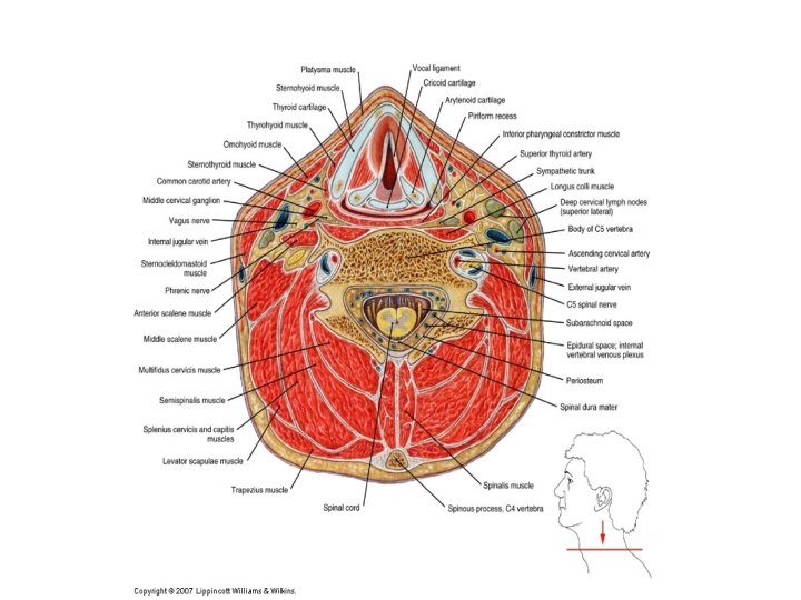

ORIGIN, INSERTION, ACTION and NERVE • Splenius Capitus and Cervicis • Origin: Spinous processes of C 7 -T 4 • Insertion: Mastoid process and lateral portion of superior nuchal line • Action: Rotate the head and neck to the same side, laterally flex and extend the head and neck • Nerve: Cervical • Levator Scapulae • Origin: Transverse processes of C 1 -4 • Insertion: Medial border of scapula between superior angle and superior portion of spine of scapula • Action: Elevate and downwardly rotate the scapula, Laterally flex, rotate and extend the head and neck • Nerve: Cervical 3 -4 and Dorsal Scapular C 4 -5 • Sternocleidomastoid • Origin: Sternal Head-Top of Manubrium; Clavicular Head-Medial 1/3 of clavicle • Insertion: Mastoid process of temporal bone and lateral portion of superior nuchal line • Action: Lateraly flex head neck to same side, Rotate head and neck to opposite side and assist in elevation of the ribcage during inhalation • Nerve: C 1 -3

MUSCLES OF THE SHOULDER: Cross Sectional View
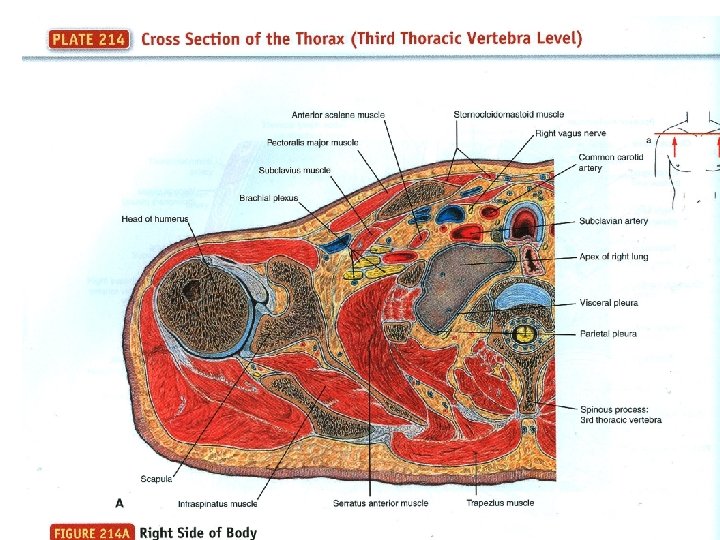

ORIGIN, INSERTION, ACTION and NERVE • Supraspinatus • • • Infraspinatus • • • Origin: Infraspinous fossa of scapula Insertion: Greater Tubercle of humerus Action: Laterally Rotate and adduct shoulder, stabilize head of humerus Nerve: Suprascapular C 4 -6 Teres Minor • • • Origin: Supraspinous fossa of scapula Insertion: Greater Tubercle of humerus Action: Adduct shoulder and stabilize head of humerus Nerve: Suprascapular C 4 -6 Origin: Upper 2/3 of lateral border of scapula Insertion: Greater Tubercle of humerus Action: Laterally Rotate and adduct shoulder, stabilize head of humerus Nerve: Axillary C 5 -6 Subscapularis • • Origin: Subscapular fossa of scapula Insertion: Lesser Tubercle of humerus Action: Medially rotate the shoulder and stabilize the head of humerus Nerve: Upper and Lower subscapular C 5 -7

MUSCLES OF THE SPINE

SPINE: Erector Spinae Group • • • Erector Spinae: Iliocostalis Cervicis, Thoracis and Lumborum • Origin: Common tendon (cervicis, thoracis and lumborum, respectively) • Insertion: Cervical, Thoracic and Lumbar, respectively • Action: Extend and Laterally flex the vertebral column to the same side • Nerve: Spinal Erector Spinae: Longissiumus Cervicis and Capitis • Origin: Same as above • Insertion: Transverse process of cervical vertebra and Lower nine ribs of transverse processes, respectively • Action: Same as above • Nerve: Spinalis: Cervicis and Capitis • Origin: Spinous processes of the upper lumbar and lower throacic vertebrae, ligamentum nuchae and spinous processes of C 7 • Insertion: Spinous processes of upper thoracic and cervicals • Action: Latterally flex and Extend the vertebral column and head • Nerve: Spinal

SPINE: Transversospinalis Group • Semispinalis Capitis, Cervicis and Thoracis • Origin: Transverse processes of C 4 -T 5 • Insertion: Between superior and inferior nuchal lines • Action: Extend the vertebral column and head • Nerve: Cervical • Multifidus and Rotatores • Origin: Sacrum and transverse processes of lumbar through cervical vertebrae • Insertion: Spinous processes of lumbar vertebrae through C 2 • Action: Rotate and Extend vertebral column • Nerve: Spinal

MUSCLES OF THE BACK: Cross Sectional View

ORIGIN, INSERTION, ACTION and NERVE • Rhomboid Minor and Major Muscles • • Origin: Spinous processes of T 2 -5 and C 7 -T 1, respectively Insertion: Medial border of scapula across spine of scapula Action: Adduct, elevate and downwardly rotate scapula Nerve: Dorsal Scapular C 4 -5 • Quadratus Lumborum • Origin: Posterior Iliac Crest • Insertion: Last rib and transverse processes of L 1 -4 • Action: Laterally tilt pelvis, laterally flex to same side and extend vertebral column • Nerve: Lumbar Plexus T 12 -L 3

Arteries, Veins, Nerves and Lymph Nodes • Accessory Nerve (Lies inferior on the trapezius muscle) • Arteries and Veins • Laterally: – External Jugular Vein – Internal Jugular Vein – Lateral cutaneous branches of Ventral Rami-Intercostal Nerves of Ribs • Posteriorly: – – Occipital Artery and Vein Great Auricular Nerve Posterior Cutnaeous Branches of Dorsal Rami (C 4 -T 6) Posterior Cutaneous Branches of Ventral Rami (T 7 -12) – Subclavian artery and vein – Superficial cervical artery and vein (descending branch) – Iliohypogastric Nerve (just superior to the Iliac Crest)


LYMPH NODES: • Occipital • Mastoid • Sternocleidomastoid • External Jugular • Inferior Deep Lateral Cervical • Thoracic Duct • Transverse Cervical Chain • Supraclavicular • Jugular Trunk • Subclavian Trunk and Node

Dissection Techniques • • • This week: Latissimus Dorsi: Identify the Thoracodorsal Nerve and Artery along the deep surface of the muscle. Locate the serratus posterior inferior, separate the Lats from it. Also locate the Teres Major muscle and follow it to the point of insertion for both the Lats and Teres Major on the intertubercular sulcus at the posterior axillary fold. Separate the two muscles from eachother. Locate T 9 and the beginning of the Thoracolumbar Fascia. Make an arcing cut from the medial to the lateral border (just above the iliac crest) separating the Latissimus Dorsi from the Fascia. Rhomboids Major and Minor: ID the two muscles and separate them from underlying tissue. ID the dorsal scapular nerve and transverse cervical artery. Sever the rhomboids from the vertebral column and reflect laterally. Serratus Posterior Superior and Inferior: ID both portions along the spine at C 7 -T 3 and T 11 -L 3 respectively. Sever them both at their attachments on the vertebral column. Reflect them laterally to expose thoracolumbar fascia. Thoracolumbar Fascia: The posterior layer is a thick aponeurosis that covers the erector spinae. A vertical incision will be made from the 12 th rib to the iliac crest, cutting medially to the midline and laterally to the border of the erector spinae muscles. A vertical cut will be made along the line fo fusionto expose the underlying Quadratus Lumborum muscle.

UPCOMING DISSECTIONS Quadratus Lumborum Splenius Capitis and Cervicis Levator Scapulae Erector Spinae Spinalis: Cervicis and Capitis Transversospinalis: Semispinalis Capitis, Cervicis and Thoracis • Multifidus • Rotatores • Interspinous and Intertransverse • • •

BIBLIOGRAPHY • Biel, Andrew. Trail Guide to the Body, 4 th Edition. Boulder, CO: Books of Discovery. 2010. 66 -85. • Clemente, Carmine D. Anatomy: A Regional Atlas of the Human Body, 6 th Edition. Los Angeles: Lippincott, Williams and Wilkins, 2011. 33 -34, 38, 40, 44 -45, 47, 53, 55, 65, 371373. • Clemente, Carmine D. Anatomy Dissector, 3 rd Edition. Los Angeles: Lippincott, Williams and Wilkins, 2011. • Netter, Frank. Atlas of Human Anatomy, 3 rd Edition. New Jersey: Novartis. 1999. 148 -150, 160 -163. • Semenow, Bluhm and Oliver. . Rapid Review: Anatomy Reference Guide, 3 rd Edition. Skokie, IL: Lippincott, Williams and Wilkins, 2010. 8 -11, 18 -21.
- Slides: 20