DEEP CERVICAL FASCIA Posses three layers from outside


 • External occipital protuberance, superior nuchal line • Mastoid process")
")
 • Spine and acromial process of scapula • Upper surface")


- Slides: 29
DEEP CERVICAL FASCIA
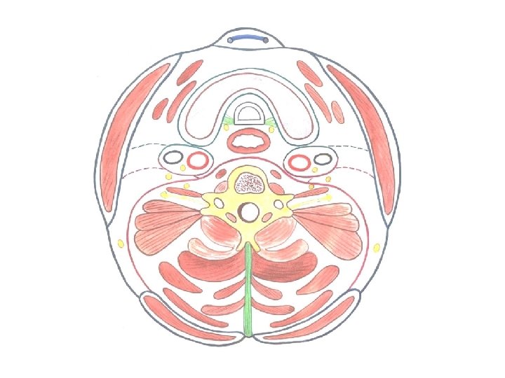
Posses three layers from outside inwards • Investing layer • Pretracheal fascia • Prevertebral fascia • Bucccopharyngeal fascia • Pharyngo-basilar fascia • Carotid sheath
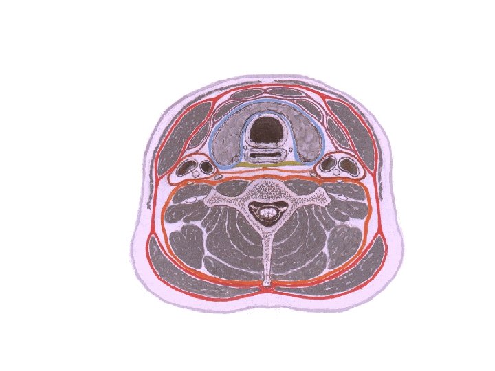
Investing layer C 7
Investing layer • Lies under cover of platysma • Attachments Behind – ligamentum nuchae and the seventh cervical spine In front – continuous with the similar layer of the opposite side across the midline and attached to the Hyoid bone and symphysis menti
Above– (behind forwards ) • External occipital protuberance, superior nuchal line • Mastoid process • Cartilagenous part of external ear • Lower margin of zygomatic arch • Lower border of mandible
Above– (behind forwards )
Below- (behind forwards ) • Spine and acromial process of scapula • Upper surface of clavicle • Manubrium
Horizontal extent • Splits twice to enclose the trapezius and sternomastoid muscles • Forms roof for posterior and anterior triangle
Investing layer - Horizontal extent Strap muscles Sternocleidomastoid At the lateral border of strap muscles it splits and covers it Trapezius 11
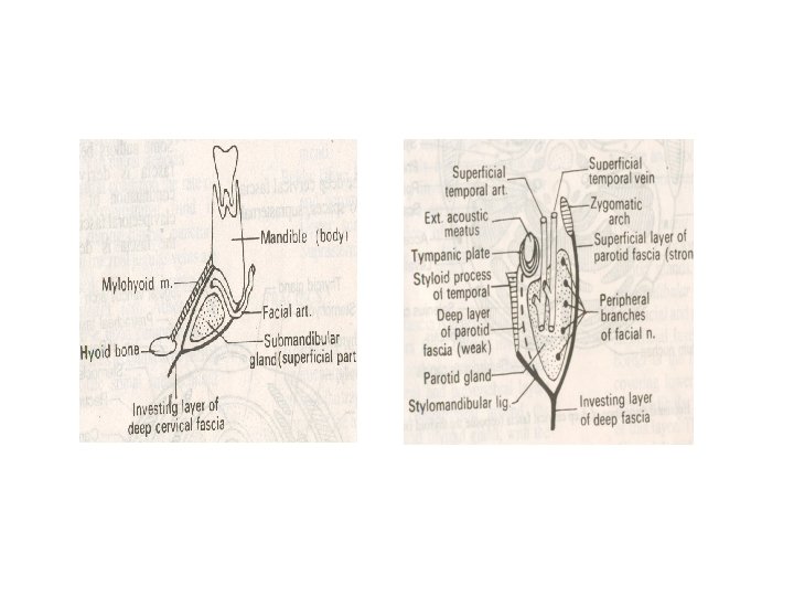
Vertical extent • Splits to enclose the submandibular and parotid glands • Cranially it forms the stylomandibular ligament • Caudally it encloses the supra sternal and supraclavicular spaces
Investing layer presents everything two • Encloses trapezius and sternomastoid muscles • Encloses two glands, parotid and submandibular glands • Splits to enclose two spaces- suprasternal and supraclavicular spaces
• Thickened twice to form parotido-masseteric fascia and stylomandibular ligament • It forms pulleys to bind the tendons of digastric and omohyoid muscle
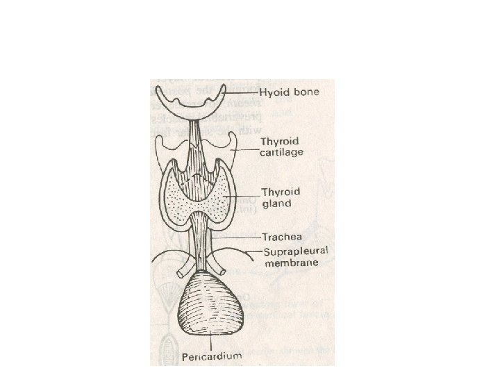
PRETRACHEAL FASCIA
Attachments • Superiorly: attaches to hyoid bone, oblique line of thyroid cartilage and cricoid cartilage • Inferiorly: blends with arch of aorta • On each side: fuses with carotid sheath
• Ligament of berry which is a fascial sling connects the thyroid to cricoid cartilage • Splits to enclose thyroid gland continuous with the layer of opposite layer • Provides slippery surface for trachea
Pretracheal: vertical extent 20
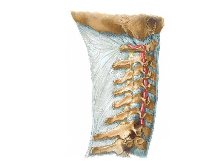
PREVERTEBRAL FASCIA
Attachments • Superiorly to the bones of base of the skull • Inferiorly to the anterior longitudinal ligament and body of fourth thoracic vertebra • Anteriorly it is related to retropharyngeal space
• Forms axillary sheath around the trunks of brachila plexus and subclavian artery • Forms floor of posterior triangle • Cervical plexus and brachial plexus lie beneath it
CAROTID SHEATH üTubular investment of deep cervical fascia üExtends from base of the skull to AOA Contents: üCommon & Int. carotid arteries- medially üInternal jugular vein – laterally üVagus nerve – b/w & behind the vessels
• Rami of ansa cervicalis is embedded in anterior wall • Thickened over arteries but ill defined over vein
• Buccopharyngeal fascia • Pharyngobasilar fascia
Applied Anatomy • Painful parotid swellings • Thyroid swellings move with deglutition • Neck infections – infront / behind prevertebral fascia - retropharyngeal abscess • Air embolism of external jugular vein
THANK YOU