DCAP PPT Chapter 6 Bone Tissue Functions of

DCAP PPT Chapter 6 Bone Tissue

Functions of Bones

Bone marrow ¨ Aka myeloid tissue ¨ Yellow bone marrow – Fat storage – Found in medullary canal of long bones ¨ Red bone marrow – Found in spongy bone (ends of long bones, flat bones, irregular bones) – Hematopoiesis (formation of all blood cells)

Types of Bone Tissue

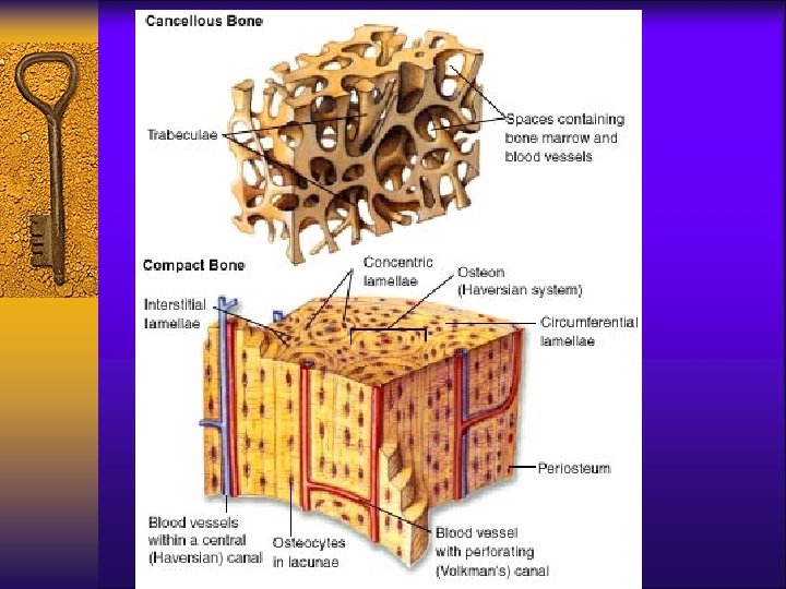
Cortical bone tissue ¨ Arranged in OSTEONS/ Haversian system – Repeated structural units ¨ Provide strength by providing a weight bearing surface ¨ Contains a series of openings that permit exchange of materials between osteocytes (& other bone cells) and the blood.

Osteon parts & functions ¨ Central/Haversian canal – central canal of each osteon – runs down the long axis of the bone ¨ Perforating/Volkmann’s canal – runs perpendicular to the Haversian canals – these are the communicating pathways from outside the bone to inside the bone ¨ Both provide passageways for BV & nerves

Types of Lamellae ¨ Lamellae – concentric rings or circles of matrix ¨ Circumferential lamellae – Rings that surround entire bone – Located deep to periosteum

Types of Lamellae ¨ Concentric lamellae: – rings of calcified extracellular matrix that surround the central canal – function to resist torsion stress (twisting) on bone tissue ¨ Interstitial lamellae: – Fragments from old osteons – located between new osteons – Function to provide strength
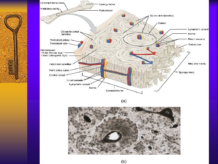
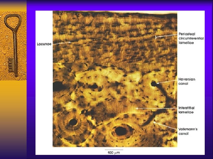

Osteon parts & functions ¨ Lacunae – tiny cavities within the matrix – location of osteocytes ¨ Canaliculi – tiny canals that radiate out from Haversian canals to all lacunae

Osteon diagram – cross section

osteocytes ¨ Lacunae, osteocytes, & canaliculi

Osteon diagram – sagittal section
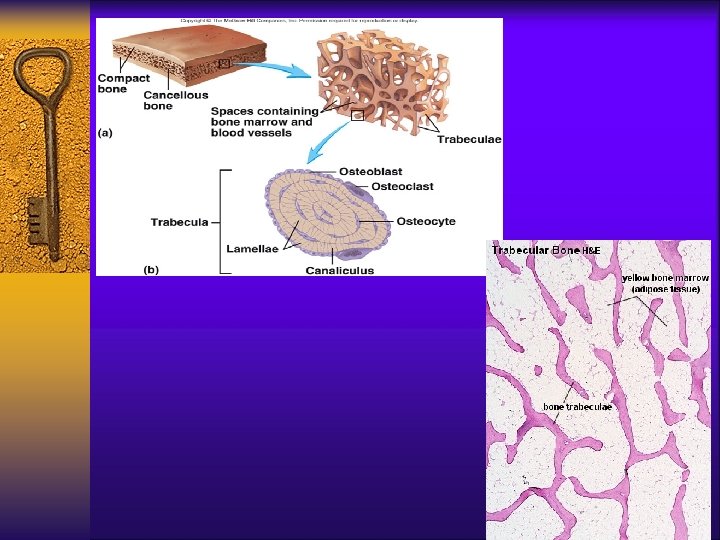
 bone tissue aka: Trabecular Bone ¨ Main structures are the trabeculae - irregularly")
Cancellous(spongy) bone tissue aka: Trabecular Bone ¨ Main structures are the trabeculae - irregularly arranged columns of lamellae – arranged along stress lines to provide strength & resist physical stress ¨ Lacunae – tiny cavities within the matrix – location of osteocytes ¨ Canaliculi – tiny canals that radiate out from lacunae & contain interstitial fluid – Location of bone marrow & BV

More dense trabeculae due to more stress on that area

Types of Bone Tissue Cortical Trabecular ¨ 80% of skeleton ¨ 20% of skeleton ¨ Strongest bone tissue – ¨ Light weight bone more dense ¨ Resists torsion stress from movement ¨ Resists mechanical stress from weight ¨ Osteons – less dense ¨ Spaces contain bone marrow & blood vessels ¨ Trabeculae

Lab Info Cortical Bone Trabecular Bone

BONE TISSUE CELLS
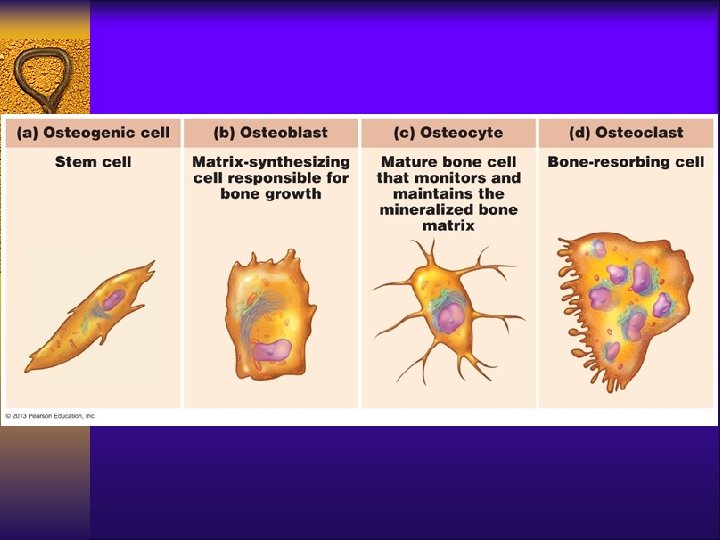

Blasts, Clasts & Cytes

BONE TISSUE COMPOSITION

Composition of bone tissue ¨ Osseous CT with hard ECM & widely distributed cells ¨ ECM – approximately 25% water, 25% collagen fibers, and 50% crystallized mineral salts (mainly Ca. PO 4) – Formed by process of calcification

Organic portion: 35% of mass ¨ The organic portion consists of the bone cells and the osteoid ¨ The Bone cells are the: – Osteocytes – Osteoblasts – Osteoclasts – FYI: There also osteoprogenitor cells that are the precursers to blasts & cytes. They are derived from mesenchyme & found on all bone surfaces.

Osteoid ¨ is produced by the osteoblasts ¨ It consists of ground substance (proteoglycans and glycoproteins) & collagen fibers produced by CT cells ¨ Its function is to provide the bone with tensile strength and resilience – in other words, to make the bone a little flexible & compressive

Inorganic matrix: 65% of mass ¨ The inorganic matrix consists of inorganic salt compounds mainly: – Hydroxyapatites: Calcium & phosphorus salt compounds ¨ Function to provide structure due to hardness that resists compression

Human Skeletons – dark color

Types of Bones

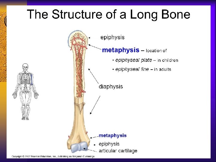
Long Bone Structure Know the descriptions & functions from your notes, as well as, the diagrams.


Lab Info Hyaline cartilage Fibrocartilage


Bone ¨ Bone is very vascular with multiple arteries, veins & capillaries ¨ Bone has an extensive nerve supply and lots of sensory nerve endings

BONE DEVELOPMENT

Terms ¨ Osteogenesis/Ossification – formation of bone – Endochondral – bone replaces cartilage – Intramembranous – bone develops directly from mesenchyme or fibrous CT ¨ Calcification – deposition of calcium salts (can occur in other tissue types)

Endochondral ossification

Intramembranous ossification

TYPES OF BONE GROWTH

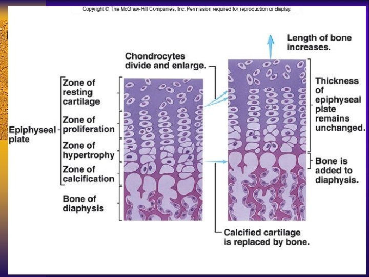
Types of growth ¨ Longitudinal growth – bone growth in length at epiphyseal plates (till plates ossify) ¨ Appositional growth – bone growth in diameter (throughout life) – Known as remodeling ¨ These 2 types work together to make the bones long enough & strong enough

Regulation of bone growth ¨Bone is a dynamic and active tissue. They are constantly being remodeled according to the activities that we do. ¨Main factor = Ca levels in blood ¨Ca imp for bone strength but also for nervous & muscular system to work correctly!!!

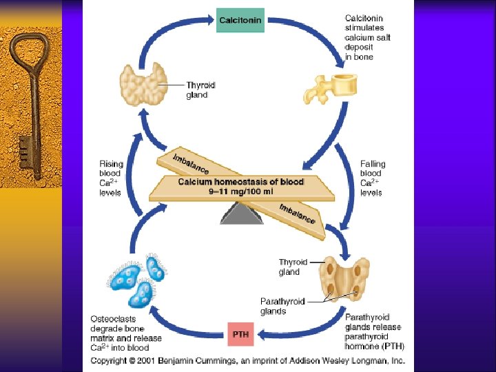
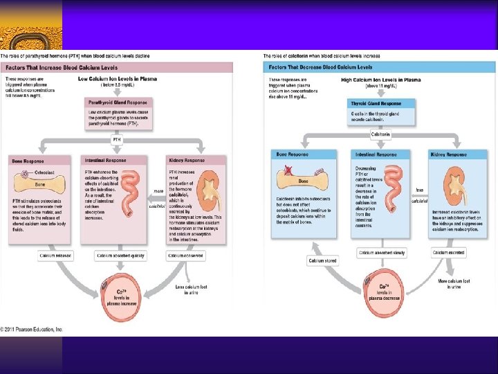
Regulation by hormonal feedback ¨ Purpose: to maintain optimal ionic calcium levels in blood – This is your body’s TOP priority!!! ¨ A main factor that affects what our bone tissue does is our blood calcium level – Optimum blood Ca 2+ level = 9 -11 mg/100 ml of blood – Calcium ions are VERY important for muscle & nervous function – our body cares more about this level than it does our bone strength!!
 – activated when Ca levels in")
Regulation by hormonal feedback ¨ PTH (parathyroid gland) – activated when Ca levels in blood are too low – (hypocalcemia) - promotes calcium reabsorption – Calcium will go from bone to blood ¨ Calcitonin (thyroid gland) activated when Ca levels in blood are too high – (hypercalcemia) - promotes calcium deposition – Calcium will go from blood to bone

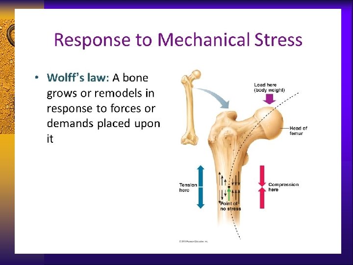
Regulation by mechanical stress ¨ Purpose: keep bones strong – This is the secondary purpose ¨ Wolff’s law states that bones will grow according to the stresses placed upon them – Simply stated, bone grows in response to mechanical stress so as to produce an anatomical structure best able to resist the applied stress. – So activities that compress bones and pull on muscles which pull on bones can make bones stronger
 determine WHEN the")
How they work together to regulate ¨ PTH & calcitonin (hormones) determine WHEN the remodeling will occur – Primary purpose = Ca 2+ regulation in blood ¨ The Mechanical stresses determine WHERE the remodeling will occur – Secondary purpose = where will the calcium ions be deposited or reabsorbed from
- Slides: 53