Day 4 THE BRAIN How many neurons About

Day 4 THE BRAIN

How many neurons? � About 100 billion multipolar neurons � Innumberable nerve fibers �Allow the neurons to communicate with one another and to other parts of nervous system.
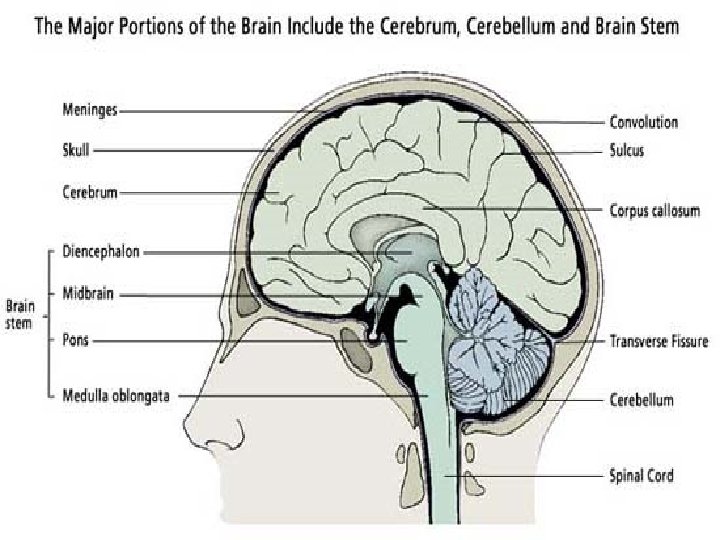

Three Major Parts � Cerebrum �Largest, contains nerve centers associated with sensory and motor functions, provides higher mental functions including memory and reasoning � Cerebellum �Center that coordinates voluntary muscular movements � Brain Stem �Includes the DIENCEPHALON ○ Processes sensory information �Connects various parts of nervous system, regulates certain visceral activities

Structure of Cerebrum

Structure of Cerebrum � Cerebral Hemispheres �Right and Left Halves �Layer of Dura Mater separates � Corpus Callosumbridge of nerve fibers �Connects hemispheres
 that are separated")
Structure of Cerebrum � Surface contains many ridges called convolutions (gyri) that are separated by grooves. � Sulcus �Shallow groove � Fissure �Deep groove � Very complex compressions but form distinct patterns in normal brains.

Important Fissures and Sulcuses � Longitudinal Fissure �Separates R and L hemispheres � Transverse Fissure �Separates cerebrum from cerebellum � Central Sulcus �Divides frontal lobe from parietal lobe

Cerebrum Lobes � Names after their skull bones they lie under � Frontal Lobe �Anterior portion of each cerebral hemisphere �Divided by the longitudinal fissure, central sulcus, and lateral sulcus. � Parietal Lobe �Posterior to the frontal lobe and separated by central sulcus.

Cerebral Lobes Cont. � Temporal Lobe �Lies below frontal lobe �Separated by lateral sulcus � Occipital Lobe �Posterior portion of cerebral hemisperes �Boundary between parietal and temporal lobe is not clear � Insula �Located deep within lateral sulcus �Covered by parts of frontal, parietal, and temporal lobes �Separated by circular sulcus

Cerebral Cortex � Thin layer of gray matter � Outer most portion of cerebrum � Covers all the convolutions and goes into the sulci and fissures � Contains 75% of all neuron cell bodies in the nervous system

Functions of Cerebrum � HIGHER BRAIN FUNCTIONS � Center for interpreting sensory impulses arriving from sense organs � Center for initiating voluntary muscular movements � Stores information of memory � Utilizes reasoning � Responsible for intelligence and personality

Functional Regions of Cerebral Cortex � Divided areas into motor, sensory, and associational

Primary Motor Areas � Frontal Lobe just in front of central sulcus � Because of the cross over of nerve tracts, right hemisphere contains skeletal muscles on left side and vice versa � Motor Speech Area=Boca’s Area �Coordinates muscular actions of mouth, tongue, and larynx

Motor Function Areas � Frontal Eye Field �Located above Boca’s area �Voluntary movements of eyes and eyelids � Other region in front of primary motor area makes movements of hands and fingers possible �Writing

Sensory Areas � Found within several lobes � Interpret impulses that arrive from sensory receptors producing, feelings and sensations. � Skin sensations arise from anterior portions of parietal lobe along central sulcus. � Occipital lobe affects vision � Temporal lobe affects hearing
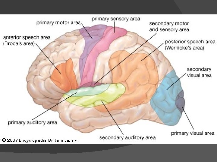

Sensory Areas � Taste is found near base of central sulci and lateral sulci � Smell comes from deep within the cerebrum � Same as motor functions, nerves cross each other causing sensation on right side of body to be interpreted in left side of brain and vice versa.

Association Areas � Neither primary sensory or motor � Connect with one another and other brain structures � Analyze and interpret sensory experiences and oversee memory, reasoning, verbalizing, judgment, and emotion. � Found on anterior portion of frontal lobe and throughout the lateral portions of parietal, temporal and occipital lobes.
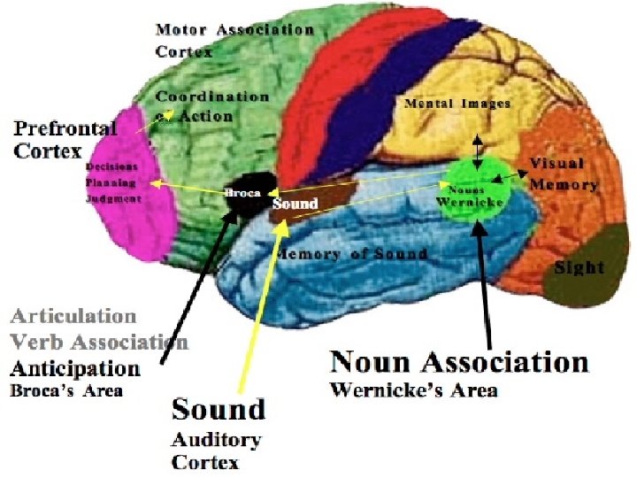

Association Areas � Frontal Lobes �Controls concentrating, planning, complex problem solving, judging the possible consequences of behavior � Parietal Lobes �Understanding speech and choosing words to express thoughts and feelings

Association Areas � Temporal Lobes �Complex sensory experiences (those needed to understand speech and to read), memory of visual scenes, music, and others. � Occipital Lobes �Analyzing visual patterns, combining visual images with other sensory experiences � General interpretative area �Where parietal, temporal and occipital areas meet �Plays primary role in complex thought procesing

Review � List the major divisions of the brain. � Describe � What the cerebral cortex are the major functions of the cerebrum?

Hemisphere Dominance � Both hemispheres receive and analyze sensory info, control skeletal muscles, and store memory. � One side just tends to do it more than the other leading to a dominant hemisphere.

Hemisphere Dominance � 90% of population is left side dominant for: �Language related activities ○ Speech, writing, and reading ○ Complex intellectual functions requiring verbal, analytical, and computational skills � Non-dominant side �Specializes in nonverbal functions such as motor tasks, understanding and interpreting musical patterns, and nonverbal visual experiences. �Emotional and intuitive thinking

Hemisphere tid-bit � The left hemisphere is dominant in 90% of righthanded adults and in 64% of left-handed ones. The right hemisphere is dominant in 10% of right-handed adults and in 20% of left-handed ones. The hemispheres are equally dominant in the remaining 16% of left-handed persons. Because of hemisphere dominance, Boca’s area on one side almost completely controls the motor activities associated with speech. For this reason, over 90% of patients with language impairment involving the cerebrum have disorders in the left hemisphere.

Hemisphere Dominance � Corpus callosum is responsible for allowing dominant hemisphere to control motor cortex of nondominant side. � Also transfer sensory info from non-dominant side to dominant side so it can be used in decision making

Basal ganglia Gray matter deep within each hemisphere � Made up of: � �Caudate nucleus, putamen, and globus pallidus Neuron bodies serve as a relay station for motor impulses � Produce inhibitory neurotransmitter dopamine � Inhibit motor functions thus controlling various skeletal muscle activities. �

Basal Ganglia

Basal Ganglia Tid. Bit � The uncontrollable movements of Parkinson disease and Huntington disease result from lesions in the basal ganglia. The lack of inhibiting impulses cause the excessive movements.

Ventricles and Cerebrospinal Fluid � Ventricles �Series of interconnected cavities within cerebral hemispheres that contains cerebrospinal fluid � Largest are the lateral ventricles(1 st and 2 nd) which extend into the frontal, temporal and occipital lobes. � 3 rd ventricle is in the midline of brain � 4 th ventricle is in brain stem

Choroid plexuses � Tiny-reddish cauliflower-like mass of specialized capillaries from the pia mater that secretes cerebrospinal fluid � Because of the ventricles allowing movement of cerebrospinal fluid, the brain is said to float. � This aids in protection to the brain and spinal cord.

Cerebrospinal Fluid Tid-Bit � Because cerebrospinal fluid is secreted and reabsorbed continuously, the fluid pressure in the ventricles normally remains relatively constant. AN infection, a tumor, or a blood clot can interfere with fluid circulation, increasing pressure within the ventricles and thus in the cranial cavity. This can injure the brain by forcing it against the rigid skull. A lumbar puncture (spinal tap) is used to measure the pressure of cerebrospinal fluid. In the procedure, a fine, hollow needle is inserted into the subarachnoid space between the 3 rd and 4 th or 4 th and 5 th lumbar vertebrae. An instrument called a manometer measures the pressure.

Review � What is hemisphere dominance? � What are the major functions of the dominate hemisphere? The nondominant one? � Where are the ventricles of the brain?
- Slides: 34