Data Interpretation a focus on ABGs Dr Budhima

Data Interpretation - a focus on ABGs Dr Budhima Nanayakkara MBBS

Ordering a test • Before you order a test, assume you know the result • Test should only confirm/ refute/ prognosticate • In essence, only order a test if you think it will change management • You are responsible for all tests ordered

Case 1 A 46 yo female post allogeneic stem cell transplantation for multiple myeloma presents with progressive chest pain, chills and a yellow productive cough. Examination reveals a cachectic female with saturations of 96% on 8 L HM, RR 32, PR 128, T 38. 3 C. Auscultation of the lungs reveals decreased air-entry on the left lower zone with bronchial breathing and inspiratory crackles

Case 1 The ED nurse performs an arterial blood gas sample for you

Case 1 Variable Measured Reference p. H 7. 48 7. 35 – 7. 45 p. CO 2 28 mm. Hg 35 - 45 p. O 2 72 mm. Hg 70 - 100 HCO 3 21 m. M 22 – 26 m. M BE +2. 4 -3 to + 3 lactate 1. 8 <2 m. M 1. What is the abnormality here? 2. How do you explain this abnormality in terms of what you think is going on?

ABG interpretation • Why? – Rapid diagnosis – Ventilation disturbances – Acid-base disorders • General approach – Acidaemia vs alkalaemia – Ventilation abnormalities – compensation
")
Part 1: Disorders of Ventilation (oxygenation)

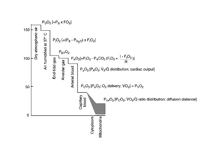
Delivery of oxygen = arterial oxygen content x cardiac output

Delivery of oxygen • Arterial oxygen content – Hemoglobin concentration • Causes of anaemia • Causes of abnormal haemoglobin – Saturation of haemoglobin • Dependent on p. O 2 and relationship with oxy-hemoglobin curve • p. O 2 dependent upon – – Ventilation Matching ventilation with perfusion Diffusion Other gases that take up room – Dissolved oxygen • Solubility co-efficient = 0. 003 • Unimportant physiologically

Cardiac Output • Stroke volume – Preload • End-diastolic volume • Compliance • frank-starling curve – Afterload • Laplace law – Contractility • Inotropic effects • Intracellular calcium

Cardiac Output • Heart rate – Autonomic stimuli – Pacemaker intrinsic rhythmicity – Arrythmias • Brady • Tachy But heart rate and stroke volume are also related to each other

A-a gradient • The difference between alveolar oxygen and arterial oxygen • Alveolar oxygen must be inferred • Can be calculated easily

 – P(H 2 O)) –")
A-a gradient • PAO 2 = Fi. O 2(P(atm) – P(H 2 O)) – Pa. CO 2/R • Most cases, let R = 0. 8, P(atm) = 760 mm. Hg, P(H 2 O) = 42 mm. Hg

Interpretation of A-a gradient • The presence of a high A-a gradient implies the presence of intrapulmonary pathology – Pus, water, blood in alveoli • Pneumonia, pneumonitis, mucus plugging, – Diffusion defect in alveoli – Perfusion defect (local, systemic) • Normal = <15

Case 1: Variable Measured Reference p. H 7. 48 7. 35 – 7. 45 p. CO 2 28 mm. Hg 35 - 45 p. O 2 72 mm. Hg 70 - 100 HCO 3 21 m. M 22 – 26 m. M BE +2. 4 -3 to + 3 lactate 1. 8 <2 m. M

Case 1 • Respiratory alkalosis • Hypoxia with increased A-a gradient – Pa. O 2 = 72 mm. Hg – Fi. O 2 = 0. 6 – PAO 2 = 0. 6(760 – 42) – 28/0. 8 – PAO 2 = 430. 8 – 35 – PAO 2 = 395. 8 – A-a = 395. 8 – 72 mm. Hg – A-a = 328. 8 mm. Hg

Respiratory failure • Type 1 respiratory failure – Hypoxia without hypercapnea • Type 2 respiratory failure – Hypoxia with hypercapnea

Concept of V/Q

Concept of V/Q RV RV 75% V/Q = 1 100% LA 75%

Dead space ventilation RV RV 75% V/Q = ++ V/Q = 1 75% 100% LA 75%

Dead space ventilation RV RV 75% V/Q = ++ V/Q = 1 75% Increased oxygen 75% 100% LA

Shunt RV RV 0 V/Q = 1 Pulmonary arteriolar vasoconstriction LA Increased oxygen does not help because V/Q units already saturated

Case 2 Met Call ward 6 A Canberra Hospital. You attend to see a 94 yo female, admitted for infective bronchiectasis, no other known co-morbidities. She is in severe respiratory distress with peripheral + Central cyanosis. JMO 30 minutes ago prescribed 5 mg temazepam over phone order

Case 2 Variable Measured Reference p. H 7. 02 7. 35 – 7. 45 p. CO 2 128 mm. Hg 35 - 45 p. O 2 72 mm. Hg 70 - 100 HCO 3 38 m. M 22 – 26 m. M BE +2. 9 -3 to + 3 lactate 2. 6 m. M <2 m. M

CO 2 – Ventilation relationship • CO 2 is inversely related to ventilation • Ventilation dependent on – RR • • Central nervous system Peripheral nervous system NMJ Diaphragm – Tidal volume • • Chest wall compliance – including burns to skin Lung compliance Pleura Diaphragm (including workload – such as exhaustion in asthma and COPD) – Dead space volume • Additional respiratory circuits • Pulmonary pathology – V = RR. (Vt – Vd)

Case 3 You are the ICU registrar who has been called in to A and E to assess a patient for ICU. There is a 32 yo female, unconscious and tubed. An Abg has been performed

Case 3 Variable Measured Reference p. H 7. 20 7. 35 – 7. 45 p. CO 2 20 mm. Hg 35 - 45 p. O 2 180 mm. Hg 70 - 100 HCO 3 18 m. M 22 – 26 m. M BE +2. 4 -3 to + 3 lactate 1. 8 <2 m. M Na+ 140 135 – 145 m. M K+ 4 3. 5 – 5. 2 m. M 1. What is the acid base disorder 2. Calculate the anion gap

Metabolic acidosis CO 2 + H 2 O H 2 CO 3 H+ + HCO 3 Le-chatelier’s principle: Any perturbation to the system results in the system attempting to restore equilibrium - This explains why CO 2 produces acidocis - This explains why decreased HCO 3 produces acidocis
 vs non-anion gap (NAGMA)")
Approach to metabolic acidosis • Anion gap (RAGMA) vs non-anion gap (NAGMA)
 D")
RAGMA • • • MUDPILES M – Methanol U – Uraemia (renal failure) D – DKA, alcoholic ketoacidocis, starvation ketoacidocis P – para-aldehyde I – isoniazid, iron L – lactic acidocis (Type A and Type B) E – ethylene glycol S – salicylates

Osmolar Gap • To further investigate RAGMA, we can – Measure lactate – BGL – Ketones • Osmolar gap – Measured osmolality (freezing point depression) – calculated osmolality (2. Na + urea + glucose) – If >10, implies that there is a significant unmeasured anion present

NAGMA • Increased H+ – TPN solutions, HCl ingestion • HCO 3 loss – Renal • Renal tubular acidocis • Acetazolamide – GI • Diarrhoea • Urinary diversion • Small bowel fistula

NAGMA and RAGMA co-exist • Delta gap: – Allows for the partitioning of metabolic acidosis into a RAGMA component and NAGMA component – Derived from the fact (and assumption) that in a pure anion gap metabolic acidocis, the increase in anion gap (delta AG) is equal to the decrease in bicarbonate (delta bicarb) – If delta bicarb decreases much more than delta AG, then there is evidence for both a RAGMA and NAGMA – For example a lactic acidosis with renal tubular acidosis

Case 4 48 yo female who loves licorice presents with nausea and vomiting that is intractable. She is hypotensive and tachycardic on arrival. A VBG is obtained prior to further resuscitation.

Case 4 Variable Measured Reference p. H 7. 64 7. 35 – 7. 45 p. CO 2 50 mm. Hg 35 - 45 p. O 2 380 mm. Hg 70 - 100 HCO 3 48 m. M 22 – 26 m. M BE +10. 4 -3 to + 3 lactate 1. 8 <2 m. M

Metabolic alkalosis CO 2 + H 2 O H 2 CO 3 H+ + HCO 3 If HCO 3 goes up, then CO 2 will also increase H+ will be used up in the equation as the equilibrium tries to restore itself

Metabolic alkalosis • Loss of H+/ Cl – Vomiting – Excessive nasogastric suction – Diuretic therapy • Increased HCO 3 reabsorption – Milk-alkali syndrome – Contraction alkalosis – Mineralocorticoid excess • Licorice – Barters syndrome – Gitlemans syndrome • Severe hypokalaemia

- Slides: 41