
Daniel J Wallace MD FACP MACR Associate Director


- Slides: 70
Daniel J. Wallace, MD, FACP, MACR Associate Director, Rheumatology Fellowship Program Board of Governors, Cedars-Sinai Medical Center Professor of Medicine, Cedars-Sinai Medical Center David Geffen School of Medicine Center at UCLA In affiliation with Attune Health
Learning Objectives • Apply the ACR diagnostic criteria to recognize patients who may have SLE • Utilize and interpret laboratory findings to investigate possible SLE • Develop SLE treatment plans based on individual patients’ disease characteristics, treatment goals, and consensus recommendations • Identify a validated SLE disease activity measure for regular patient monitoring
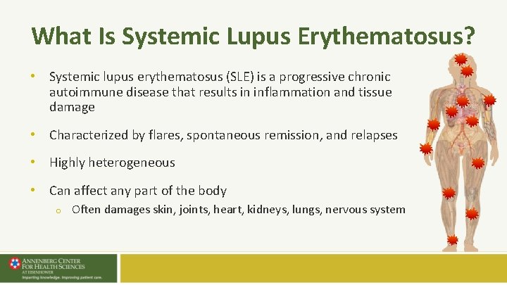
What Is Systemic Lupus Erythematosus? • Systemic lupus erythematosus (SLE) is a progressive chronic autoimmune disease that results in inflammation and tissue damage • Characterized by flares, spontaneous remission, and relapses • Highly heterogeneous • Can affect any part of the body o Often damages skin, joints, heart, kidneys, lungs, nervous system
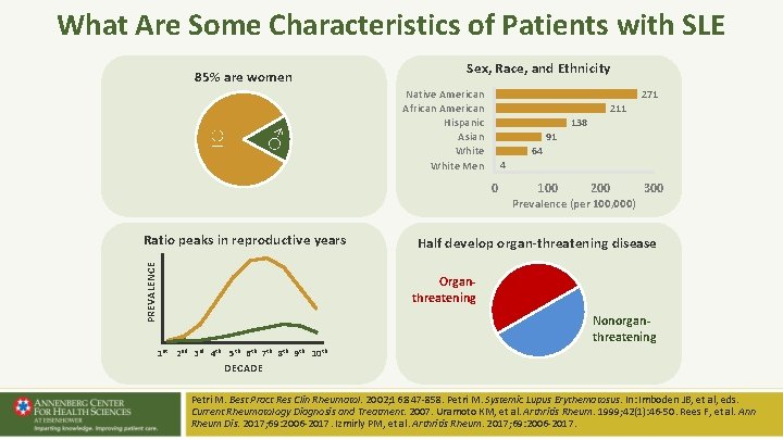
What Are Some Characteristics of Patients with SLE 85% are women ♀ ♂ Sex, Race, and Ethnicity Native American African American Hispanic Asian White Men 271 211 138 91 64 4 0 PREVALENCE Ratio peaks in reproductive years 100 200 Prevalence (per 100, 000) 300 Half develop organ-threatening disease Organthreatening Nonorganthreatening 1 st 2 nd 3 rd 4 th 5 th 6 th 7 th 8 th 9 th 10 th DECADE Petri M. Best Pract Res Clin Rheumatol. 2002; 16: 847 -858. Petri M. Systemic Lupus Erythematosus. In: Imboden JB, et al, eds. Current Rheumatology Diagnosis and Treatment. 2007. Uramoto KM, et al. Arthritis Rheum. 1999; 42(1): 46 -50. Rees F, et al. Ann Rheum Dis. 2017; 69: 2006 -2017. Izmirly PM, et al. Arthritis Rheum. 2017; 69: 2006 -2017.
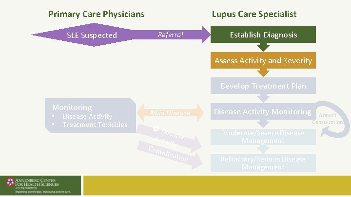
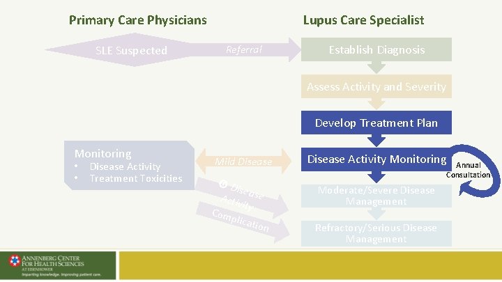
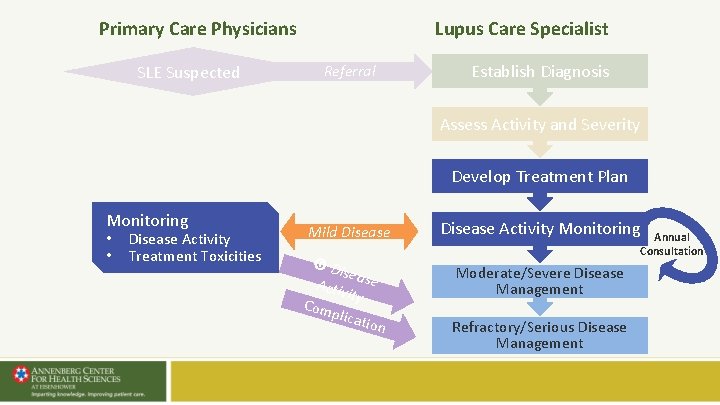
Lupus Care Specialist Primary Care Physicians SLE Suspected Referral Establish Diagnosis Assess Activity and Severity Develop Treatment Plan Monitoring • • Disease Activity Treatment Toxicities Mild Disease Di sea Activ se ity Com plica tion Disease Activity Monitoring Moderate/Severe Disease Management Refractory/Serious Disease Management Annual Consultation
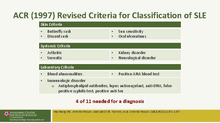
ACR (1997) Revised Criteria for Classification of SLE Skin Criteria • • Butterfly rash Discoid rash • • Sun sensitivity Oral ulcerations • • Kidney disorder Neurological disorder • Positive ANA blood test Systemic Criteria • • Arthritis Serositis Laboratory Criteria • Blood abnormalities • Immunologic disorder o Antiphospholipid antibodies, lupus anticoagulant, anti-DNA, falsepositive syphilis test, positive anti-Sm 4 of 11 needed for a diagnosis Hochberg MC. Arthritis Rheum. 1997; 40: 1725. Tan EM, et al. Arthritis Rheum. 1982; 25(11): 1271 -1277.
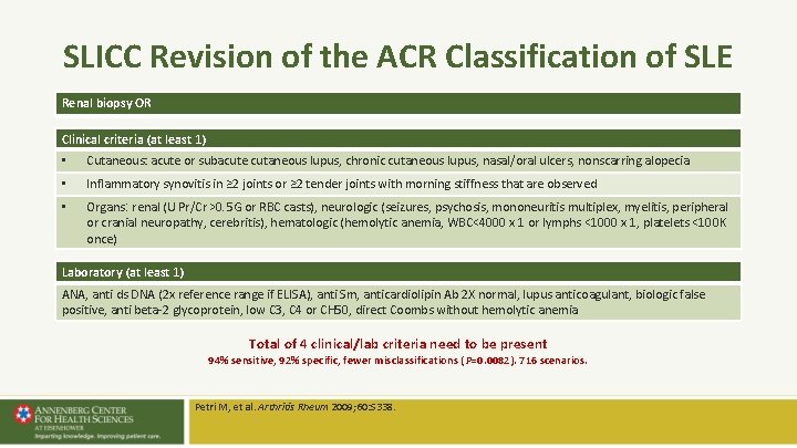
SLICC Revision of the ACR Classification of SLE Renal biopsy OR Clinical criteria (at least 1) • Cutaneous: acute or subacute cutaneous lupus, chronic cutaneous lupus, nasal/oral ulcers, nonscarring alopecia • Inflammatory synovitis in ≥ 2 joints or ≥ 2 tender joints with morning stiffness that are observed • Organs: renal (U Pr/Cr >0. 5 G or RBC casts), neurologic (seizures, psychosis, mononeuritis multiplex, myelitis, peripheral or cranial neuropathy, cerebritis), hematologic (hemolytic anemia, WBC<4000 x 1 or lymphs <1000 x 1, platelets <100 K once) Laboratory (at least 1) ANA, anti ds DNA (2 x reference range if ELISA), anti Sm, anticardiolipin Ab 2 X normal, lupus anticoagulant, biologic false positive, anti beta-2 glycoprotein, low C 3, C 4 or CH 50, direct Coombs without hemolytic anemia Total of 4 clinical/lab criteria need to be present 94% sensitive, 92% specific, fewer misclassifications ( P=0. 0082). 716 scenarios. Petri M, et al. Arthritis Rheum 2009; 60: S 338.
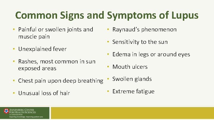
Common Signs and Symptoms of Lupus • Painful or swollen joints and muscle pain • Unexplained fever • Rashes, most common in sun exposed areas • Raynaud’s phenomenon • Sensitivity to the sun • Edema in legs or around eyes • Mouth ulcers • Chest pain upon deep breathing • Swollen glands • Unusual loss of hair • Extreme fatigue
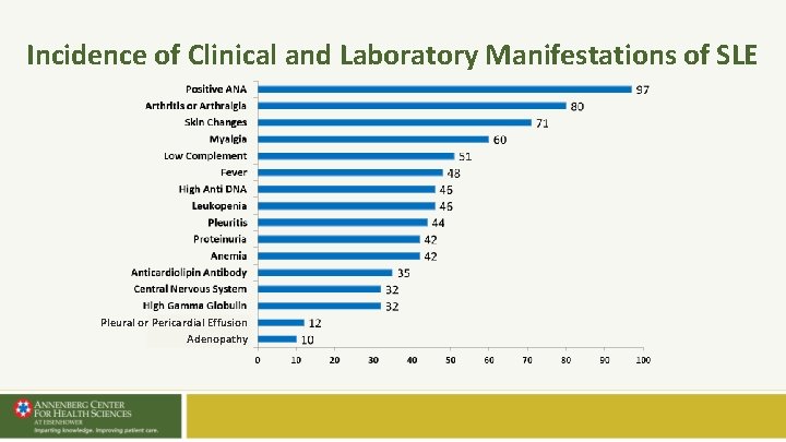
Incidence of Clinical and Laboratory Manifestations of SLE Pleural or Pericardial Effusion Adenopathy
Mucocutaneous Features of SLE • • Malar (butterfly) rash Discoid lupus (DLE) Mucosal ulcers Alopecia Subacute cutaneous lupus (SCLE) Cutaneous vasculitis Bullous lupus Panniculitis
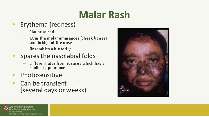
• Erythema (redness) Malar Rash – Flat or raised – Over the malar eminences (cheek bones) and bridge of the nose – Resembles a butterfly • Spares the nasolabial folds – Differentiates from rosacea which has a similar appearance • Photosensitive • Can be transient (several days or weeks)
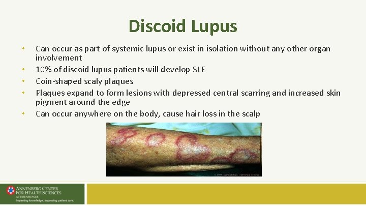
Discoid Lupus • • • Can occur as part of systemic lupus or exist in isolation without any other organ involvement 10% of discoid lupus patients will develop SLE Coin-shaped scaly plaques Plaques expand to form lesions with depressed central scarring and increased skin pigment around the edge Can occur anywhere on the body, cause hair loss in the scalp
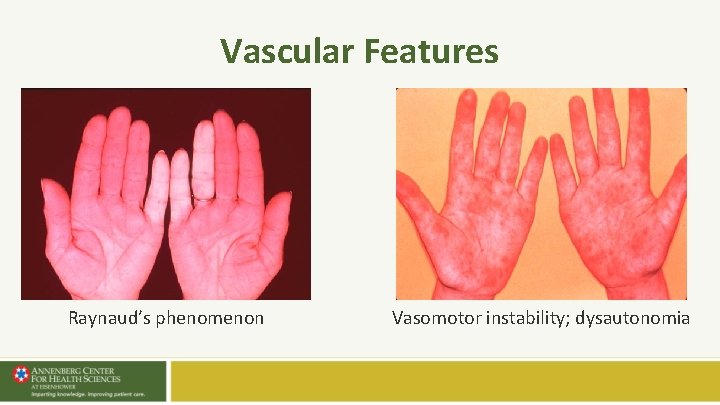
Vascular Features Raynaud’s phenomenon Vasomotor instability; dysautonomia
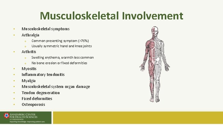
Musculoskeletal Involvement • • Musculoskeletal symptoms Arthralgia o o • Arthritis o o • • Common presenting symptom (>76%) Usually symmetric hand knee joints Swelling erythema, warmth less common No bone erosion or fixed deformities Myositis Inflammatory tendonitis Myalgia Musculoskeletal system organ damage Tendon degeneration Fixed deformities Osteoporosis
Serositis • Lining of heart, lungs, abdomen o Pleuritis is inflammation of the lining of the lungs o Pericarditis is inflammation of the lining of the heart • Symptoms include pain in chest with deep breathing or when lying flat • Fluid may accumulate (effusion) causing increased shortness of breath
Blood Abnormalities • Anemia o Decreased red blood cells due to immune destruction/hemolysis • Thrombocytopenia o Platelets < 100 k • Lymphopenia/neutropenia o Decreased white blood cells < 4 k, particularly lymphocytes < 1. 5 k • Exclude other reasons o Medications o Blood loss o Infection Dooley MA. In: Wallace DJ, Hahn BH, eds. Dubois’ Lupus Erythematosus. 8 th Ed. Philadelphia, PA: Elsevier; 2012: 526.
Differential Diagnosis of SLE Autoimmune Rheumatoid arthritis, scleroderma, myositis, vasculitis, spondyloarthropathies, inflammatory bowel disorder, Behçet's disease, sarcoidosis, Sjogren’s syndrome, thyroiditis, polymyalgia rheumatica, undifferentiated connective tissue disease Infections Tuberculosis, Lyme, Bacterial endocarditis, HIV, CMV, EBV Fibromyalgia Allergies Neurologic disorders (esp myasthenia gravis, multiple sclerosis) Malignancy (esp, lymphoproliferative disorders) Drug-induced lupus Chlorpromazine, methyldopa, isoniazid, hydralazine, procainamide, quinidine Psychiatric disorders Bipolar illness, malnutrition, substance abuse Wallace DJ. The Lupus Book. New York, NY: Oxford University Press; 2012. Manson JJ, et al. Orphanet Journal of Rare Diseases. 2006, 1: 6, http: //bestpractice. bmj. com/best-practice/monograph/103/diagnosis/differential. html.
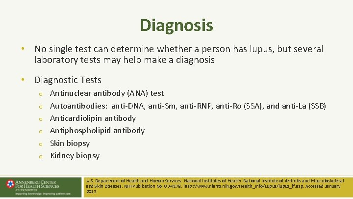
Diagnosis • No single test can determine whether a person has lupus, but several laboratory tests may help make a diagnosis • Diagnostic Tests o o o Antinuclear antibody (ANA) test Autoantibodies: anti-DNA, anti-Sm, anti-RNP, anti-Ro (SSA), and anti-La (SSB) Anticardiolipin antibody Antiphospholipid antibody Skin biopsy Kidney biopsy U. S. Department of Health and Human Services. National Institutes of Health. National Institute of Arthritis and Musculoskeletal and Skin Diseases. NIH Publication No. 03 -4178. http: //www. niams. nih. gov/Health_Info/Lupus/lupus_ff. asp. Accessed January 2013.
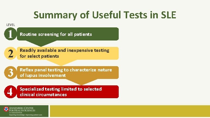
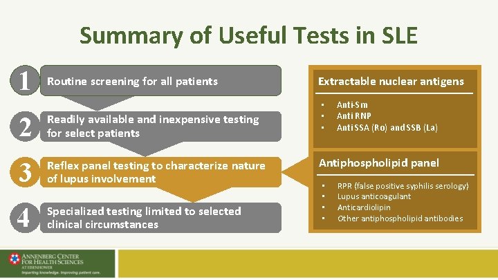
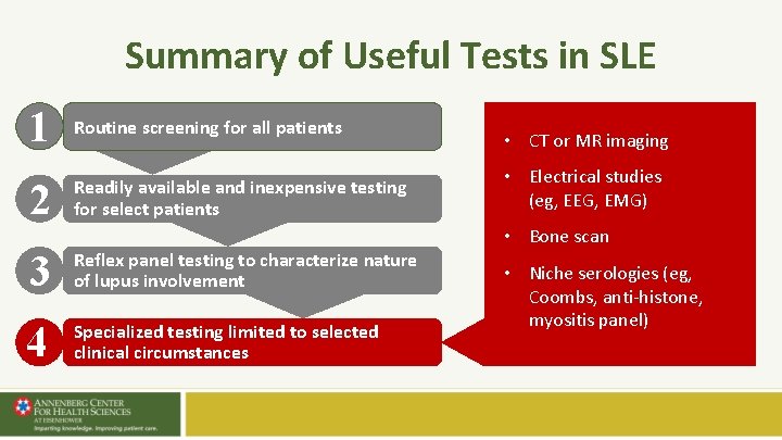
Summary of Useful Tests in SLE LEVEL 1 Routine screening for all patients 2 Readily available and inexpensive testing for select patients 3 Reflex panel testing to characterize nature of lupus involvement 4 Specialized testing limited to selected clinical circumstances
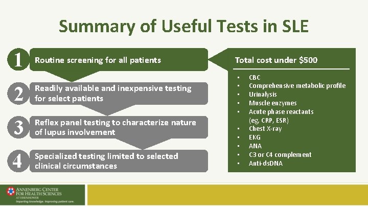
Summary of Useful Tests in SLE 1 Routine screening for all patients 2 Readily available and inexpensive testing for select patients 3 Reflex panel testing to characterize nature of lupus involvement 4 Specialized testing limited to selected clinical circumstances Total cost under $500 • • • CBC Comprehensive metabolic profile Urinalysis Muscle enzymes Acute phase reactants (eg, CRP, ESR) Chest X-ray EKG ANA C 3 or C 4 complement Anti-ds. DNA
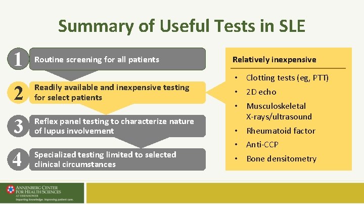
Summary of Useful Tests in SLE 1 Routine screening for all patients 2 Readily available and inexpensive testing for select patients 3 Reflex panel testing to characterize nature of lupus involvement 4 Specialized testing limited to selected clinical circumstances Relatively inexpensive • Clotting tests (eg, PTT) • 2 D echo • Musculoskeletal X-rays/ultrasound • Rheumatoid factor • Anti-CCP • Bone densitometry
Summary of Useful Tests in SLE 1 Routine screening for all patients 2 Readily available and inexpensive testing for select patients 3 Reflex panel testing to characterize nature of lupus involvement 4 Specialized testing limited to selected clinical circumstances Extractable nuclear antigens • • • Anti-Sm Anti RNP Anti SSA (Ro) and SSB (La) Antiphospholipid panel • • RPR (false positive syphilis serology) Lupus anticoagulant Anticardiolipin Other antiphospholipid antibodies
Summary of Useful Tests in SLE 1 Routine screening for all patients 2 Readily available and inexpensive testing for select patients 3 Reflex panel testing to characterize nature of lupus involvement 4 Specialized testing limited to selected clinical circumstances • CT or MR imaging • Electrical studies (eg, EEG, EMG) • Bone scan • Niche serologies (eg, Coombs, anti-histone, myositis panel)
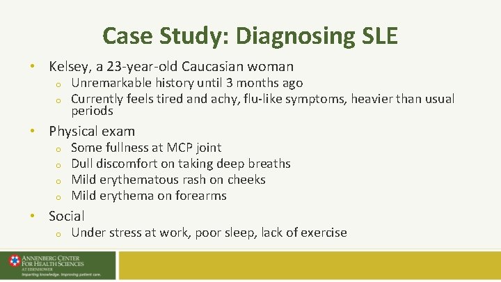
Case Study: Diagnosing SLE • Kelsey, a 23 -year-old Caucasian woman o o Unremarkable history until 3 months ago Currently feels tired and achy, flu-like symptoms, heavier than usual periods • Physical exam o o Some fullness at MCP joint Dull discomfort on taking deep breaths Mild erythematous rash on cheeks Mild erythema on forearms • Social o Under stress at work, poor sleep, lack of exercise
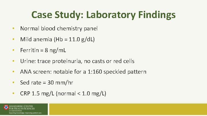
Case Study: Laboratory Findings • Normal blood chemistry panel • Mild anemia (Hb = 11. 0 g/d. L) • Ferritin = 8 ng/m. L • Urine: trace proteinuria, no casts or red cells • ANA screen: notable for a 1: 160 speckled pattern • Sed rate = 30 mm/hr • CRP 1. 5 mg/L (normal < 1. 0 mg/L)
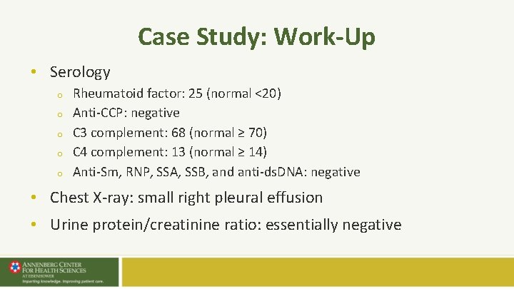
Case Study: Work-Up • Serology o o o Rheumatoid factor: 25 (normal <20) Anti-CCP: negative C 3 complement: 68 (normal ≥ 70) C 4 complement: 13 (normal ≥ 14) Anti-Sm, RNP, SSA, SSB, and anti-ds. DNA: negative • Chest X-ray: small right pleural effusion • Urine protein/creatinine ratio: essentially negative
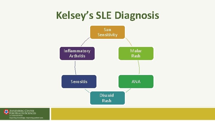
Kelsey’s SLE Diagnosis Sun Sensitivity Malar Rash Inflammatory Arthritis Serositis ANA Discoid Rash
Lupus Care Specialist Primary Care Physicians SLE Suspected Referral Establish Diagnosis Assess Activity and Severity Develop Treatment Plan Monitoring • • Disease Activity Treatment Toxicities Mild Disease Di sea Activ se ity Com plica tion Disease Activity Monitoring Moderate/Severe Disease Management Refractory/Serious Disease Management Annual Consultation
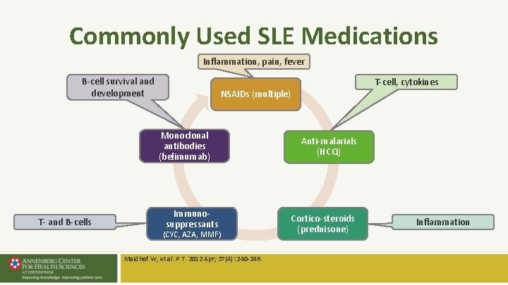
Commonly Used SLE Medications Inflammation, pain, fever B-cell survival and development NSAIDs (multiple) Monoclonal antibodies (belimumab) T- and B-cells T-cell, cytokines Immunosuppressants (CYC, AZA, MMF) Maidhof W, et al. P T. 2012 Apr; 37(4): 240 -246. Anti-malarials (HCQ) Cortico-steroids (prednisone) Inflammation
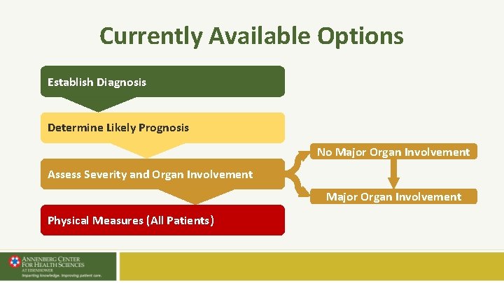
Currently Available Options Establish Diagnosis Determine Likely Prognosis No Major Organ Involvement Assess Severity and Organ Involvement Major Organ Involvement Physical Measures (All Patients)
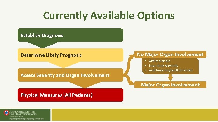
Currently Available Options Establish Diagnosis Determine Likely Prognosis Assess Severity and Organ Involvement No Major Organ Involvement • Antimalarials • Low-dose steroids • Azathioprine/methotrexate Major Organ Involvement Physical Measures (All Patients)
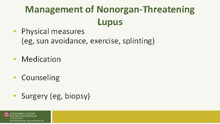
Management of Nonorgan-Threatening Lupus • Physical measures (eg, sun avoidance, exercise, splinting) • Medication • Counseling • Surgery (eg, biopsy)
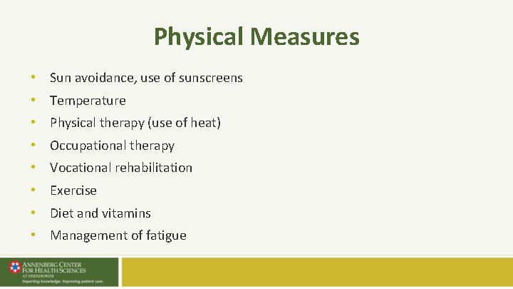
Physical Measures • Sun avoidance, use of sunscreens • Temperature • Physical therapy (use of heat) • Occupational therapy • Vocational rehabilitation • Exercise • Diet and vitamins • Management of fatigue
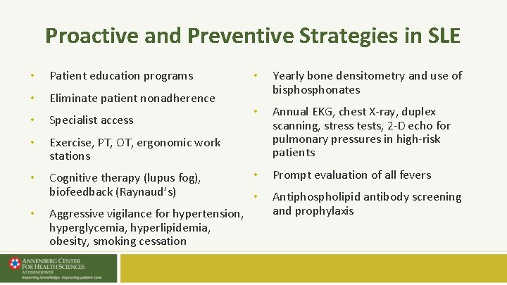
Proactive and Preventive Strategies in SLE • Patient education programs • Eliminate patient nonadherence • Specialist access • Exercise, PT, OT, ergonomic work stations • Cognitive therapy (lupus fog), biofeedback (Raynaud’s) • Aggressive vigilance for hypertension, hyperglycemia, hyperlipidemia, obesity, smoking cessation • Yearly bone densitometry and use of bisphonates • Annual EKG, chest X-ray, duplex scanning, stress tests, 2 -D echo for pulmonary pressures in high-risk patients • Prompt evaluation of all fevers • Antiphospholipid antibody screening and prophylaxis
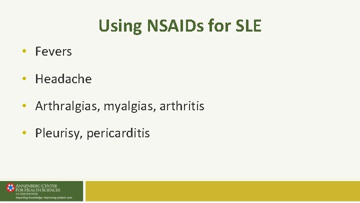
Using NSAIDs for SLE • Fevers • Headache • Arthralgias, myalgias, arthritis • Pleurisy, pericarditis
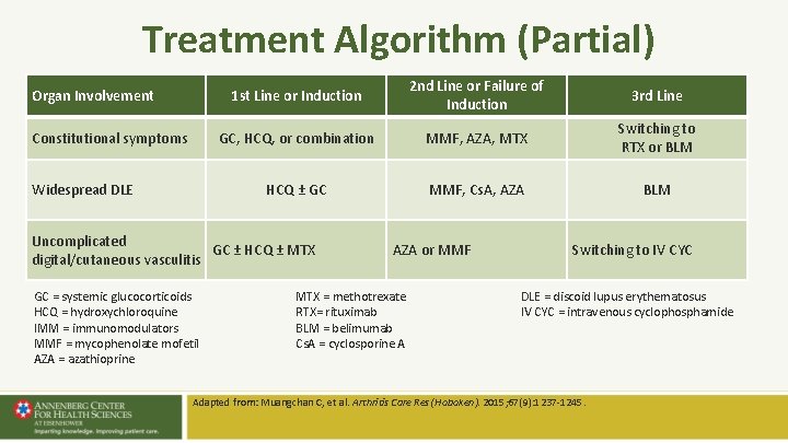
Treatment Algorithm (Partial) Organ Involvement Constitutional symptoms Widespread DLE 1 st Line or Induction 2 nd Line or Failure of Induction 3 rd Line GC, HCQ, or combination MMF, AZA, MTX Switching to RTX or BLM HCQ ± GC MMF, Cs. A, AZA BLM Uncomplicated GC ± HCQ ± MTX digital/cutaneous vasculitis GC = systemic glucocorticoids HCQ = hydroxychloroquine IMM = immunomodulators MMF = mycophenolate mofetil AZA = azathioprine AZA or MMF MTX = methotrexate RTX= rituximab BLM = belimumab Cs. A = cyclosporine A Switching to IV CYC DLE = discoid lupus erythematosus IV CYC = intravenous cyclophosphamide Adapted from: Muangchan C, et al. Arthritis Care Res (Hoboken). 2015; 67(9): 1237 -1245.
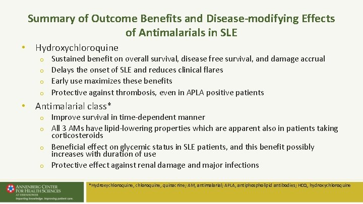
Summary of Outcome Benefits and Disease-modifying Effects of Antimalarials in SLE • Hydroxychloroquine o o Sustained benefit on overall survival, disease free survival, and damage accrual Delays the onset of SLE and reduces clinical flares Early use maximizes these benefits Protective against thrombosis, even in APLA positive patients • Antimalarial class* o o Improve survival in time-dependent manner All 3 AMs have lipid-lowering properties which are apparent also in patients taking corticosteroids Beneficial effect on glycemic status in SLE patients, and this benefit possibly increases with duration of use Protective effect against renal damage and major infections *Hydroxychloroquine, quinacrine; AM, antimalarial; APLA, antiphospholipid antibodies; HCQ, hydroxychloroquine
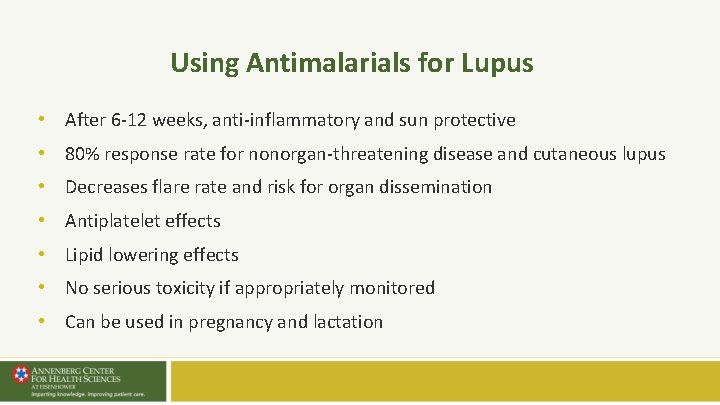
Using Antimalarials for Lupus • After 6 -12 weeks, anti-inflammatory and sun protective • 80% response rate for nonorgan-threatening disease and cutaneous lupus • Decreases flare rate and risk for organ dissemination • Antiplatelet effects • Lipid lowering effects • No serious toxicity if appropriately monitored • Can be used in pregnancy and lactation
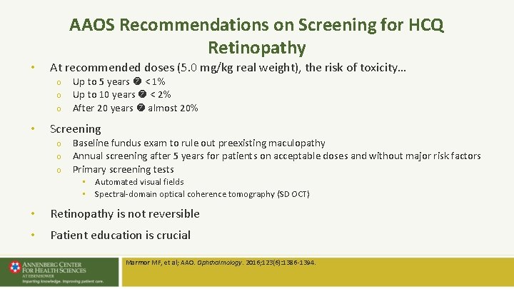
AAOS Recommendations on Screening for HCQ Retinopathy • At recommended doses (5. 0 mg/kg real weight), the risk of toxicity… o o o • Up to 5 years < 1% Up to 10 years < 2% After 20 years almost 20% Screening o o o Baseline fundus exam to rule out preexisting maculopathy Annual screening after 5 years for patients on acceptable doses and without major risk factors Primary screening tests • Automated visual fields • Spectral-domain optical coherence tomography (SD OCT) • Retinopathy is not reversible • Patient education is crucial Marmor MF, et al; AAO. Ophthalmology. 2016; 123(6): 1386 -1394.
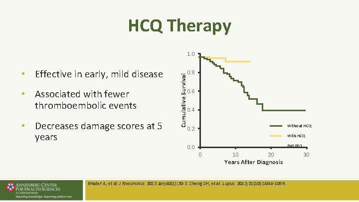
HCQ Therapy • Effective in early, mild disease • Associated with fewer thromboembolic events • Decreases damage scores at 5 years Cumulative Survival 1. 0 0. 8 0. 6 0. 4 Without HCQ 0. 2 With HCQ P=0. 003 0. 0 0 10 20 Years After Diagnosis Broder A, et al. J Rheumatol. 2013 Jan; 40(1): 30 -3. Zheng ZH, et al. Lupus. 2012; 21(10): 1049 -1056. 30
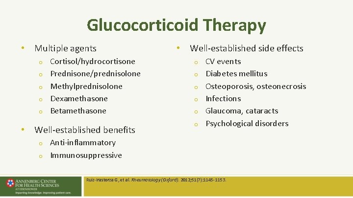
Glucocorticoid Therapy • Multiple agents o o o Cortisol/hydrocortisone Prednisone/prednisolone Methylprednisolone Dexamethasone Betamethasone • Well-established benefits o o • Well-established side effects o o o CV events Diabetes mellitus Osteoporosis, osteonecrosis Infections Glaucoma, cataracts Psychological disorders Anti-inflammatory Immunosuppressive Ruiz-Irastorza G, et al. Rheumatology (Oxford). 2012; 51(7): 1145 -1153.
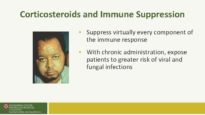
Corticosteroids and Immune Suppression • Suppress virtually every component of the immune response • With chronic administration, expose patients to greater risk of viral and fungal infections
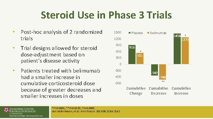
Steroid Use in Phase 3 Trials • Post-hoc analysis of 2 randomized trials • Trial designs allowed for steroid dose-adjustment based on patient’s disease activity • Patients treated with belimumab had a smaller increase in cumulative corticosteroid dose because of greater decreases and smaller increases in doses 1500 Placebo Belimumab 1200 900 600 300 1458 † 1272 916 * 531 0 -300 -600 -900 *P<0. 0001; **P=0. 0165; †P=0. 0005 van Vollenhoven, et al. Arth Rheum. 2016; 68: 2184 -2192 -542 -740 ** Cumulative Change Decrease Increase
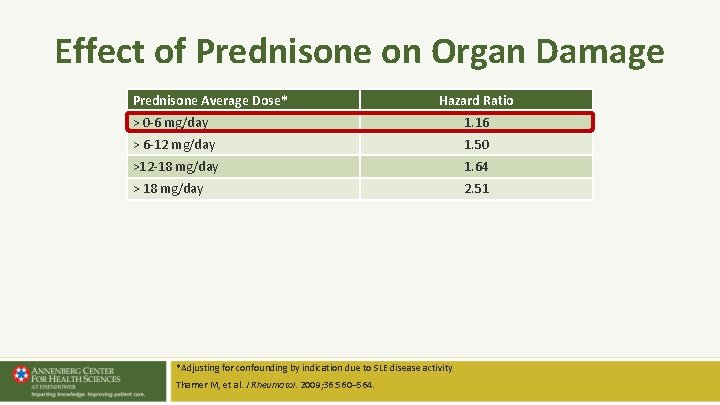
Effect of Prednisone on Organ Damage Prednisone Average Dose* Hazard Ratio > 0 -6 mg/day 1. 16 > 6 -12 mg/day 1. 50 >12 -18 mg/day 1. 64 > 18 mg/day 2. 51 *Adjusting for confounding by indication due to SLE disease activity Thamer M, et al. J Rheumatol. 2009; 36: 560– 564.
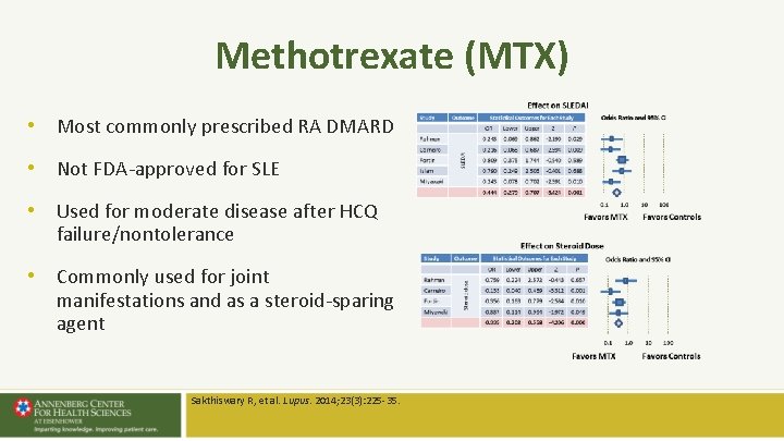
Methotrexate (MTX) • Most commonly prescribed RA DMARD • Not FDA-approved for SLE • Used for moderate disease after HCQ failure/nontolerance • Commonly used for joint manifestations and as a steroid-sparing agent Sakthiswary R, et al. Lupus. 2014; 23(3): 225 -35.
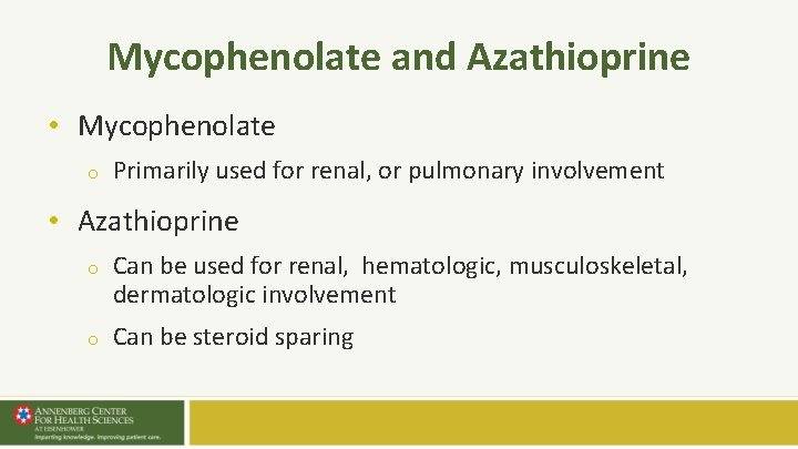
Mycophenolate and Azathioprine • Mycophenolate o Primarily used for renal, or pulmonary involvement • Azathioprine o Can be used for renal, hematologic, musculoskeletal, dermatologic involvement o Can be steroid sparing
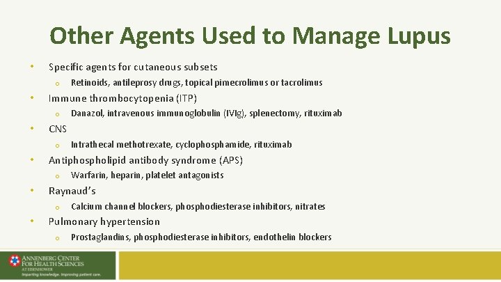
Other Agents Used to Manage Lupus • Specific agents for cutaneous subsets o • Immune thrombocytopenia (ITP) o • Warfarin, heparin, platelet antagonists Raynaud’s o • Intrathecal methotrexate, cyclophosphamide, rituximab Antiphospholipid antibody syndrome (APS) o • Danazol, intravenous immunoglobulin (IVIg), splenectomy, rituximab CNS o • Retinoids, antileprosy drugs, topical pimecrolimus or tacrolimus Calcium channel blockers, phosphodiesterase inhibitors, nitrates Pulmonary hypertension o Prostaglandins, phosphodiesterase inhibitors, endothelin blockers
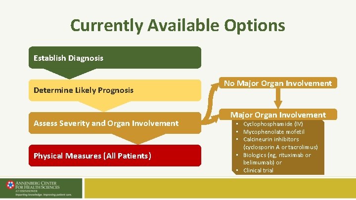
Currently Available Options Establish Diagnosis Determine Likely Prognosis Assess Severity and Organ Involvement Physical Measures (All Patients) No Major Organ Involvement • Cyclophosphamide (IV) • Mycophenolate mofetil • Calcineurin inhibitors (cyclosporin A or tacrolimus) • Biologics (eg, rituximab or belimumab) or • Clinical trial
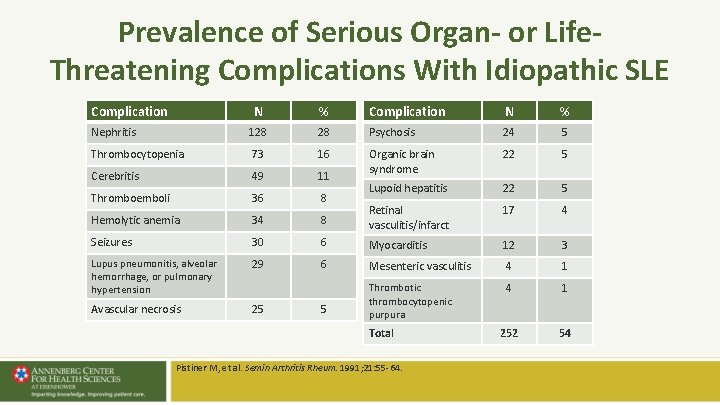
Prevalence of Serious Organ- or Life. Threatening Complications With Idiopathic SLE Complication N % Nephritis 128 28 Psychosis 24 5 Thrombocytopenia 73 16 22 5 Cerebritis 49 11 Organic brain syndrome Thromboemboli 36 8 Lupoid hepatitis 22 5 Hemolytic anemia 34 8 Retinal vasculitis/infarct 17 4 Seizures 30 6 Myocarditis 12 3 Lupus pneumonitis, alveolar hemorrhage, or pulmonary hypertension 29 6 Mesenteric vasculitis 4 1 Avascular necrosis 25 5 Thrombotic thrombocytopenic purpura 252 54 Total Pistiner M, et al. Semin Arthritis Rheum. 1991; 21: 55 -64.
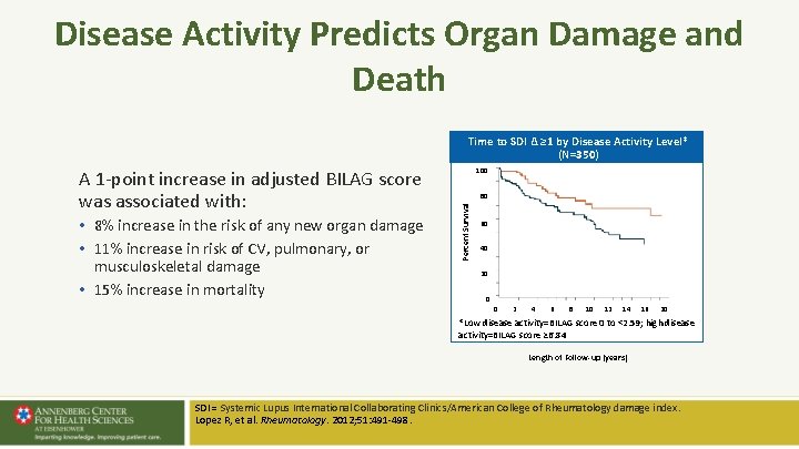
Disease Activity Predicts Organ Damage and Death Time to SDI Δ ≥ 1 by Disease Activity Level* (N=350) • 8% increase in the risk of any new organ damage • 11% increase in risk of CV, pulmonary, or musculoskeletal damage • 15% increase in mortality 100 80 Percent Survival A 1 -point increase in adjusted BILAG score was associated with: 60 40 20 0 0 2 4 6 8 10 12 14 16 20 *Low disease activity=BILAG score 0 to <2. 59; high disease activity=BILAG score ≥ 6. 84 Length of Follow-up (years) SDI = Systemic Lupus International Collaborating Clinics/American College of Rheumatology damage index. Lopez R, et al. Rheumatology. 2012; 51: 491 -498.
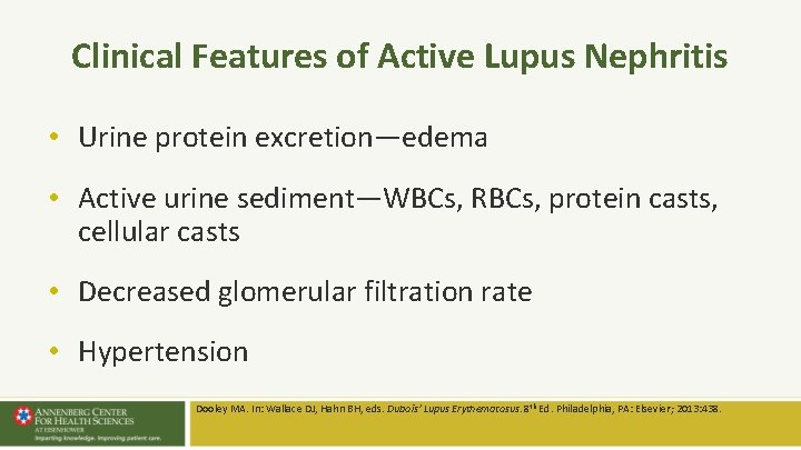
Clinical Features of Active Lupus Nephritis • Urine protein excretion—edema • Active urine sediment—WBCs, RBCs, protein casts, cellular casts • Decreased glomerular filtration rate • Hypertension Dooley MA. In: Wallace DJ, Hahn BH, eds. Dubois’ Lupus Erythematosus. 8 th Ed. Philadelphia, PA: Elsevier; 2013: 438.
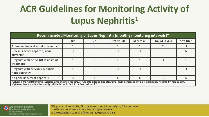
ACR Guidelines for Monitoring Activity of Lupus Nephritis 1 Recommended Monitoring of Lupus Nephritis (monthly monitoring intervals)* BP UA Protein: CR Serum CR C 3/C 4 Levels Anti-DNA 3 Active nephritis at onset of treatment 1 1 2† Previous active nephritis, none currently 3 3 3 6 Pregnant with active GN at onset of treatment 1 1 1 Pregnant with previous nephritis, none currently 1 1 3 3 No prior or current nephritis 3 6 6 6 *Values are the monthly intervals suggested as the minimum frequency at which the indicated laboratory tests should be measured in the SLE scenarios shown in the left-hand column. †Opinion of the authors based on a study published after the Task Force Panel had voted. 2 GN, glomerulonephritis; BP, blood pressure; UA, urinalysis; CR, creatinine. 1. Hahn BH, et al. Arthrit Care Res. 2012; 64: 797 -808. 2. Grootscholten C, et al. Kidney Int. 2006; 70: 732 -742.
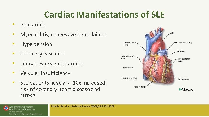
Cardiac Manifestations of SLE • Pericarditis • Myocarditis, congestive heart failure • Hypertension • Coronary vasculitis • Libman-Sacks endocarditis • Valvular insufficiency • SLE patients have a 7– 10 x increased risk of coronary heart disease and stroke Esdaile JM, et al. Arthritis Rheum. 2001; 44: 2331 -2337.
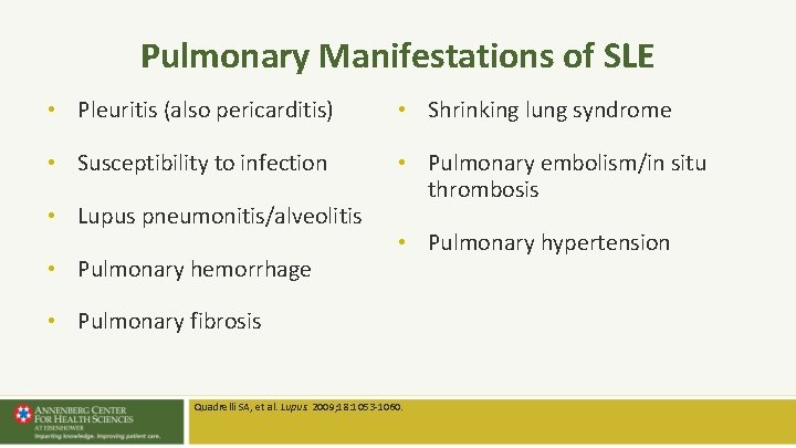
Pulmonary Manifestations of SLE • Pleuritis (also pericarditis) • Shrinking lung syndrome • Susceptibility to infection • Pulmonary embolism/in situ thrombosis • Lupus pneumonitis/alveolitis • Pulmonary hemorrhage • Pulmonary hypertension • Pulmonary fibrosis Quadrelli SA, et al. Lupus. 2009; 18: 1053 -1060.
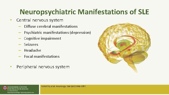
Neuropsychiatric Manifestations of SLE • Central nervous system – – – Diffuse cerebral manifestations Psychiatric manifestations (depression) Cognitive impairment Seizures Headache Focal manifestations • Peripheral nervous system Futrell N, et al. Neurology. 1992; 42: 1649 -1657.
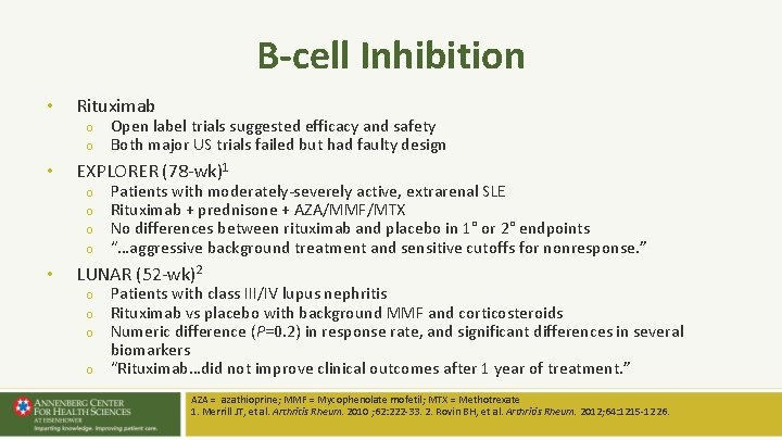
B-cell Inhibition • Rituximab o o • EXPLORER (78 -wk)1 o o • Open label trials suggested efficacy and safety Both major US trials failed but had faulty design Patients with moderately-severely active, extrarenal SLE Rituximab + prednisone + AZA/MMF/MTX No differences between rituximab and placebo in 1° or 2° endpoints “…aggressive background treatment and sensitive cutoffs for nonresponse. ” LUNAR (52 -wk)2 o o Patients with class III/IV lupus nephritis Rituximab vs placebo with background MMF and corticosteroids Numeric difference (P=0. 2) in response rate, and significant differences in several biomarkers “Rituximab…did not improve clinical outcomes after 1 year of treatment. ” AZA = azathioprine; MMF = Mycophenolate mofetil; MTX = Methotrexate 1. Merrill JT, et al. Arthritis Rheum. 2010 ; 62: 222 -33. 2. Rovin BH, et al. Arthritis Rheum. 2012; 64: 1215 -1226.
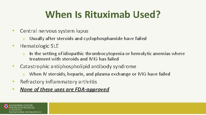
When Is Rituximab Used? • Central nervous system lupus o Usually after steroids and cyclophosphamide have failed • Hematologic SLE o In the setting of idiopathic thrombocytopenia or hemolytic anemias where treatment with steroids and IVIG has failed • Catastrophic antiphospholipid antibody syndrome o When IV steroids, heparin, and plasma exchange or IVIG have failed • Refractory inflammatory arthritis • None of these uses are FDA-approved
Belimumab Is a BLy. S-Specific Inhibitor • Indicated for the treatment of adult patients with active, autoantibody-positive SLE who are receiving standard therapy • Only biologic approved for SLE • First drug approved for SLE (2011) since 1957 BENLYSTA [package insert]. Rockville, MD: Human Genome Sciences, Inc. 2011.
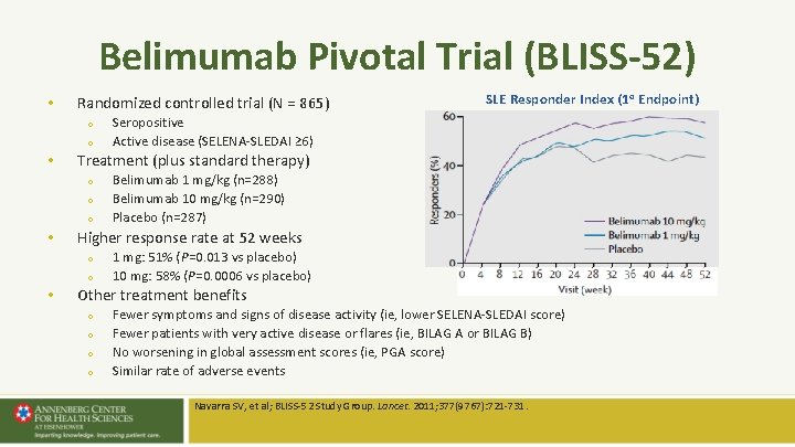
Belimumab Pivotal Trial (BLISS-52) • Randomized controlled trial (N = 865) o o • o o Belimumab 1 mg/kg (n=288) Belimumab 10 mg/kg (n=290) Placebo (n=287) Higher response rate at 52 weeks o o • Seropositive Active disease (SELENA-SLEDAI ≥ 6) Treatment (plus standard therapy) o • SLE Responder Index (1 o Endpoint) 1 mg: 51% (P=0. 013 vs placebo) 10 mg: 58% (P=0. 0006 vs placebo) Other treatment benefits o o Fewer symptoms and signs of disease activity (ie, lower SELENA-SLEDAI score) Fewer patients with very active disease or flares (ie, BILAG A or BILAG B) No worsening in global assessment scores (ie, PGA score) Similar rate of adverse events Navarra SV, et al; BLISS-52 Study Group. Lancet. 2011; 377(9767): 721 -731.
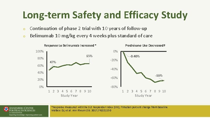
Long-term Safety and Efficacy Study o o Continuation of phase 2 trial with 10 years of follow-up Belimumab 10 mg/kg every 4 weeks plus standard of care Response to Belimumab Increased * 100% 80% 60% Prednisone Use Decreased† 0% 65% 47% -20% -0. 40% -40% -66% -60% 20% 0% -80% 1 2 3 4 5 6 7 8 9 10 Study Year *Response measured with the SLE Responder Index (SRI); †Median percent change from baseline Wallace DJ, et al. Ann Rheum Dis. 2017; 76(S 2): 150
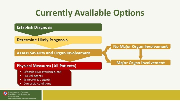
Currently Available Options Establish Diagnosis Determine Likely Prognosis No Major Organ Involvement Assess Severity and Organ Involvement Physical Measures (All Patients) • • Lifestyle (sun avoidance, etc) Topical agents Symptomatic agents Comorbid conditions Major Organ Involvement
Case Study: Treatment for Kelsey • Therapy should begin with prednisone and HCQ o Also consider NSAIDs • Treat associated comorbidities (eg, heavy periods with iron-deficiency anemia)
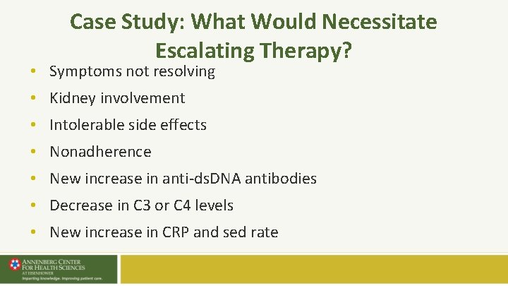
Case Study: What Would Necessitate Escalating Therapy? • Symptoms not resolving • Kidney involvement • Intolerable side effects • Nonadherence • New increase in anti-ds. DNA antibodies • Decrease in C 3 or C 4 levels • New increase in CRP and sed rate
Lupus Care Specialist Primary Care Physicians SLE Suspected Referral Establish Diagnosis Assess Activity and Severity Develop Treatment Plan Monitoring • • Disease Activity Treatment Toxicities Mild Disease Di sea Activ se ity Com plica tion Disease Activity Monitoring Moderate/Severe Disease Management Refractory/Serious Disease Management Annual Consultation
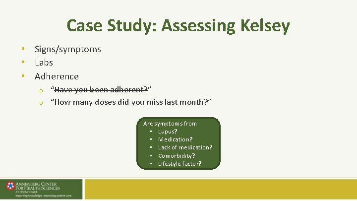
Case Study: Assessing Kelsey • Signs/symptoms • Labs • Adherence o o “Have you been adherent? ” “How many doses did you miss last month? ” Are symptoms from • Lupus? • Medication? • Lack of medication? • Comorbidity? • Lifestyle factor?
Medication Nonadherence is a Problem • • US Medicaid data from 2000 -2006 Adults ages 18 -64 years with SLE New users of HCQ (n = 9600) or ISMs (n = 3829) Outcomes o All-cause and SLE-related ED visits o Hospitalizations • Nonadherence o 79% of HCQ users o 83% of ISM users Feldman CH, et al. Arthritis Care Res (Hoboken). 2015 Dec; 67(12): 1712 -21.
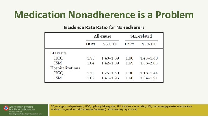
Medication Nonadherence is a Problem Incidence Ratio for Nonadherers ED, emergency department; HCQ, hydroxychloroquine; IRR, incidence ratio; ISM, immunosuppressive medications Feldman CH, et al. Arthritis Care Res (Hoboken). 2015 Dec; 67(12): 1712 -21.
Case Study: Monitoring Kelsey • Screen for organ-threatening disease • Patient education, disease monitoring, and adherence assessment should be reinforced in the next office visits • Benefits of educational sessions are documented in the literature o Review benefits, side effects, and complications of NSAIDs, antimalarials, corticosteroids
Key Points • • • Diagnosis by rheumatology is the gold standard Screen early for organ-threatening disease Early treatment o o o • Adherence is critical at all disease stages o • NSAIDs should be considered Steroids may be useful in early, active disease, if used with caution Patients should receive an antimalarial Patient education, disease monitoring, and adherence assessment Even mild disease should be overseen by a lupus care specialist at least annually or by phone consultation o Flares happen and can be subtle