Damage to Brain Areas Aphasia impaired ability to


Damage to Brain Areas Aphasia = impaired ability to use language, as a result of damage to brain 2 types of aphasias 1. Broca’s aphasia -caused by damage to left frontal lobe 2. Wernicke’s aphasia -caused by damage to left temporal lobe

Read aloud. Then complete the chart that follows, describing those language tasks that the patient is able to accomplish, and those tasks which are difficult for him.

Now, watch each of the video clips below, and add to the chart the capabilities and challenges of the Broca’s patients in each clip. https: //www. youtube. com/watch? v=12 d. O 78 c 6 -q 8&feature=related https: //www. youtube. com/watch? v=f 2 Ii. MEb. Mn. PM&feature=related Based on these three patients, write a description of Broca’s aphasia.

Examine these diagrams and f. MRI scans, and use them to address the questions Language mapping f. MRI overlaid on axial T 2 -weighted image. The active area in the inferior left frontal lobe (arrow) represents Broca’s expressive speech area. Source: https: //radiologykey. com/magnetic-resonance-imaging-2/ Source: http: //163. 178. 103. 176/Casos. Berne/2 b. SNervioso/Casox 111/HTMLC/Casos. B 2/Brocas 3. html

Brain Damage Questions – Broca’s Aphasia Examine the diagrams and f. MRI scans provided, and use them to address the following questions: 1. Patients with Broca’s aphasia are able to understand language and know what they want to say, but struggle to physically communicate their answers. How might the location of Broca’s area explain that difficulty? 2. Paul Broca found that while patients with damage to the Broca’s area struggled to form words in conversation, they were often able to sing familiar songs without difficulty. What factor(s) do you think might explain this discrepancy?

Read aloud. Then complete the chart that follows, describing those language tasks that the patient is able to accomplish, and those tasks which are difficult for him.

Now, watch each of the video clips below, and add to the chart the capabilities and challenges of the Wernicke’s patients in each clip. https: //www. youtube. com/watch? v=B-LD 5 jz. Xp. LE&feature=related https: //www. youtube. com/watch? v=67 HMx-Td. AZI Based on these three patients, write a description of Wernicke’s aphasia.

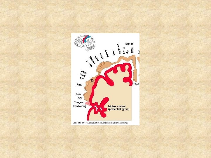
Examine these diagrams and f. MRI scans, and use them to address the questions Copyright 2004, Pearson Education, Inc. , publishing as Benjamin Cummings Source: http: //163. 178. 103. 176/Casos. Berne/2 b. SNervioso/Casox 111/HTMLC/Casos. B 2/Brocas 3. html

Brain Damage Questions – Wernicke’s Aphasia Examine the diagram and f. MRI scan provided, and use them to address the following questions: 1. Patients with Wernicke’s aphasia are able to produce language readily, though they struggle with meaning and coherent conversation. How might the location of Wernicke’s area explain that difficulty? 2. While patients with damage to the Wernicke’s area readily initiate speech, they seem to have difficulty stopping speech. However, often a gentle touch of the hand or arm seem to, at least briefly, signal them to stop. What factor(s) do you think might explain this discrepancy?
- Slides: 11