Cytological findings Moderate to low cellularity Bloody inflammatory

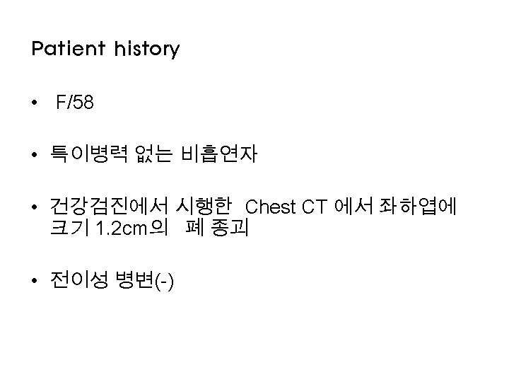
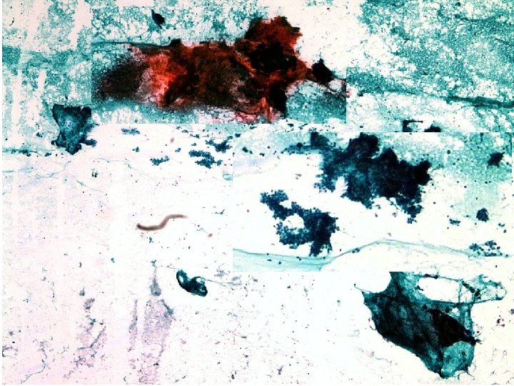
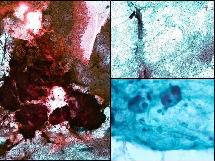
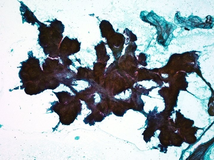
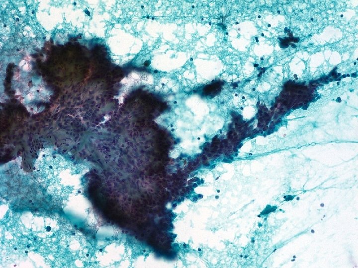
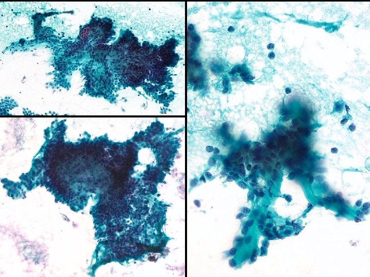
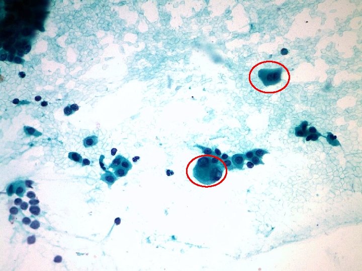
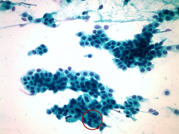
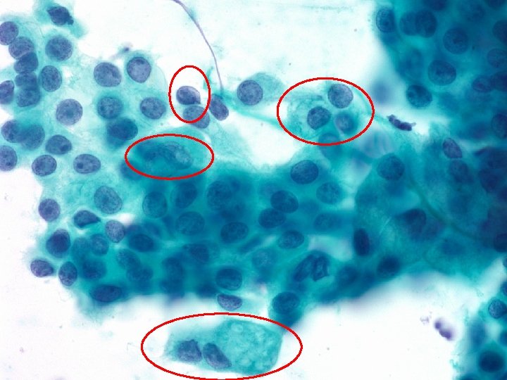
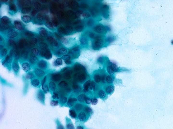


Cytological findings • Moderate to low cellularity • Bloody inflammatory background • Atypical cellular clusters with papillary features and monolayer sheets • Fibrous cores within papillary clusters • Rare mitosis • Mild atypical epithelial cell clusters with - indistinct cell borders and irregular nuclear membrane -occasionally spindle elongated cells - nuclei: small nucleoli, bi-nucleation , intra-nuclear groove & inclusion - variable cytoplasm from delicate clear, vacuolated to dense area

Differential diagnosis Neoplasm Lung mass 1. 2 cm Malignant tumor Primary tumor Mild atypical epithelial cell clusters with papillary feature Metastatic tumor Benign tumor

Differential diagnosis -primary malignant lung tumor with mild nuclear atypia and papillary feature Adenocarcinoma with papillary pattern Adenocarcinoma, lepidic pattern Papillary adenocarcinoma Adenocarcinoma with micropapillary pattern Mucoepidermoid carcinoma with papillary variant

1. Adenocarcinoma with papillary pattern Papillae and glandular differentiation-glands Prominent nucleoli with irregular nuclear membrane Binucleation or multinucleation Intranuclear cytoplasmic invagination Delicate & finely vacuolated cytoplasm Mitosis, necrosis, and apoptosis are common Lack of cytologic atypia and mitosis

2. Adenocarcinoma, lepidic pattern Abundance of monolayered sheet and occasional papillae of cells Mild nuclear crowding or overlap Round and relatively uniform enlarged nuclei with nuclear groove and irregular nuclear membranes Intranuclear cytoplasmic invagination Finely granular chromatin Clean background without necrosis Lack of monolayer sheet

3. Papillary adenocarcinoma Pure true papillae with fibrovascular core Large cells with significant cytologic atypia Mitosis, necrosis, and psammoma bodies can occur Mild cytological atypia and rare mitosis

4. Other tumors 1. Adenocarcinoma, micropapillatry variant Not true papillae small papillae 2. Mucoepidermoid carcinoma with papillary feature Three cell type: mucus-secreting glandular cells, squamous cells, and intermediate cells 3. Sclerosing hemangiona (Pneumocytoma) Dual population of epithelial cells and stromal cells flat sheet, papillary aggregates cytologically bland with rare mitosis

KCP 773 character Papillae with fibrous core and sheet Adenocarcinoma , lepidic pattern Adenocarcinoma, papillary pattern Papillary adenocarcinoma Monotonous monolayer cell population occasional papillae Glandular differentiation-glands, papillae or mucin production Pure papillae with fibrovascular core Cytological atypia mild Moderated to marked Nucleoli inconspicuous prominent Chromatin pattern dedicate to coarse finely granular relatively coarse Nuclear inclusion and groove + + Cytoplasm variable clear vacuolated to dense foamy granular finely vacuolated delicate and finely vacuolated and often contain mucin Clear to dense Mitosis rare frequent Necrosis rare frequent

KCP 773 character Papillae with fibrous core and sheet Adenocarcinoma , lepidic pattern Adenocarcinoma, papillary pattern Papillary adenocarcinoma Monotonous monolayer cell population occasional papillae Glandular differentiation-glands, papillae or mucin production Pure papillae with fibrovascular core Cytological atypia mild Moderated to marked Nucleoli inconspicuous prominent Chromatin pattern dedicate to coarse finely granular relatively coarse Nuclear inclusion and groove + + Cytoplasm variable clear vacuolated to dense foamy granular finely vacuolated delicate and finely vacuolated and often contain mucin Clear to dense Mitosis rare frequent Necrosis rare frequent

Final diagnosis Suspicious for Adenocarcinoma, lepidic pattern
- Slides: 22