Cytologic findings Lymphoid background Cell clusters papillary solid

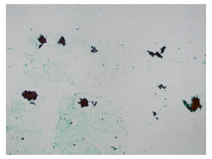
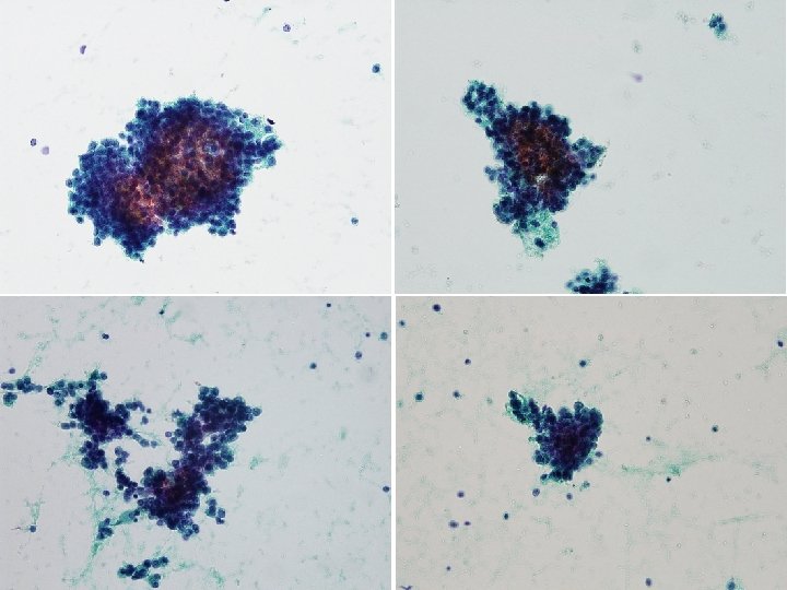
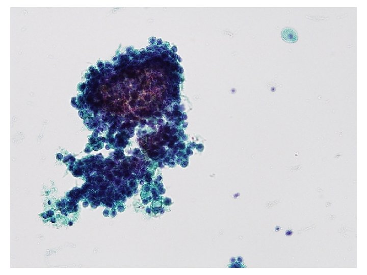

Cytologic findings • Lymphoid background • Cell clusters: papillary, solid, acinar, and microacinar arrangement • Single scattered cells • Small uniform round cells • Overlapping • High N/C ratio • Hyperchromatic • Prominent nucleoli • Delicate, fine and focal vacuolated cytoplasm

Differential diagnosis • Lymphoma • Metastatic carcinoma – Well differentiated adenocarcinoma • Biliary tract/stomach/colon/lung etc. – Prostatic adenocarcinoma – Breast lobular carcinoma – Neuroendocrine tumor

• Lymphoma – Monotonous, equal size의 lymphoid cell – Cluster를 이루지 않고 single cell로 나옴 • Well differentiated adenocarcinoma – – (biliary tract/lung/stomach/colon etc. ) Many individual cells + 3 -D clusters Cohesiveness↓, polarity↓, nuclear enlargement, high N/C ratio, prominent nucleoli, hyperchromasia, variable mucin Biliary tract: Large 3 -D cluster, coarse chromatin clumping Lung: 3 -D cluster (acini, tubule, papillae), fine granular to powdery chromatin, round to oval nuclei, variable cell size
 Carcinoid • Dispersed or small groups (forming flat, loosely")
• Neuroendicrine tumor 1) Carcinoid • Dispersed or small groups (forming flat, loosely structured gland like clusters) • Uniform appearance: cuboidal or rectangular with faintly basophilic transparent cytoplasm and eccentric nuclei, plasmacytoid nuclei, fine granular chromatin (“salt and pepper”), tiny nucleoli, occasional giant cells 2) Well differentiated neuroendocrine carcinoma (atypical carcinoid) • Organoid arrangement, high mitotic rate, prominent nucleoli (single or multiple) and focal necrosis • variable nuclear size, hyperchromasia, abundant cytoplasm, nonpyknotic ovoid nuclei, clear cytoplasm

• Breast lobular carcinoma – – Small cell size, minimal atypia, intracytoplasmic vacuole Indistinct nucleoli, intracytoplasmic neolumina (12 -57%) Dispersed cells, no acini Extremely rare (1% of male breast cancer) • Prostatic adenocarcinoma – 비교적 uniform한 cell들의 small cluster or acini – Small, round to oval cells – Enlarged, round nuclei, increased N/C ratio, moderate amount of cytoplasm – Microacinar pattern과 prominent nucleoli가 가장 특징적인 소견

Differential diagnosis • Lymphoma • Metastatic carcinoma – Well differentiated carcinoma • Biliary tract/stomach/colon/lung etc. – Prostatic adenocarcinoma – Breast lobular carcinoma – Neuroendocrine tumor

Diagnostic Cytopathology 2007 2011

• Small- to medium sized relatively uniform cells • Small clusters forming acini, solid, and cirbriform microacinar + scattered single cells • Loss of honeycomb • Moderately enlarged, hyperchromatic nuclei with mild nuclear membrane irregularities • Nuclear membrane irregularity, hyperchromasia와 chromatin clumping 은 다양하게 나타나지만 often minimal • Prominent nucleoli (single or multiple) • Delicate and finely vacuolated cytoplasm • Neuroendocrine differentiation (10 -33%) • Immunohistochemical pannel – PSA (prostate-specific antigen), PSAP (prostatic acid phosphatase), and AMACR (alphamethylacyl-Co. A racemase)
 1. The possibility of")
Diagnosis • Positive for malignancy, metastatic carcinoma, see note. Note) 1. The possibility of metastatic adenocarcinoma from prostate can be suggested. 2. Further evaluation is recommended.

- Slides: 16