Cytologic Findings Clusters of hyperchromatic crowded columnar cells


- Slides: 22
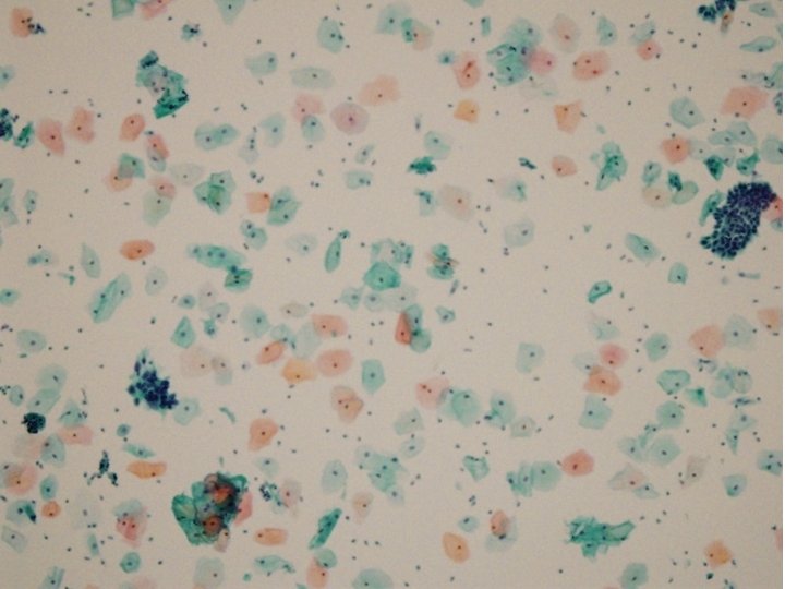
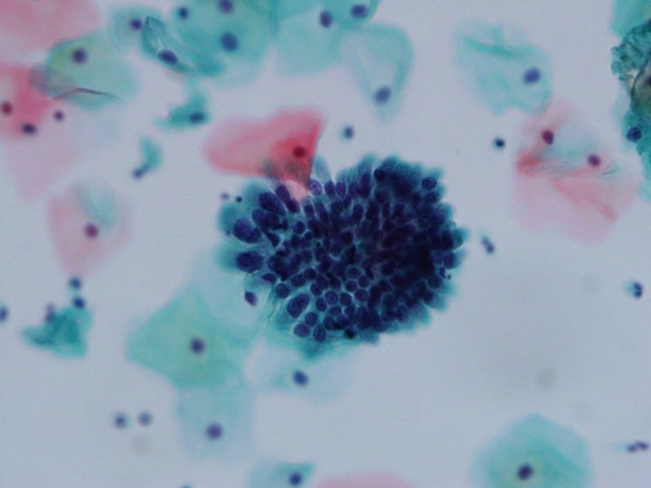
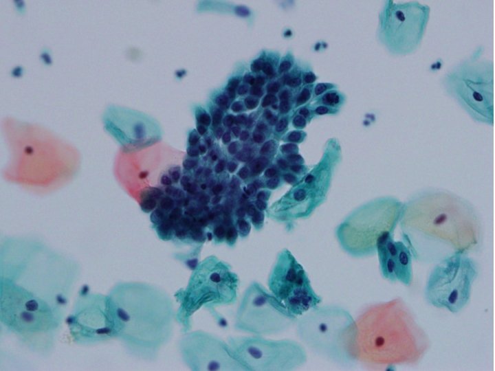
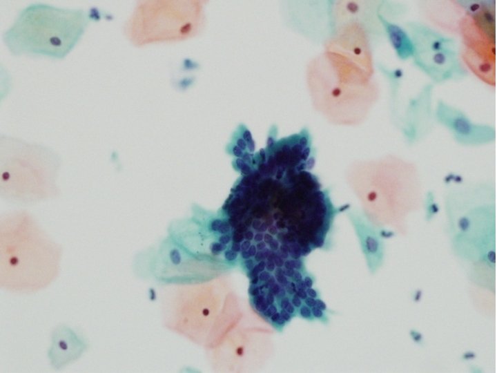
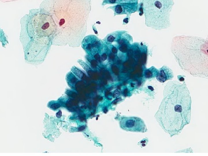
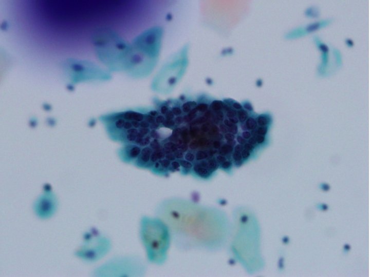
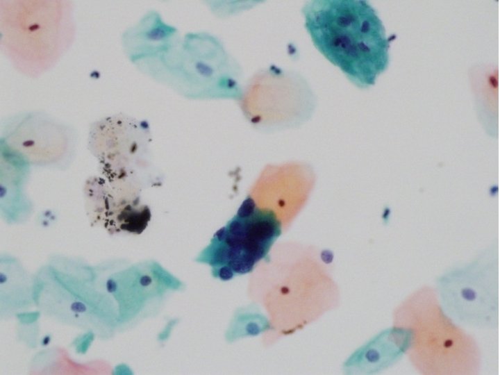
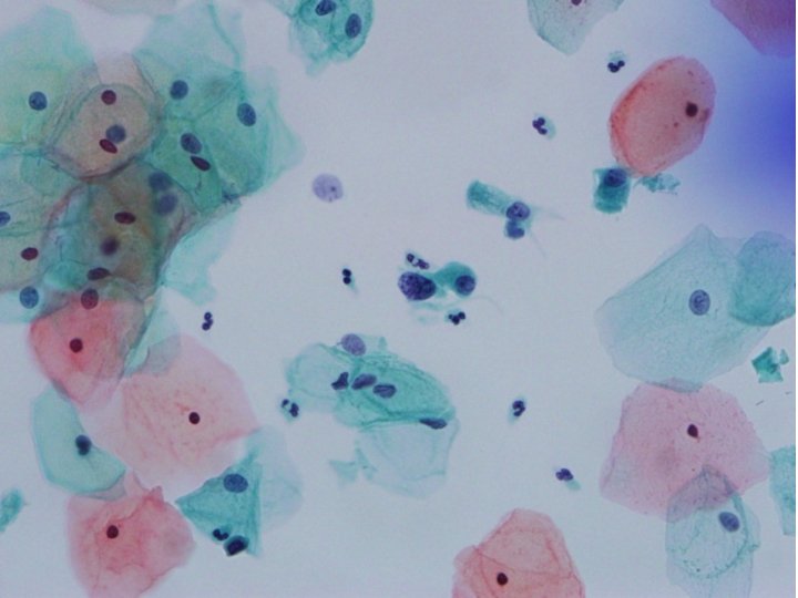
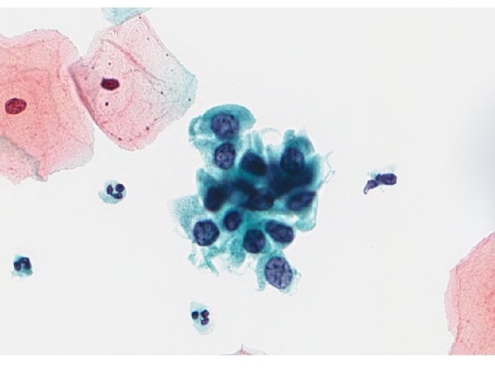
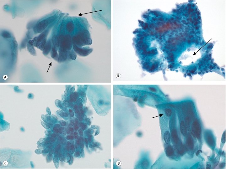
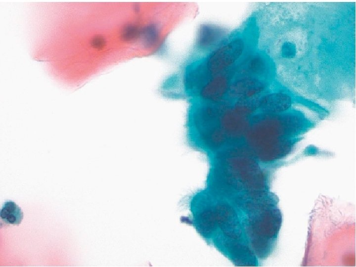
Cytologic Findings • • Clusters of hyperchromatic crowded columnar cells Disorganized honeycombed arrangements Nuclear overlapping Feathering Bird tail Rosettes A few isolated atypical cells • Enlarged nuclei • Coarsely granular chromatin patterns • Small and inconspicuous nucleoli • Clean backgrounds with scattered inflammatory cells
Differential Diagnosis • Endocervical Adenocarcinoma in Situ • Endocervical Adenocarcinoma • Tubal Metaplasia • High-Grade Squamous Dysplasias • Abraded endometrial cells
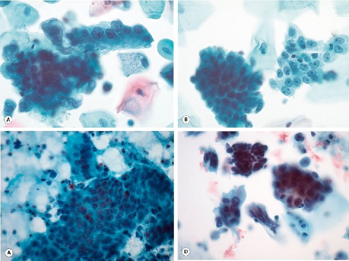
Endocervical Adenocarcinoma in Situ • Hyperchromatic crowded groupings of cells • Pseudostratified strips of columnar cells • Epithelial rosettes (gland formations) • Nuclear and cytoplasmic “feathering” • Nuclear size twofold greater than the intermediate squamous cell nucleus • N: C increased beyond normal endocervical cells • Coarsely granular, evenly distributed hyperchromatic chromatin • Possible presence of small nucleoli • Presence of mitotic figures and apoptotic bodies • Not associated with a background “tumor” diathesis
Endocervical Adenocarcinoma • Architectural features of AIS may be retained in well differentiated lesions: – Feathered group edges – Rosette formation – Pseudostratified strips • Large numbers of abnormal cells present with many isolated cells • Granular clinging, necrotic and bloody tumor diathesis • Nuclei enlarged (greater than 2– 3 times the size of an intermediate squamous cell nucleus) • Chromatin is coarsely granular with heterogeneity and clearing • Macronucleoli are common • Mitoses and apoptotic bodies are commonly seen • Cytoplasm is generally granular and finely vacuolated
Tubal Metaplasia • Incomplete features of AIS • Pseudostratified strips of cells • Nuclear feathering • Rosettes • Finely granular, evenly distributed chromatin • “Washed-out” appearance to nuclei • Enlarged nuclei with pleomorphism • Nucleus-to-cytoplasmic ratio increased • Occasional mitoses and rare apoptotic bodies; and • Cilia/terminal bars
Differential Points Endocervical Adenocarcinoma in Situ Adenocarcinoma Tubal Metaplasia Pseudostratified strips of cells Nuclear feathering Common features Rosettes Nucleoli Small Macronucleoli Chromatin Coarsely granular, evenly distributed Coarsely granular with Finely granular, heterogeneity and evenly distributed clearing Diathesis Bloody Necrotic Granular clinging Cilia Isolated cells present Many
Diagonosis Endocervical adenocarcinoma in situ
Endocervical Adenocarcinoma In Situ • Normal endocervical cytology • The incidence of AIS is a mere 0. 61/100, 000 which is 2% that of CIN 3 • In practice, one is likely to see one case of AIS for every 50 cases of HSIL