CYTOGENETICS Dr Mohammed Hussein Introduction Numerical Chromosomal Abnormalities

CYTOGENETICS Dr. Mohammed Hussein

• Introduction • Numerical Chromosomal Abnormalities • Structural Chromosomal Abnormalities • Advances in Molecular Cytogenetics

• Structural alterations of chromosomes occur when chromosomes are broken by agents termed clastogens, example o. Radiation o. Some viruses o. Some chemicals

Balanced & Unbalanced alterations • Balanced alterations: do not result in a gain or loss of genetic material and usually have fewer clinical consequences • Unbalanced alterations: alterations result in a loss or gain of genetic material

Germ cell & Somatic cell alterations • Germ line alterations can be transmitted to offspring. • Somatic cells alterations not transmitted to offspring, but can alter genetic material such that the cell can give rise to cancer.
 2. Deletions (del) Common 3. Inversions")
Types of Structural Chromosome Abnormalities 1. Translocations (t) 2. Deletions (del) Common 3. Inversions (inv) 4. Ring Chromosome (r) 5. Isochromosome (i) Less Common

Translocation s

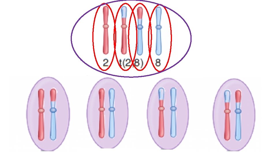
Translocations • Translocations occur when chromosomes are broken and the broken elements reattach to other chromosomes. • Translocations can be classified into two major types: 1. Reciprocal 2. Robertsonian
 Reciprocal")
• Reciprocal translocations occur in non homologous chromosomes (other than acrocentric chromosomes) Reciprocal vs. Robertsonian • Robertsonian translocations occur only in acrocentric chromosomes (13, 14, 15, 21, and 22). the

Reciprocal translocation Homologs Non homologs

Reciprocal translocation
")
karyotype 46, XY, t(2 p; 8 p)

Consequences of a Reciprocal Translocation
 4 Spermatocytes 4 Oocytes (Haploid)")
Spermatogonium Oogonium (Diploid) 4 Spermatocytes 4 Oocytes (Haploid)

Fertilization with normal egg/sperm
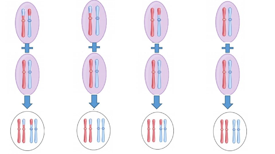
 Normal 50% Translocation carrier 50% 25% Pregnancy loss (50%) Partial trisomy")
25% Survive (50%) Normal 50% Translocation carrier 50% 25% Pregnancy loss (50%) Partial trisomy 2 Partial trisomy 8 Partial monosomy 2 Partial monosomy 8

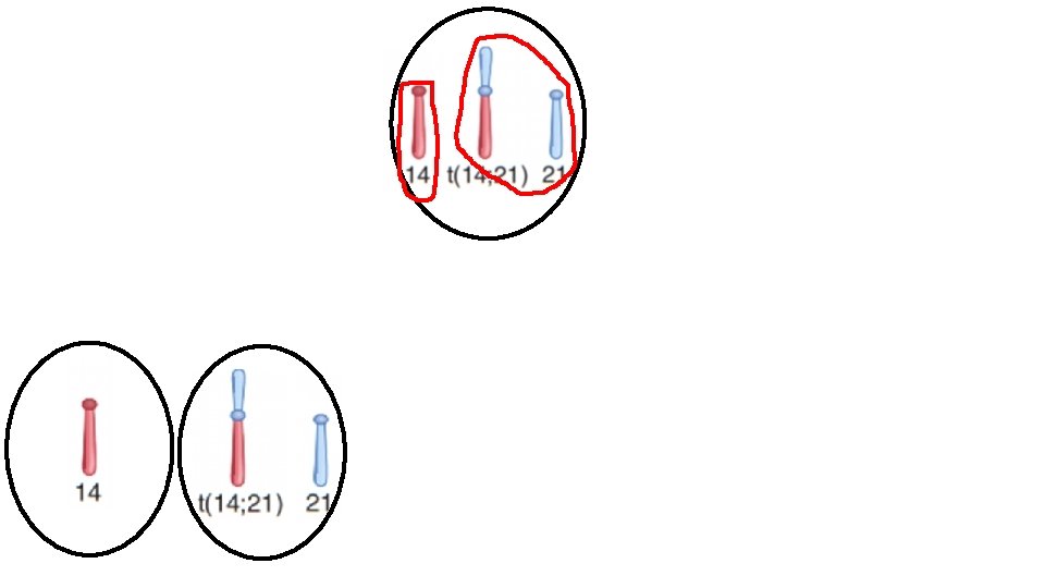
Robertsonian translocations

• Much more common than reciprocal translocations • occur in approximately 1 in 1, 000 live births. • They occur only in the acrocentric chromosomes. • Involve the loss of the short arms of two of the chromosomes and subsequent fusion of the long arms.

q. Acrocentric chromosomes: have the centromere far toward one end
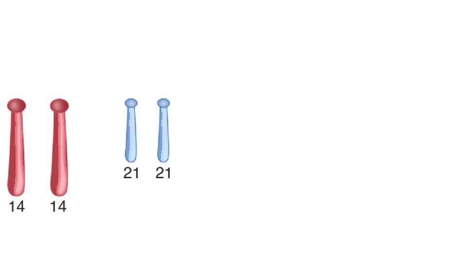
 14 t(14 q; 21")
45, XY, – 14, – 21, +t(14 q; 21 q) 14 t(14 q; 21 q) 21 14 45 21 44 + t(14 q; 21 q) 45
")
Down Syndrome Cytogenetics (Karyotyping)

Down Syndrome Cytogenetics • The extra chromosome 21 may result from 1. Meiotic non disjunction (aneuploidy) 94% 2. Robertsonian translocation 5% 3. Mosaicism 1%

Meiotic non disjunction karyotype 47, XX, +21 47, XY, +21

Robertsonian translocation • About 5% of Down syndrome cases are the result of a Robertsonian translocation affecting chromosome 14 and chromosome 21.
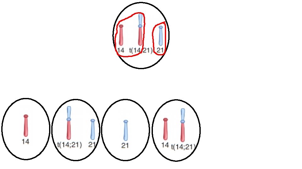
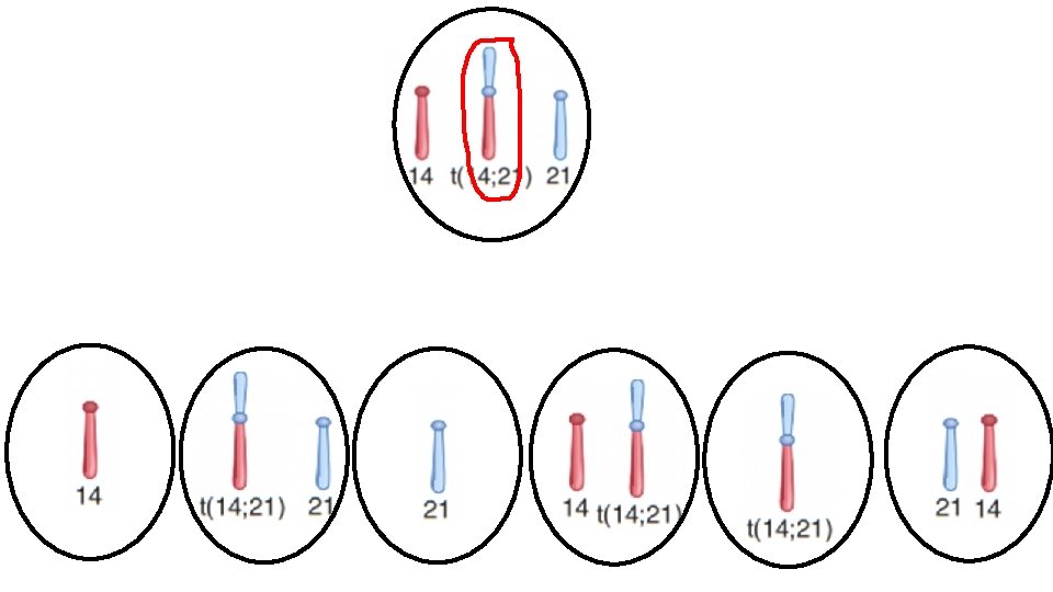

Fertilization with normal egg/or sperm
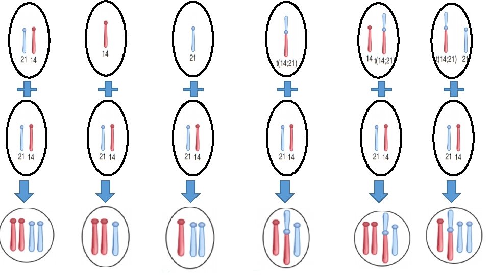

Survive Pregnancy loss Monosomy 14 21 Trisomy 14 Trisomy 21 Translocation Down carrier 1/3 1/3 of children – Down syndrome 2/3 of children – Phenotypically normal Normal Diploid 1/3
")
21 46, XY, – 14, +t(14 q; 21 q)

Three copies of chromosome 21 = Down syndrome 46, XY, – 14, +t(14 q; 21 q)
 Down syndrome (Parent carries Robertsonian translocation) 47, XX, +21")
Down syndrome (Nondisjunction during meiosis) Down syndrome (Parent carries Robertsonian translocation) 47, XX, +21 or 47, XY+21 46, XX, – 14, +t(14 q; 21 q), or 46, XY, – 14, +t(14 q; 21 q) No association with prior pregnancy loss May be associated with prior pregnancy loss Older mother May be a younger mother Very low recurrence rate Recurrence rate 10– 15% if the mother is translocation carrier; 1– 2% if the father is translocation carrier Parental chromosomal analysis is not recommended Parental chromosomal analysis is recommended

Deletions

• A deletion occurs when a chromosome loses some of its genetic information. • Two types: 1. Terminal deletions (the end of the chromosome is lost) 2. Interstitial deletions (material within the chromosome is lost)

Interstitial deletion Normal chromosome 5 Terminal deletion
 46, XY, del(5 p) Cri-du-chat syndrome")
46, XX, del(5 p) 46, XY, del(5 p) Cri-du-chat syndrome

Microdeletions

Microdeletions Some deletions may be so small that they are not readily apparent microscopically without special fluorescent probes (FISH)

Microdeletions Examples: • Prader-Willi: 15 q 11 13 • Angelman syndromes: 15 q 11 13 • Williams syndrome: 7 q 11 • Di. George syndrome: 22 q 11

Other Structural Chromosomal Abnormalities • Their frequency and clinical consequences tend to be less severe than those of translocations and deletions. 1. Inversions (inv) 2. Ring Chromosome (r) 3. Isochromosome (i)

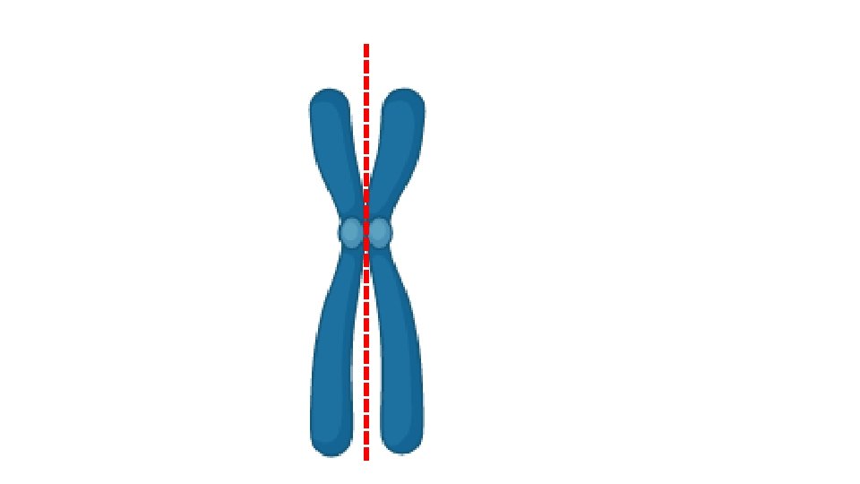
Inversions • Inversions occur when the chromosome segment between two breaks is reinserted in the same location but in reverse order. 1. Pericentric inversions include the centromere 2. Paracentric inversions do not include the centromere
 Pericentric inversion")
Normal chromosome 3 46, XY, inv(3) Pericentric inversion

Consequences • Inversion carriers till retain all of their genetic material, so they are usually unaffected • However if inversion interrupt or otherwise affect a specific gene and thus cause disease

Ring Chromosome • A ring chromosome can form when a deletion occurs on both tips of a chromosome and the remaining chromosome ends fuse together
 Ring chromosome X r(X)")
Normal chromosome X 46, X, r(X) Ring chromosome X r(X)

Consequences • Autosomal ring chromosomes are lethal • Ring X chromosome results in Turner syndrome

Isochromosome • When a chromosome divides along the axis perpendicular to its normal axis of division, an isochromosome is created (i. e. , two copies of one arm but no copy of the other).
 Two long arms i(q)")
Two short arms i(p) Two long arms i(q)
 Normal chromosome X Xi(p) 46, X, i(Xq) 46, X, i(Xp)")
Xi(q) Normal chromosome X Xi(p) 46, X, i(Xq) 46, X, i(Xp)

Consequences • Autosomal isochromosomes are lethal • X chromosome isochromosome results in Turner syndrome

Advances in Molecular Cytogenetics
 • Spectral Karyotyping")
Advances in Molecular Cytogenetics • Fluorescence in situ Hybridization (FISH) • Spectral Karyotyping
 • A chromosome-specific DNA segment is labelled with a")
Fluorescence in situ Hybridization (FISH) • A chromosome-specific DNA segment is labelled with a fluorescent tag to create a probe. • This probe is then hybridized with the patient’s chromosomes, which are visualized under a fluorescence microscope. • Because the probe will hybridize only with a complementary DNA sequence, the probe will mark the presence of the chromosome segment being tested.

Down syndrome

Spectral Karyotyping • Spectral karyotyping involves the use of five different fluorescent probes that hybridize differentially to different sets of chromosomes. • In combination with special cameras and image-processing software, this technique produces a karyotype in which every chromosome is “painted” a different colour.

Spectral Karyotyping • This allows the ready visualization of chromosome rearrangements, such as small translocations • e. g. , the Philadelphia chromosome rearrangement t(9; 22) involved in chronic myelogenous leukaemia (CML).
")
Spectral Karyotyping shows chromosome rearrangement t(2; 9)
,")
Thank You Dr. Mohammed Hussein M. B. Ch. B, MSC, Ph. D, DCH (UK), MRCPCH
- Slides: 64