
Cystic Lesions Of The Jaws Radiological Interpretation Dr


- Slides: 87
Cystic Lesions Of The Jaws Radiological Interpretation Dr ASHRAF ABU KARAKY Assistant Professor in Oral and Maxillofacial Surgery The University of Jordan
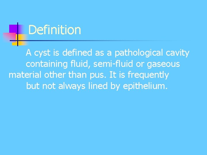
Definition A cyst is defined as a pathological cavity containing fluid, semi-fluid or gaseous material other than pus. It is frequently but not always lined by epithelium.
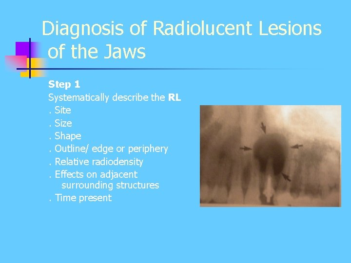
Diagnosis of Radiolucent Lesions of the Jaws Step 1 Systematically describe the RL. Site. Size. Shape. Outline/ edge or periphery. Relative radiodensity. Effects on adjacent surrounding structures. Time present
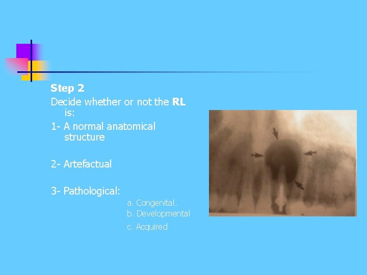
Step 2 Decide whether or not the RL is: 1 - A normal anatomical structure 2 - Artefactual 3 - Pathological: a. Congenital. b. Developmental c. Acquired
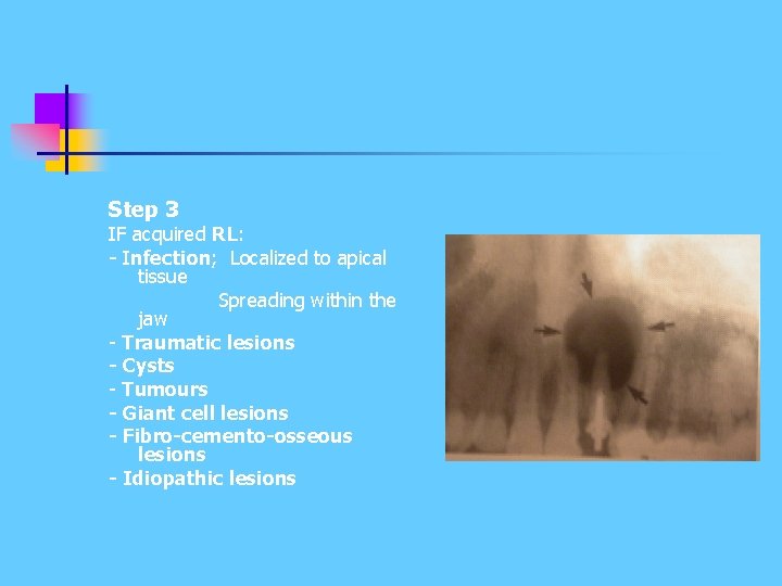
Step 3 IF acquired RL: - Infection; Localized to apical tissue Spreading within the jaw - Traumatic lesions - Cysts - Tumours - Giant cell lesions - Fibro-cemento-osseous lesions - Idiopathic lesions
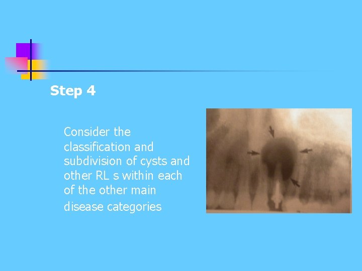
Step 4 Consider the classification and subdivision of cysts and other RL s within each of the other main disease categories
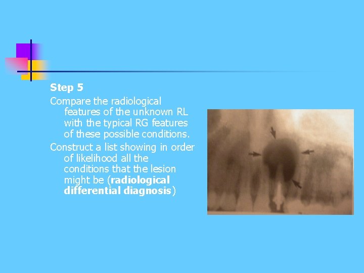
Step 5 Compare the radiological features of the unknown RL with the typical RG features of these possible conditions. Construct a list showing in order of likelihood all the conditions that the lesion might be (radiological differential diagnosis)
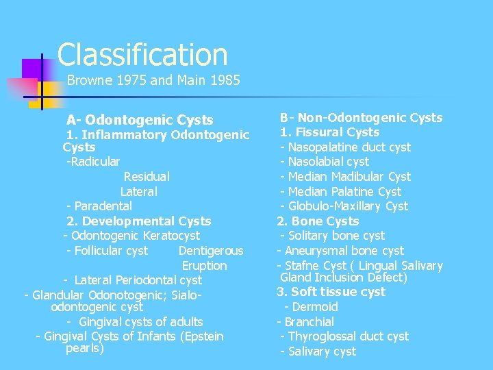
Classification Browne 1975 and Main 1985 A- Odontogenic Cysts 1. Inflammatory Odontogenic Cysts -Radicular Residual Lateral - Paradental 2. Developmental Cysts - Odontogenic Keratocyst - Follicular cyst Dentigerous Eruption - Lateral Periodontal cyst - Glandular Odonotogenic; Sialoodontogenic cyst - Gingival cysts of adults - Gingival Cysts of Infants (Epstein pearls) B- Non-Odontogenic Cysts 1. Fissural Cysts - Nasopalatine duct cyst - Nasolabial cyst - Median Madibular Cyst - Median Palatine Cyst - Globulo-Maxillary Cyst 2. Bone Cysts - Solitary bone cyst - Aneurysmal bone cyst - Stafne Cyst ( Lingual Salivary Gland Inclusion Defect) 3. Soft tissue cyst - Dermoid - Branchial - Thyroglossal duct cyst - Salivary cyst
Inflammatory Odontogenic Cysts Radicular Residual Lateral Paradental
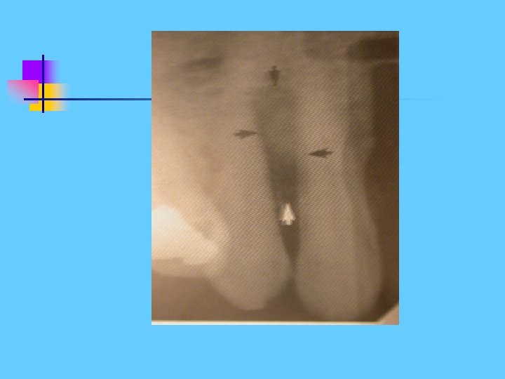
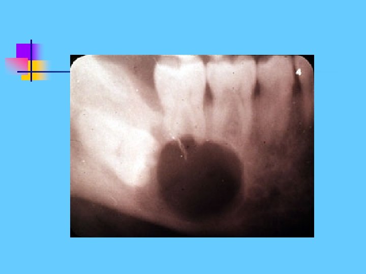
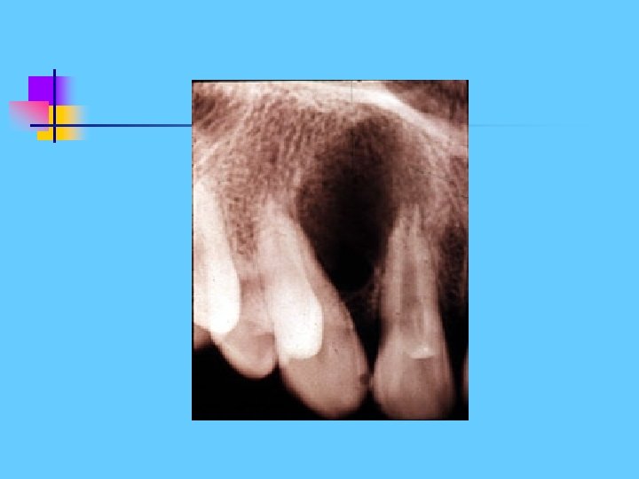
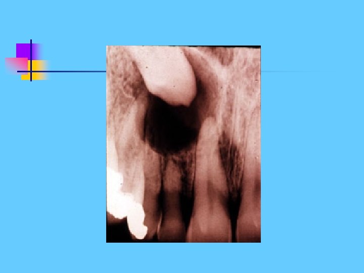
Radicular Cyst Develops from the epithelial remnants of Hertwig s sheath- the cell rests of Malassez Age usually adults, 20 -50 yrs Frequency: most common of all jaw cysts (70%)
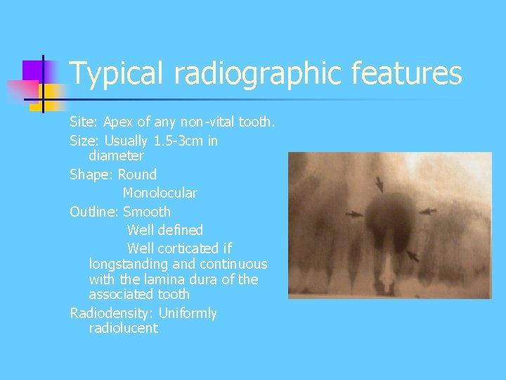
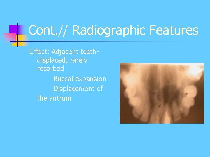
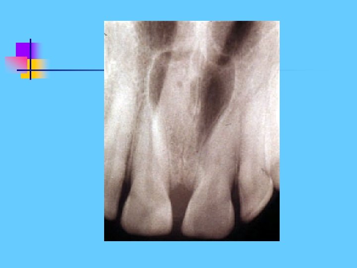
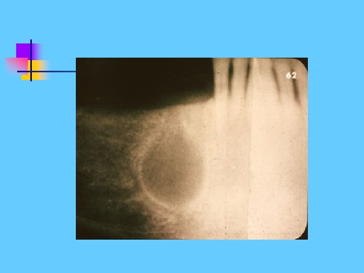
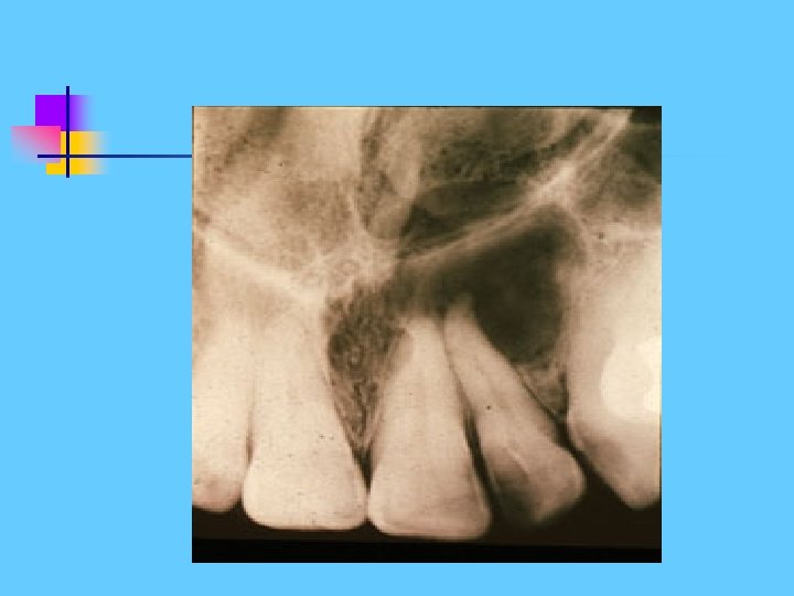
Typical radiographic features Site: Apex of any non-vital tooth. Size: Usually 1. 5 -3 cm in diameter Shape: Round Monolocular Outline: Smooth Well defined Well corticated if longstanding and continuous with the lamina dura of the associated tooth Radiodensity: Uniformly radiolucent
Cont. // Radiographic Features Effect: Adjacent teethdisplaced, rarely resorbed Buccal expansion Displacement of the antrum
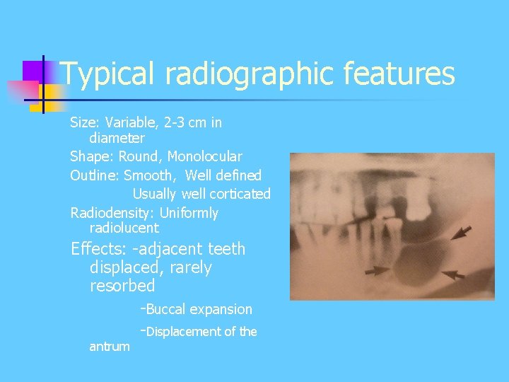
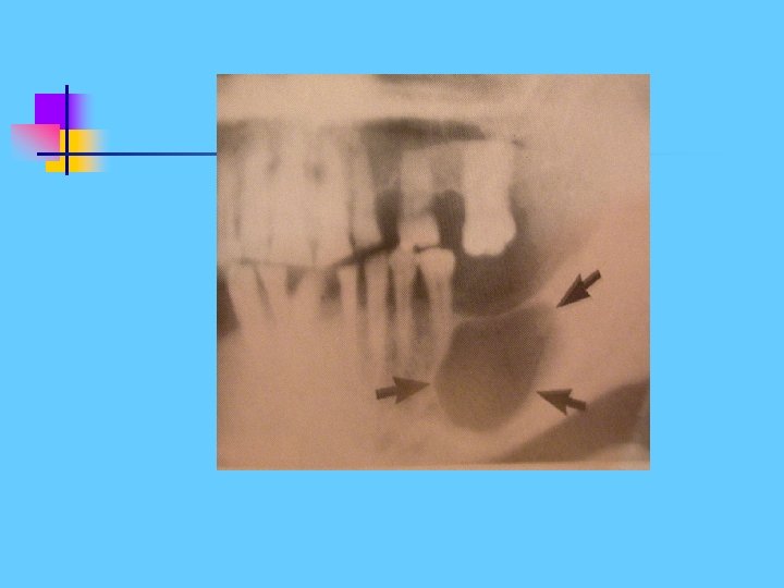
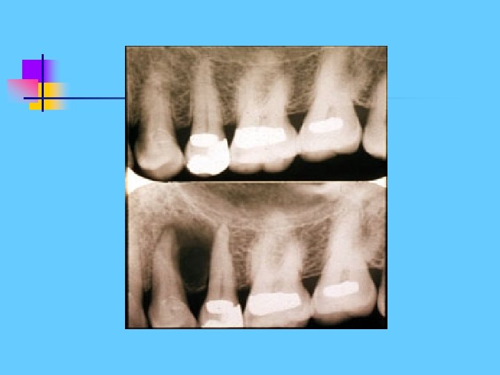
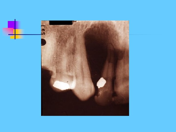
Residual Cyst This term refers to radicular (dental) cyst remaining after the causative tooth has been extracted Age: Adults > 20 yrs Site: Apical regions of tooth bearing portion of the jaws
Typical radiographic features Size: Variable, 2 -3 cm in diameter Shape: Round, Monolocular Outline: Smooth, Well defined Usually well corticated Radiodensity: Uniformly radiolucent Effects: -adjacent teeth displaced, rarely resorbed -Buccal expansion -Displacement of the antrum
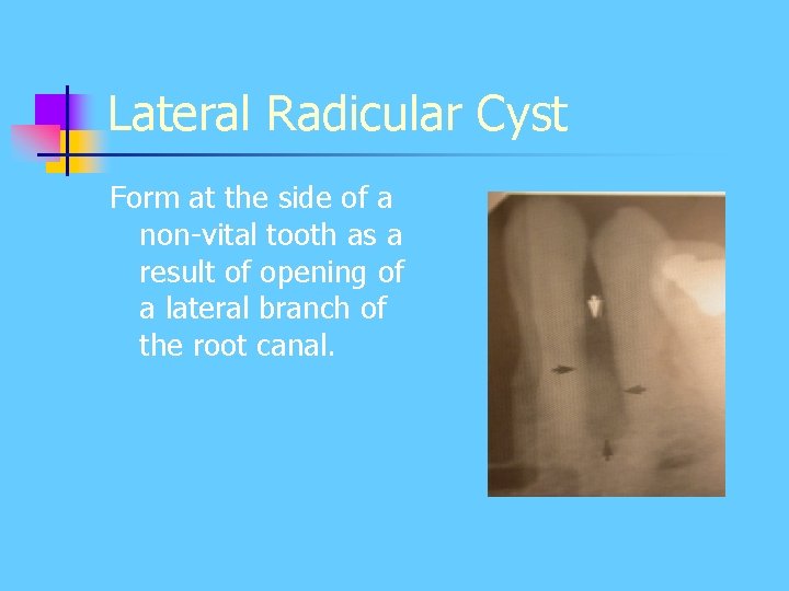
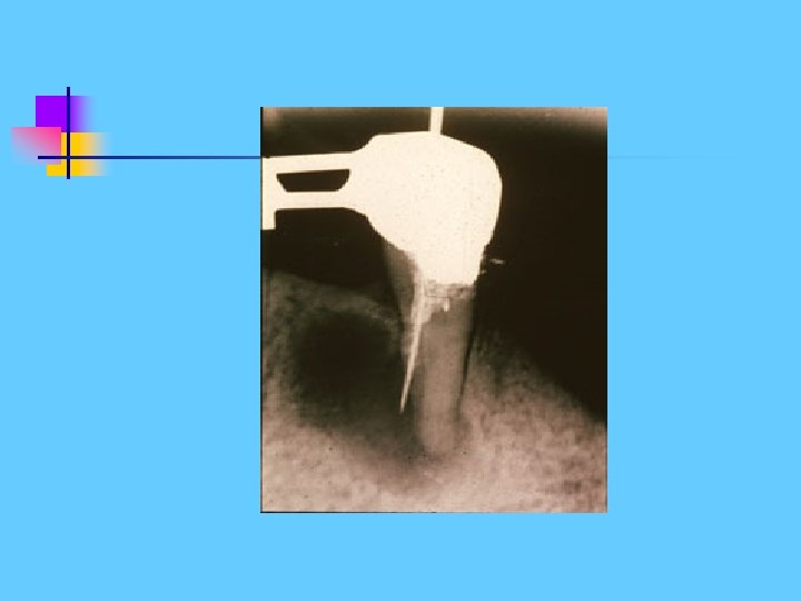
Lateral Radicular Cyst Form at the side of a non-vital tooth as a result of opening of a lateral branch of the root canal.
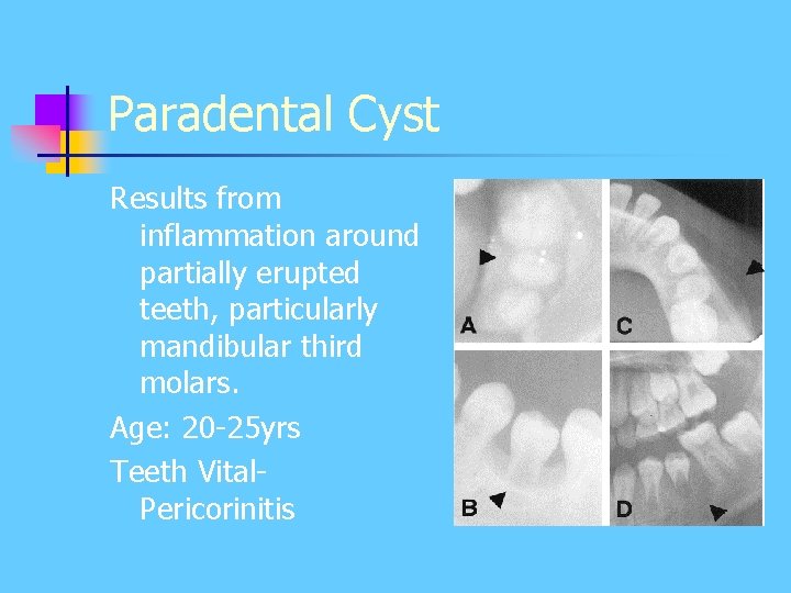
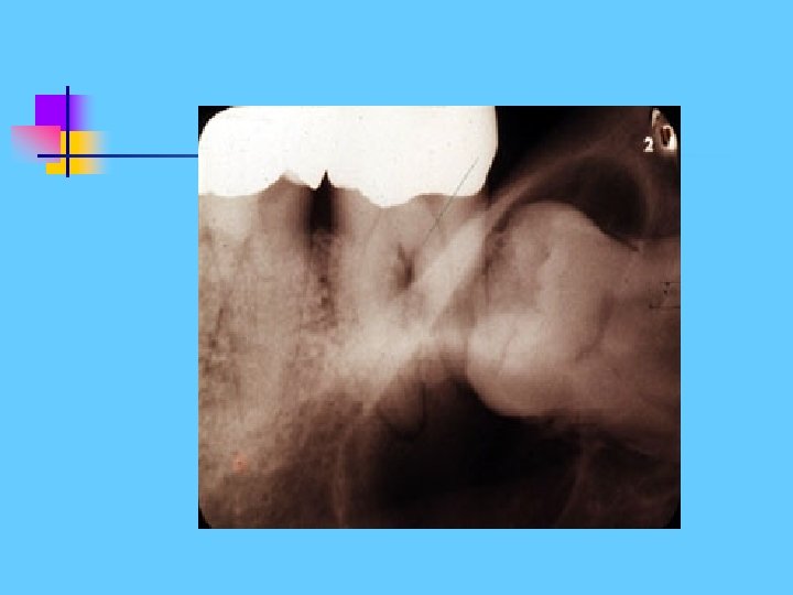
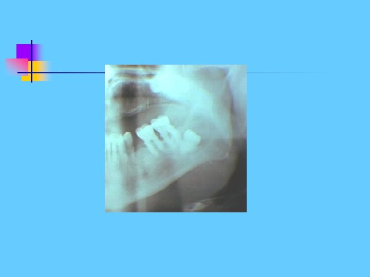
Paradental Cyst Results from inflammation around partially erupted teeth, particularly mandibular third molars. Age: 20 -25 yrs Teeth Vital. Pericorinitis
Developmental Cysts - Odontogenic Keratocyst - Follicular cyst Dentigerous cyst Eruption Cyst - Lateral Periodontal cyst - Glandular Odontogenic Cyst - Gingival Cyst of Adults - Gingival Cyst of Newborn (Epstein Pearls)
Dentigerous (follicular cyst) Develop from the remnants of the reduced dental epithelium Age: Usually adolescents or young adults (2040 yrs), occasionally the elderly. Frequency: About 20% of all Cysts
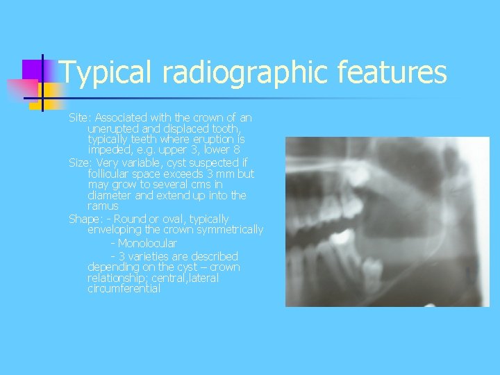
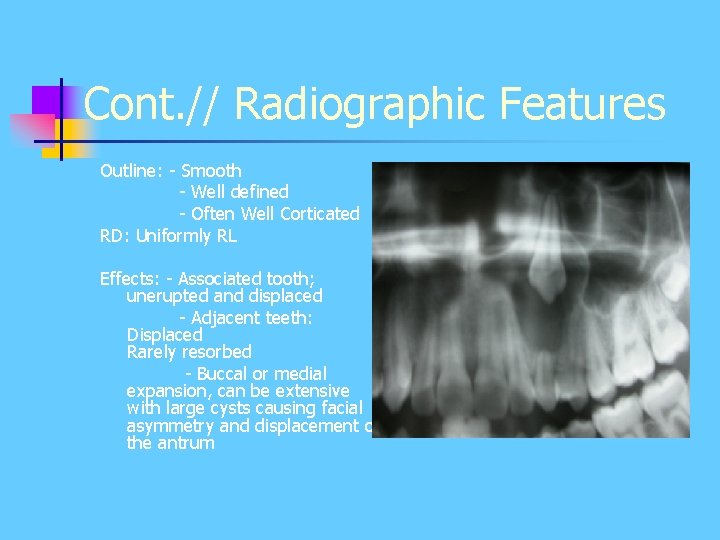
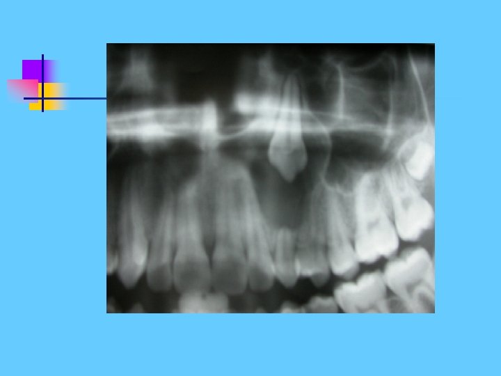
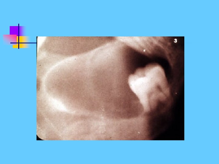
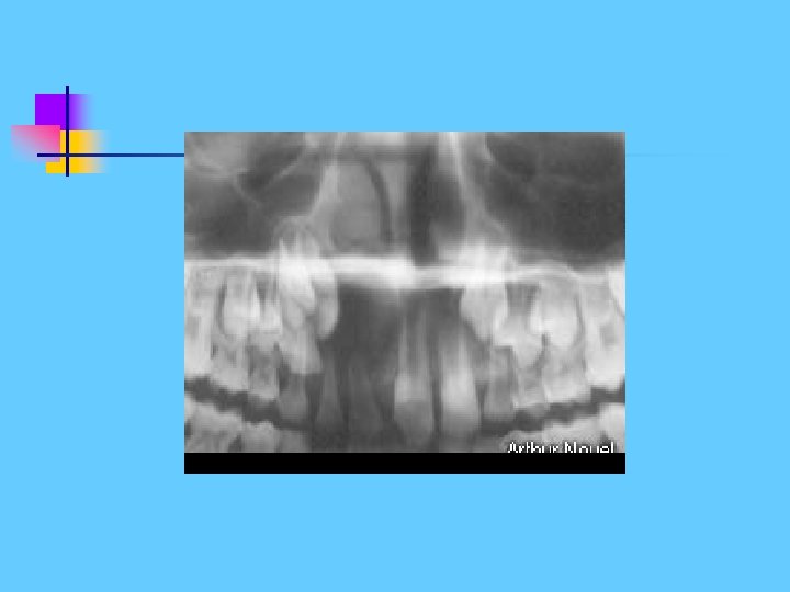
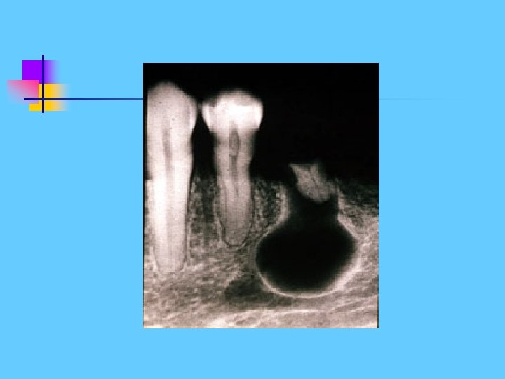
Typical radiographic features Site: Associated with the crown of an unerupted and displaced tooth, typically teeth where eruption is impeded, e. g. upper 3, lower 8 Size: Very variable, cyst suspected if follicular space exceeds 3 mm but may grow to several cms in diameter and extend up into the ramus Shape: - Round or oval, typically enveloping the crown symmetrically - Monolocular - 3 varieties are described depending on the cyst – crown relationship; central, lateral circumferential
Cont. // Radiographic Features Outline: - Smooth - Well defined - Often Well Corticated RD: Uniformly RL Effects: - Associated tooth; unerupted and displaced - Adjacent teeth: Displaced Rarely resorbed - Buccal or medial expansion, can be extensive with large cysts causing facial asymmetry and displacement of the antrum
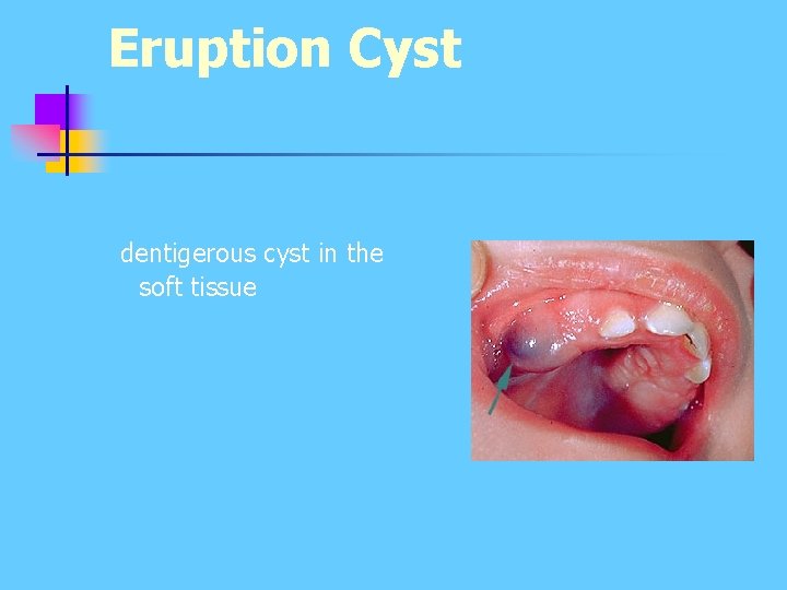
Eruption Cyst dentigerous cyst in the soft tissue
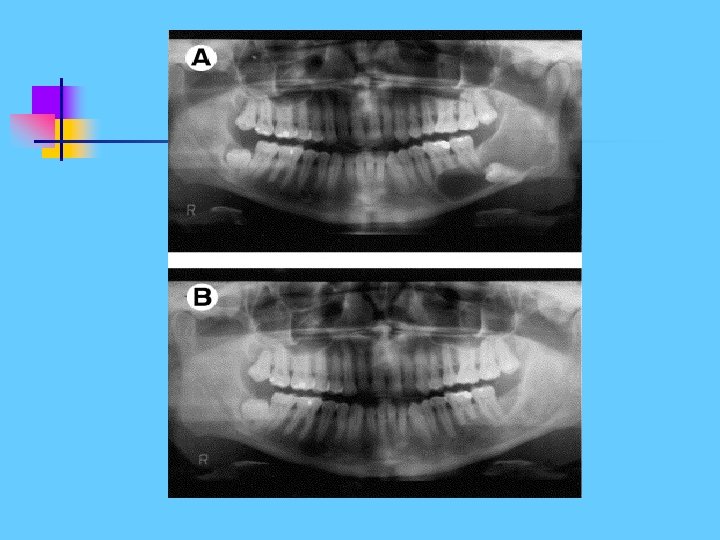
Odontogenic Keratocyst Develop from the epithelium of the dental lamina (the cell rests of Serres) Age: Very variable, 2 nd and 4 th decade Frequency : less than 5% of all odontogenic cysts
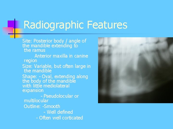
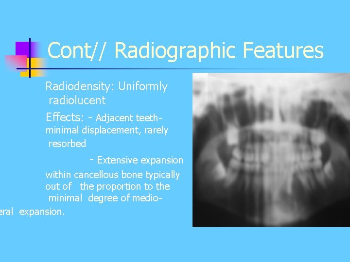
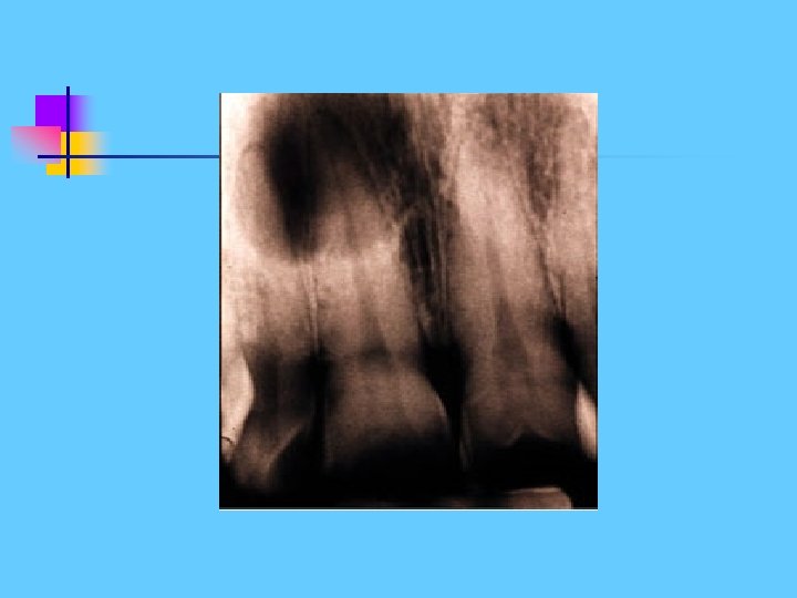
Radiographic Features Site: Posterior body / angle of the mandible extending to the ramus Anterior maxilla in canine region Size: Variable, but often large in the mandible Shape: - Oval, extending along the body of the mandible with little mediolateral expansion - Pseudolocular or multilocular Outline: -Smooth - Well defined - Often well corticated
Cont// Radiographic Features Radiodensity: Uniformly radiolucent Effects: - Adjacent teeth- minimal displacement, rarely resorbed - Extensive expansion within cancellous bone typically out of the proportion to the minimal degree of medioeral expansion.
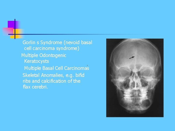
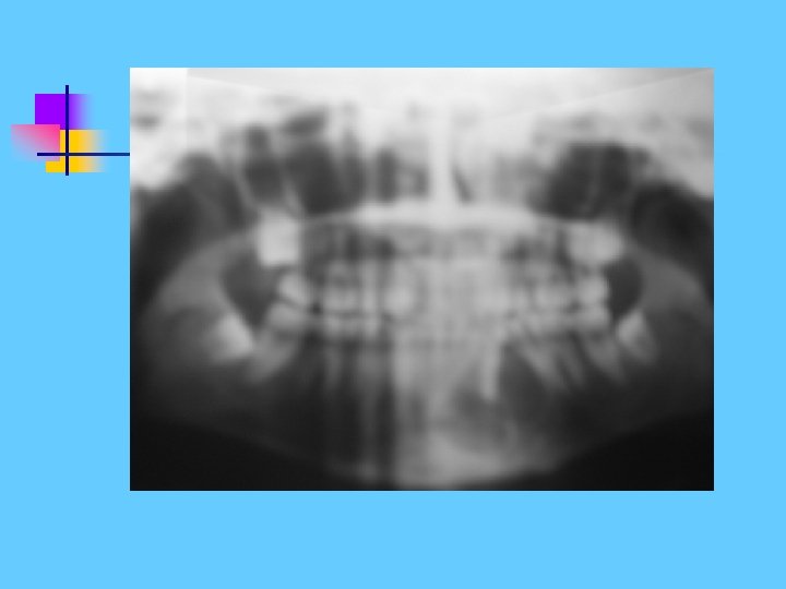
Gorlin s Syndrome (nevoid basal cell carcinoma syndrome) Multiple Odontogenic Keratocysts Multiple Basal Cell Carcinomas Skeletal Anomalies, e. g. bifid ribs and calcification of the flax cerebri.
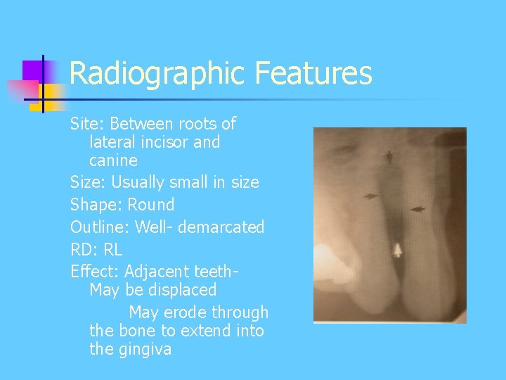
Developmental Lateral Periodontal Cyst Uncommon developmental intraosseous cysts form beside a vital tooth. Age: Variable Frequency: Uncommon
Radiographic Features Site: Between roots of lateral incisor and canine Size: Usually small in size Shape: Round Outline: Well- demarcated RD: RL Effect: Adjacent teeth. May be displaced May erode through the bone to extend into the gingiva
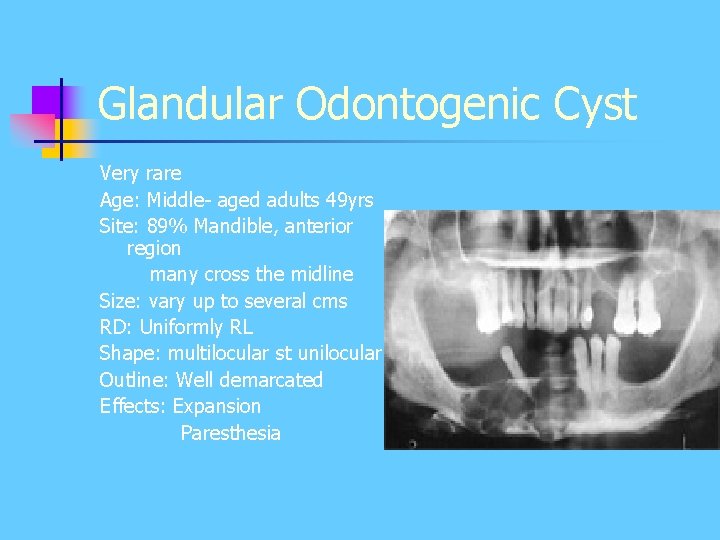
Glandular Odontogenic Cyst Very rare Age: Middle- aged adults 49 yrs Site: 89% Mandible, anterior region many cross the midline Size: vary up to several cms RD: Uniformly RL Shape: multilocular st unilocular Outline: Well demarcated Effects: Expansion Paresthesia
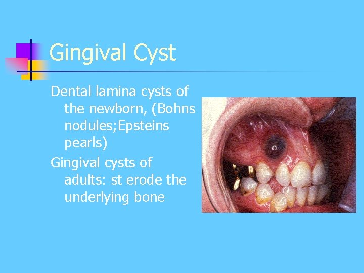
Gingival Cyst Dental lamina cysts of the newborn, (Bohns nodules; Epsteins pearls) Gingival cysts of adults: st erode the underlying bone
Non-Odontogenic Cysts Developmental Cysts Nasopalatine duct cyst Nasolabial cyst Median Palatine Cyst Globulo-Maxillary Cyst Median Mandibular Cyst
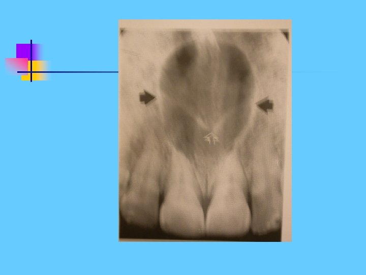
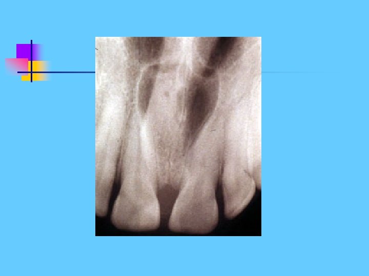
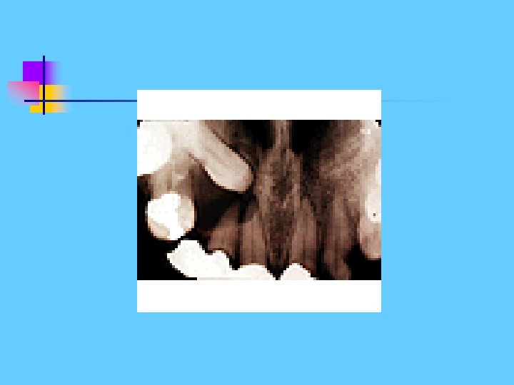
Nasopalatine Duct / Incisive Canal Cyst Develop from epithelial remnants of Nasopalatine Duct or Incisive Canal. Age: Variable, but most frequently detected in middle age (40 -60 yrs olds). Frequency: Most Common of all nonodontogenic cysts, 1% of total population
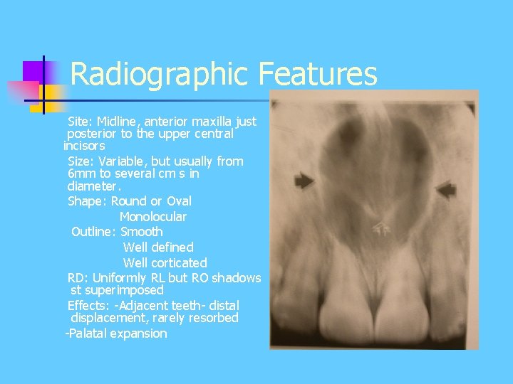
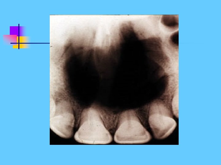
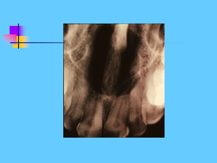
Radiographic Features Site: Midline, anterior maxilla just posterior to the upper central incisors Size: Variable, but usually from 6 mm to several cm s in diameter. Shape: Round or Oval Monolocular Outline: Smooth Well defined Well corticated RD: Uniformly RL but RO shadows st superimposed Effects: -Adjacent teeth- distal displacement, rarely resorbed -Palatal expansion
Differentiation between Nasopalatine Duct Cyst and a large normal Naopalatine foramen? . Size. Outline. Relative RD. Shape?
Median mandibular cyst Develop from embryonic epithelial remnants in the symphyseal region of the mandible
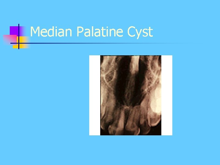
Median Palatine Cyst
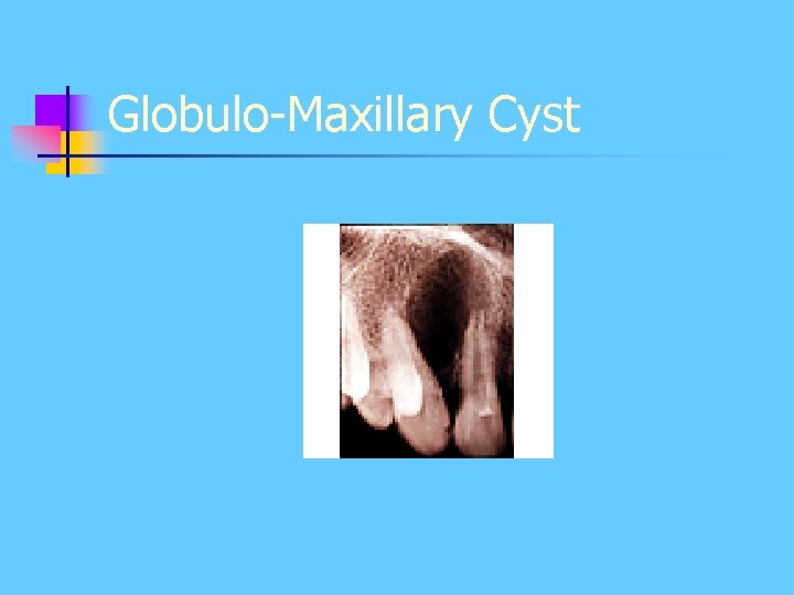
Globulo-Maxillary Cyst
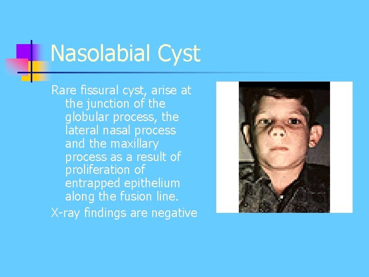
Nasolabial Cyst Rare fissural cyst, arise at the junction of the globular process, the lateral nasal process and the maxillary process as a result of proliferation of entrapped epithelium along the fusion line. X-ray findings are negative
2. Bone Cysts - Solitary bone cyst - Aneurysmal bone cyst - Stafne Cyst ( Lingual Salivary Gland Inclusion Defect)
Solitary (simple) bone cyst Unknown aetiology, may be associated with trauma. Age: Children and young adults < 20 yrs
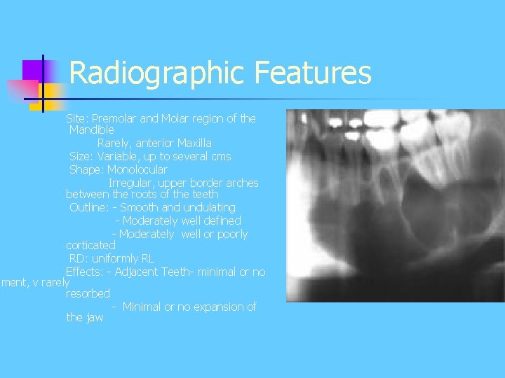
Radiographic Features Site: Premolar and Molar region of the Mandible Rarely, anterior Maxilla Size: Variable, up to several cms Shape: Monolocular Irregular, upper border arches between the roots of the teeth Outline: - Smooth and undulating - Moderately well defined - Moderately well or poorly corticated RD: uniformly RL Effects: - Adjacent Teeth- minimal or no ement, v rarely resorbed - Minimal or no expansion of the jaw
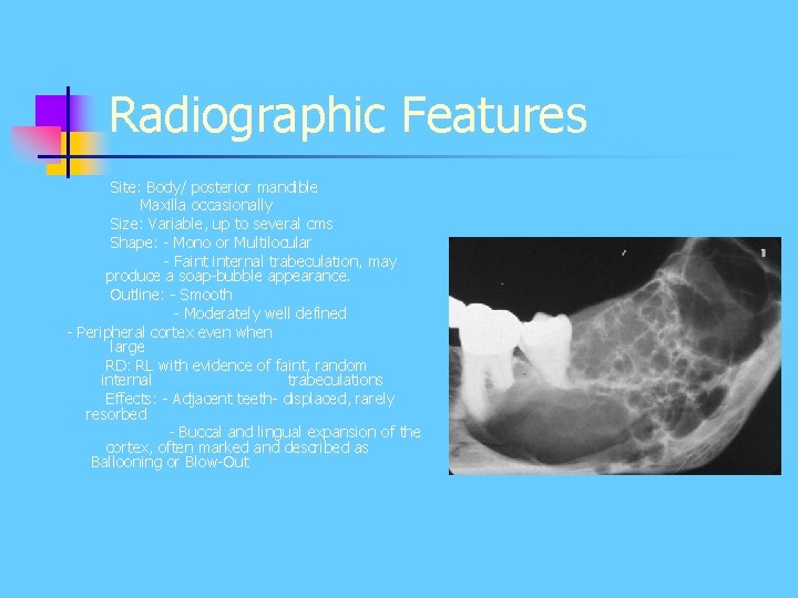
Aneurysmal Bone Cyst More accurately classified as Giant Cell Lesion Localized non-neoplastic proliferative lesion of vascular tissue, containing Giant Cells. Age: Usually < 20 yrs old Frequency: Rare.
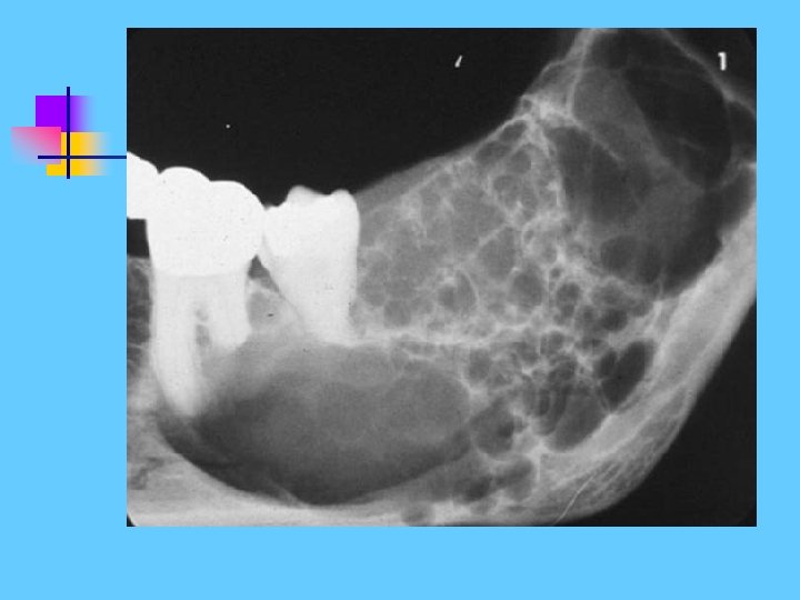
Radiographic Features Site: Body/ posterior mandible Maxilla occasionally Size: Variable, up to several cms Shape: - Mono or Multilocular - Faint internal trabeculation, may produce a soap-bubble appearance. Outline: - Smooth - Moderately well defined - Peripheral cortex even when large RD: RL with evidence of faint, random internal trabeculations Effects: - Adjacent teeth- displaced, rarely resorbed - Buccal and lingual expansion of the cortex, often marked and described as Ballooning or Blow-Out
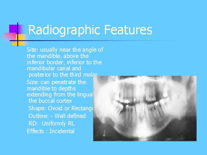
Stafne Cyst ( Lingual Salivary Gland Inclusion Defect) Well defined depression in the lingual surface of the posterior body of the mandible Usually asymptomatic and are incidental RG finding
Radiographic Features Site: usually near the angle of the mandible, above the inferior border, inferior to the mandibular canal and posterior to the third molar Size: can penetrate the mandible to depths extending from the lingual to the buccal cortex Shape: Ovoid or Rectangular Outline: - Well defined RD: Uniformly RL Effects : Incidental
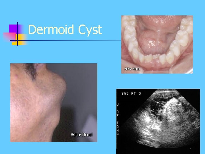
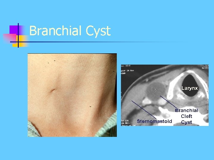
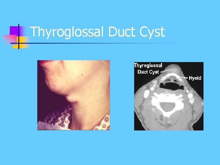
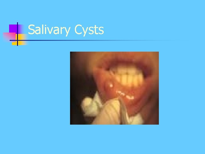
3. Soft tissue cyst - Dermoid - Branchial - Thyroglossal duct cyst - Salivary cyst
Dermoid Cyst
Branchial Cyst
Thyroglossal Duct Cyst
Salivary Cysts
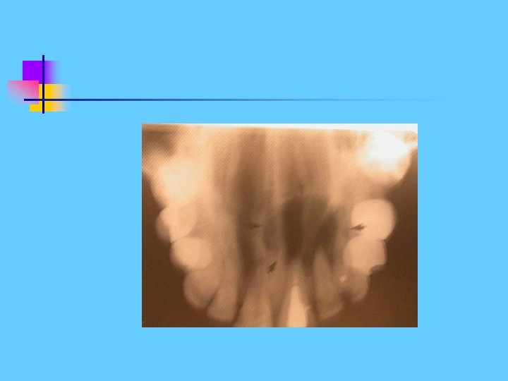
Calcifying Odontogenic Cyst (Gorlin Cyst) Classified by WHO as odontogenic tumour Presents typically as radiolucency resembling other odontogenic cysts As it develops, a variable amount of calcified material becomes evident, scattered throughout the RL. The RO can range from small flecks to large masses. Age: Variable, usually adults < 40 yrs old
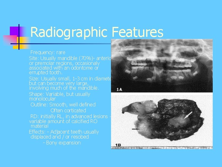
Radiographic Features Frequency: rare Site: Usually mandible (70%)- anterior or premolar regions, occasionaly associated with an odontome or errupted tooth. Size: Usually small, 1 -3 cm in diameter but can become very large, involving much of the mandible. Shape: Variable, but usually monolocular Outline: Smooth, well defined Often corticated RD: initially RL, in advanced lesions – variable amount of calcified RO material Effects: - Adjacent teeth usually displaced and / or resobed - Bony expansion
calcifying n
Thank you