CVS Scintigraphy Myocardial perfusion scans Gated blood pool

CVS Scintigraphy. Myocardial perfusion scans. Gated blood pool scans. Right ventricular studies

Myocardial Perfusion Scan Definition: is the study of myocardial perfusion, viability and infarction imaging. Identical images of the walls of LV are obtained and compared both in stress and rest. Preparation: Fasting 4 -6 hr, drug withdrawal for 48 hr (B blockers and theophilline). Technique: Exercise: physical by a treadmill or simulated exercise by IV dipyridamole or adenosine (vd of normal coronary arteries, but do not increase flow with 50 % or more stenosis). CI in active bronchospasm. IV dobutamine has direct inotropic and chronotropic effects.

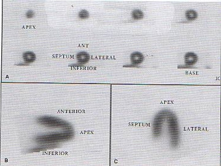
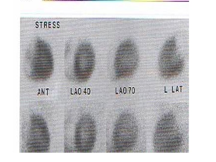
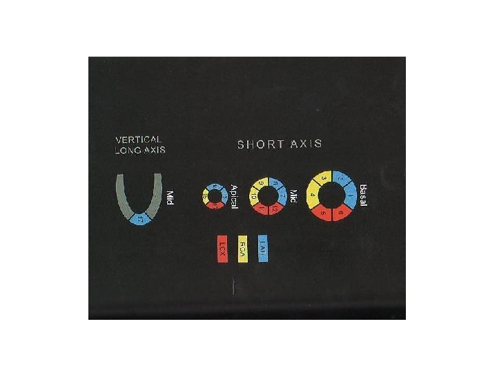
• Sequential ECG, BP, pulse measurements, ask patient about anginal pain. At least 85% of MPHR should be reached to achieve adequate exercise that is estimated by Double product (DP i. e systolic pressure x HR), should double or better triple from rest to peak exercise. • Inject radiopharmaceutical at peak exercise and exercise is continued for 30 -60 s after injection to obtain optimal mapping of stress perfusion. • SPECT images are obtained (180 o acquisition > 360 o) with reconstruction of LV into short-axis, vertical long-axis and horizontal long-axis. • Planar imaging ( ant, LAO 45 o 75 o) is used in over-weight patients. • Prone imaging after standard supine is useful in reducing breast and diaphragmatic attenuation.

Radiopharmaceuticals • Thallium-201: . Potassium analog, actively pumped into viable myocytes by Na/K ATP pump. . Cyclotron produced, low energy, long physical half-life(73 hr), relatively high energy absorbed. . Effective half-life 4 hr. . It redistributes within the myocardium, so rest scans (redistribution imaging) slowly fill in ischemic type defects. Thus, it can differentiate between infarcted and hibernated (viable but temporarily lost function). 1 m. Ci th 201 is reinjected before 24 hr imaging to detect ischemic areas rather than infarcts in high grades stenoses.
 and Tc-99 m tetrofosmin: . Taken")
• Tc-99 m Sestamibi (methoxy isobutyl isonitile) and Tc-99 m tetrofosmin: . Taken up by passive diffusion mostly within myocardial mitochondria. . No redistribution, no significant washout. . Good penetration power 140 Kev. . With same inj gated studies can be obtained to evaluate LV functional parameters. . One day protocol: 8 m. Ci for initial rest scan followed 4 hr later by stress scan with 20 m. Ci. . Two day protocol: we start with stress images, the next day 15 -20 m. Ci inj to obtain rest scan.
 uses Tc-99 m labelled RBCs to")
Gated blood pool scans • Radionuclide ventriculogram (RVG) uses Tc-99 m labelled RBCs to evaluate the size, wall motion and functional parameters of LV. • 20 -30 m. Ci in adults • ECG leads placed to obtain suitable gating signal (R wave) for the computer. • Cardiac cycle is divided into a min of 16 frames for analysis of systolic function and 32 for diastolic function. So, series of images representing the patients cardiac cycle are obtained. Acquisition time of 5 -20 min /view. The best is LAO view. • Computer processing.

Right Ventricular Studies • Images are analysed from the first pass of a radionuclide bolus through the right-sided chambers and lungs before the overlapping leftsided chambers are seen. • RAO • 30 m. Ci bolus of high-specific activity isotope is rapidly inj, followed by a non-radioactive flush dose. • In 3 -8 heartbeats, the activity will pass through the RV and a region of interest is established around it. Time-activity curve allows RV ejection fraction to calculated for each beat.

Thank YOU
- Slides: 11