CUTANEOUS MANIFESTATIONS OF SYSTEMIC DISEASES Dr Saleh Alrasheed


 u Acrodermatitis entropathica u Peutz Jeghers Syndrome u Pyoderma")


 u Usually ANA Negative u round scarring lesions on light exposed")
 papulosqamous or annular presentation u Photosensitivity u Dose not cause")


: Heliotrope : Violeceous color over the upper eyelids")
 u u u Thickened & tight skin Sclerodactyl Face: loss of")
 C=Calcification u R =Raynand’s u E =Eosopheagal dysfunction u")

 Antibody Clinical significance ANA Screening for SLE and other")


 Neurofibromas(soft pink")
 : v Epi = Epilepsy v Loi = Low intellegence v")


 u Genital ulcers (mainly scrotal) u Iritis")
 u u Easy bruishing Diagnosis : Low ascorbic acid (Vit-C) level in Leukocyte")


 Claudication, blindness, stroke -Medium-vessel")
 • Also called Leukocytoclastic vasculitis. • characterized by neutrophilic infiltration")
")


 Complements (decreased) Urinalysis")
: rest , NSAIDS")

 • In children, the onset is mostly after upper respiratory infection;")
 • Treatment by bed rest, pain relief and antibiotics if strep")
")


- Slides: 101
CUTANEOUS MANIFESTATIONS OF SYSTEMIC DISEASES Dr. Saleh Alrasheed Assistant Professor - Dermatology Department College of Medicine KSU , Riyadh. PDF created with Fine. Print pdf. Factory trial version http: //www. fineprint. com
Objectives To highlight the relation between skin manifestations and common systemic disorders. To understands various skin clues and their importance in investigating and managing different systemic diseases
Contents: Introduction: u Connective. Tissue Diseases v Lupus v Dermatomyositis v Scleroderma u Endocrinological Diseases v Diabetes Mellitus v Hyperthyrodism v Hypothyeodism v Cushing’s Syndrome v Addison’s disease PDF created with Fine. Print pdf. Factory trial version http: //www. fineprint. com
GIT: Chronic Liver Disease (CLD) u Acrodermatitis entropathica u Peutz Jeghers Syndrome u Pyoderma Gangrenosum u Hereditary hemaorrghic Telangectasia u Metabolic: u Hyperlipidemia PDF created with Fine. Print pdf. Factory trial version http: //www. fineprint. com
Neurocutaneous diseases 1. Neurofibromatosis 2. Tuberous Sclerosis Behcet’s Syndrome Nutritional deficiency disorders 1 Scurvy 2 Pellagra Causes of Generalized pruritus without skin lesions PDF created with Fine. Print pdf. Factory trial version http: //www. fineprint. com
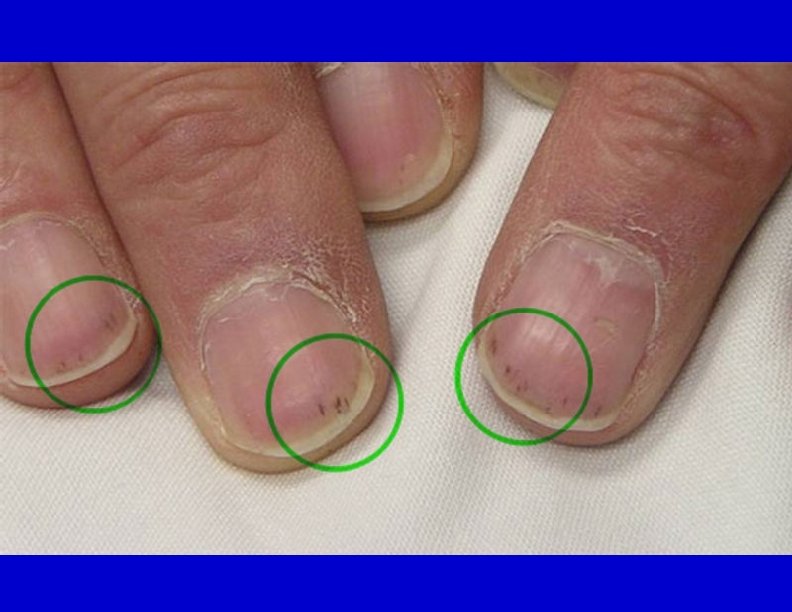
Cutaneous manifestations of internal malignancy. Acanthosis Nigricans u Dermatomyositis u Nails: Clubbing u Koilonychia u Splinter haemorrhages u When to do HIV testing for skin Disease ? PDF created with Fine. Print pdf. Factory trial version http: //www. fineprint. com
SLE: Facial Photosensitivity u Butterfly Erythema u Multisystem disease (Renal, CNS, Cardic, Blood, etc…. ) u Positive ANA and anti-ds DNA u PDF created with Fine. Print pdf. Factory trial version http: //www. fineprint. com
Discoid Lupus (DLE) u Usually ANA Negative u round scarring lesions on light exposed areas u No Systemic involvement PDF created with Fine. Print pdf. Factory trial version http: //www. fineprint. com
Subacute Cutaneous Lupus (SCLE) papulosqamous or annular presentation u Photosensitivity u Dose not cause scarring u Usually ANA negative but anti Ro positive. u Mild systemic involvement u PDF created with Fine. Print pdf. Factory trial version http: //www. fineprint. com
Neonatal Lupus: u. Appears in the first month in a photo-distribution u. Patterns : Papulosqamous and annular u. Congenital heart block (complete & permanent) usually needs pacemaker u anti Ro positive PDF created with Fine. Print pdf. Factory trial version http: //www. fineprint. com
Drug - Induced Lupus: u Cause: v Procainamide v Hydralazine v Others u Antihistone positive PDF created with Fine. Print pdf. Factory trial version http: //www. fineprint. com
Dermatomyositis (Skin Rash + Muscle Weakness): Heliotrope : Violeceous color over the upper eyelids u Gottoron’s papules: Flat- topped violaceous papules over knuckles of hands u Calcifications especially in kids u Bilateral proximal muscle weakness (with high CPK, Positive EMG and Muscle biopsy) u In adults (especially over 50 yrs) associated with internal malignancy (e. g. GI, Prostrate, ovary & breast) u PDF created with Fine. Print pdf. Factory trial version http: //www. fineprint. com
Scleroderma (Systemic Sclerosis) u u u Thickened & tight skin Sclerodactyl Face: loss of forehead lines beaked nose, small mouth, radial furrowing around the mouth) Telangectasia and calcification Systemic involvement: v Lung, GI, Kidneys v Positive anti scl-70 PDF created with Fine. Print pdf. Factory trial version http: //www. fineprint. com
CREST (milder variant of scleroderma) C=Calcification u R =Raynand’s u E =Eosopheagal dysfunction u S= Sclerodactasia u T=Telangectasia u Positive anti- centromere u Less systemic involvement. u PDF created with Fine. Print pdf. Factory trial version http: //www. fineprint. com
Morphea: u Localized scleroderma without systemic involvement. v Firm, white patch of skin surrounded by violaceous ring En coup de sabre u Linear scleroderma on the scalp and face which may give scarring alopecia + it may affect muscle or even bone PDF created with Fine. Print pdf. Factory trial version http: //www. fineprint. com
Antibody testing for CTD (table) Antibody Clinical significance ANA Screening for SLE and other CTD Anti-Centromere Marker for CREST Anti-histone Marker for Drug-Induced Lupus Anti-Smith Specific for SLE Anti - RNP For MCTD Anti - Ro Neonatal Lupus, SCLE Scl - 70 antibody For Scleroderma Anti ds- DNA For SLE PDF created with Fine. Print pdf. Factory trial version http: //www. fineprint. com
Skin and endocrine system Diabetes mellitus. Thyroid diseases. Cushing’s syndrome. Addison’s disease.
Diabetes mellitus • • Skin tags. Acanthosis nigricans. Diabetic dermopathy. Bullous diabeticorum. Thickening of skin. Necrobiosis lipoidica diabeticorum. Bacterial and fungal infections. Perforating dermatosis.
Skin tags • small, pedunculated, soft papules on the eyelids, the neck, and the axillae. • Mostly associated with obesity and insulin resistance. • If numerous usually on top of acantohsis nigricans.
Acanthosis nigricans • hyperpigmented, velvety plaques in body folds. • Increased insulin, which binds to insulin-like growth factor receptors to stimulate the growth of keratinocytes and dermal fibroblasts. • Treatment is by weight reduction and decrease insulin resistance.
Diabetic dermopathy • Very common. • Atrophic, hyperpigmented papules and plaques on shins. • Men are affected more often than women. • Possibly related to diabetic neuropathy and vasculopathy. • No effective treatment, but it does improve with diabetic control.
Bullous diabeticorum • Rare. • Spontaneous blistering of the hands and feet. • Heals without scaring.
Thickening of skin • Thickening of the hands with tiny papules on fingers and stiff joints. • Pebbled knuckles (or Huntley papules) are multiple minute papules, grouped on the extensor side of the fingers, on the knuckles, or on the periungual surface • Generalized asymptomatic thickening of the skin (diabetic stiff skin) • Scleredema on upper back and neck.
Necrobiosis lipoidica diabeticorum • Yellow atrophic plaques on the shins. • Sometimes they ulcerate. • Histopathology shows tiered granulomatous reaction. • Treatment with topical, intralesional steroids, tacrolimus, phototherapy, cyclosporine, and rarely surgery.
Bacterial and fungal infections • Pyodermic infections such as impetigo, folliculitis, carbuncles, furunculosis, ecthyma, and erysipelas can be more severe and widespread in diabetic patients. • Erythrasma, caused by Corynebacterium minutissimum mostly on axillae and groin. • malignant otitis externa, often caused by Pseudomonas aeruginosa. • Tinea pedis and onychomycosis. • Candidal infections like perleche on corners of mouth, and on vulva. • Rare infections like mucormycosis by Phycomycetes and anaerobic cellulitis by Clostridium species.
Perforating dermatosis • Pruritic hyperkeratotic papules on the legs and trunk. • Histopathology shows transepidermal elimination of collagen and/or elastin. • Common in patients with diabetes and renal failure. • treatments include topical keratolytics, phototherapy, topical and systemic retinoids, topical and intralesional steroids, oral antihistamines, and cryotherapy.
Hyperthyroidism • Pretibial myxedema: is the most characteristic features of thyrotoxicosis appearing as shiny waxy papules and plaques having orange-skin appearance on the chin of the tibia. • Warm skin and increased sweating. • Pruritus • Premature hair graying. • Alopecia with fine soft thinned scalp hair. • Hyperpigmentation or vitiligo. • Brittle nails.
Hypothyroidism • Cold, pale and dry skin. • Pruritus. • A yellowish hue to the skin due to carotenaemia. • Slow growing ridged and brittle nails. • Delayed wound healing.
Cushing’s syndrome • caused by prolonged exposure to high levels of plasma glucocorticoid. • Adrenocortical hyperplasia. • Benign or malignant adrenal tumours. • Ectopic ACTH syndrome – secretion of ACTH by malignant or benign tumours arising in structures other than the pituitary or adrenal glands. • Exogenous steroid administration • Acne and hirsutism. • Clitromegaly and male pattern alopecia. • Striae. • Easy bruising and purpura. • Moon face and buffalo hump with fat redistribution. • Telangectasia on face. • Poor wound healing.
Addison’s disease: Hyperpigmentation: at Sun exposed skin, sites of trauma, axillae, palmar creases, old scars , nevi and mucous membranes Adrenocortical hypofunction. Diffuse pigmentation on skin and mucous membranes. Melanocytes stimulation by ACTH PDF created with Fine. Print pdf. Factory trial version http: //www. fineprint. com
Gastrointestinal disease • • Dermatitis herpetiformis. Acrodermatits enteropathica. Pyoderma gangrenosum. Peutz Jeghers syndrome. Porphyria cutanea tarda. Hemochromatosis. Liver cirrhosis.
Dermatitis herpetiformis • Small severely pruritic vesicular lesions found in a symmetric distribution of both upper and lower extensor surfaces, buttocks and the scalp. • direct immunofluorescence finding is granular deposition of Ig. A within the dermal papillae. • celiac disease (also known as gluten-sensitive enteropathy and celiac sprue) are caused by the inability to absorb gluten from the diet. • Treatment: gluten-free diet and dapsone.
Acrodermatits enteropathica • a rare autosomal recessive disorder that impairs dietary zinc absorption in the jejunum and ileum. • presents in infants several weeks after breastfeeding is discontinued. • characterized by diarrhea, inflammatory rash, and hair loss. • scaly, erythematous patches and plaques similar to atopic dermatitis, but progress to vesicles, crusts, erosions, and pustules on acral areas, perioral and perianal areas. • Treatment by zinc supplementation for life.
Peutz Jeghers syndrome • autosomal dominant disorder. • mucocutaneous hyperpigmentation together with GI polyposis. • The skin findings first appear in infancy or early childhood and involve brown macules on the lips and buccal mucosa. • multiple hamartomatous polyps occurring most commonly in the jejunum. • 2 -3% of patients develop GI carcinoma during their lifetimes.
Pyoderma gangrenosum • a painful, ulcerative lesion with a well-defined, undermined violaceous border. • start as small pustules, which subsequently burst and expand to form the larger noninfectious ulcer. • Positive pathergy test. • Mostly associated with ulcerative colitis. Also with Crohn’s disease, rheumatoid arthritis, and leukemia. • Surgery is contraindicated.
Porphyria cutanea tarda • most common porphyria occurring in adults. • skin photosensitivity with increased skin fragility, facial hypertrichosis, blisters, scarring with milia formation, and skin hyperpigmentation on the hands and other sun-exposed areas. • results from the decreased activity of the enzyme uroporphyrinogen decarboxylase. • Associated with Hep C virus. • Treatment by removal of possible triggers, including iron supplementation, alcohol, and estrogens. Also by phlebotomy and hydroxycholorquine.
Hemochromatosis • a disorder of iron overload leading to excess deposition in multiple body organs. • metallic gray or bronze-brown color that is generally diffuse. • skin atrophy, ichthyosis, partial hair loss (most often in the pubic region), and koilonychia. • cirrhosis may develop, and might lead to hepatocellular carcinoma. • treatment involves phlebotomy and chelating agents.
Renal diseases • Xerosis occurs in 50 -92% of the • Pruritus affects 15 -49% of dialysis population. patients with chronic renal failure and 50 -90% of the • Some patients may develop dialysis population. acquired ichthyosis. • Uremia is the most common • the exact cause of xerosis in metabolic cause of pruritus. ESRD remains unknown. • Cutaneous manifestations of • Many patients respond to pruritus include excoriations, routine use of emollients. prurigo nodularis, and lichen simplex chronicus. • Pruritus typically resolves after transplantation. • Treatment include sedating antihistamines, emollients, phototherapy, thalidomide, and gabapentin.
Renal diseases • Half and half nails occur in around 40% of patients on dialysis. • Kidney transplant usually resolve this sign. • Usually involve fingernails.
Renal diseases • Nephrogenic systemic fibrosis mostly seen in ESRD and dialysis patients. • Presents as thick, indurated plaques on the extremities and the trunk similar to scleroderma. • gadolinium might have a role in the pathogenesis of this condition. • Treatment includes immunosuppressive agents, phototherapy, topical steroids, retinoids, and photophoresis.
hyperlipidemia • Xanthelasma palpebrarum is the most common of the xanthomas. • asymptomatic and usually bilateral and symmetric. • Can be associated with any type of primary hyperlipoproteinemia. And could be without hyperlipidemia. • often treated with topical trichloroacetic acid, electrodesiccation, laser therapy, and surgical excision.
Hyperlipidemia • Tendinous xanthomas commonly seen on the Achilles tendon followed by the hands, feet, elbows, and knees. • The least responsive xanthoma to treatment. • Mostly seen in patients with familial hypercholesterolemia.
hyperlipidemia • Tuberous xanthomas are firm and nontender cutaneous and subcutaneous yellowish nodules on extensor surfaces. • Mostly associated with familial dysbetalipoproteinemia. • May resolve after months of treatment with lipid lowering agents.
Hyperlipidemia • Eruptive xanthomas are painless, yellowish papules on an erythematous base that present as grouped lesions on trunk, elbows and buttocks. • Usually associated with hypertriglyceridemia. • Could be seen in poorly controlled diabetes and acute pancreatitis. • Usually resolve in few weeks after therapy.
Hyperlipidemia • Planar xanthomas are elevated cutaneous yellowish-orange deposits on palmar creases. • Usually associated with familial dysbetalipoprotei nemia.
Types of Xanthomas Eruptive: small papules appear in crops over buttocks & extensors u Tendinous: Nodules over tendons e. g. extensor tendons of hands & feet and Achilles tendon. u Palmar crease xanthoma: on palms u Tuberous: Papules & nodules over knees and elbows u Xanthelasma: Bilateral symmetrical over both eyelids. u PDF created with Fine. Print pdf. Factory trial version http: //www. fineprint. com
Neurocutaneous Disorders Neurofibromatosis: u u u u Autosomal dominant Café-au-lait macules(light brown) Neurofibromas(soft pink or skin- colored papules and nodules) Axillary freckling Optic glioma Lisch nodules (iris hamartoma, seen by slit-lamp examination) Associated with Neurological complications e. g. tumors, seizures and mental retardation. PDF created with Fine. Print pdf. Factory trial version http: //www. fineprint. com
Tuberous Sclerosis (Epiloia) : v Epi = Epilepsy v Loi = Low intellegence v a = adenoma sebaceoum Skin features 1. Adenoma sebaceoum (anigofibroma): red papules around the nose and on chin 2. Ash-leaf hypopigmention: oval area of hypopigmention This is the earliest sign of TS 3. Periungal fibroma: multiple papules & nodules around the nail 4. Shagreen patch: skin colored plaque on the trunk with “orangepeel” surface PDF created with Fine. Print pdf. Factory trial version http: //www. fineprint. com
PDF created with Fine. Print pdf. Factory trial version http: //www. fineprint. com
PDF created with Fine. Print pdf. Factory trial version http: //www. fineprint. com
Behcet’s Syndrome: Oral ulcer (the most common) u Genital ulcers (mainly scrotal) u Iritis and arthoropathy u May have CNS involvement u Scurvy : Vitamin C deficiency u Bleeding gums u Can cause teeth loss (permanent complication) u PDF created with Fine. Print pdf. Factory trial version http: //www. fineprint. com
Scurvy(cont’d) u u Easy bruishing Diagnosis : Low ascorbic acid (Vit-C) level in Leukocyte Pellagra: u u Nictonic acid deficiency 4 “D”s Dermatitis (Photodermatitis) Diarrhea Dementia Death (if not treated) PDF created with Fine. Print pdf. Factory trial version http: //www. fineprint. com
PDF created with Fine. Print pdf. Factory trial version http: //www. fineprint. com
Pellagra PDF created with Fine. Print pdf. Factory trial version http: //www. fineprint. com
Causes of generalized pruritus without skin lesions: 1. 2. 3. 4. 5. Endocrine: DM, hypo& hyperthyroidsm Haematological: polycythemia rubra vera, iron def anemia Malignancy; e. g. Lymphoma Hepatic: primary biliary cirrhosis Renal: CRF The commonest manifestation of CRF is pruritus 6. 7. Neurological : e. g. Tabes dorialis Others: v Psychognic v Drugs v Idiopthaic PDF created with Fine. Print pdf. Factory trial version http: //www. fineprint. com
Nails Clubbing : v Exaggeration of the normal nail curve associated with loss of the normal angle between nail and posterior nail fold v Causes: 1. 2. 3. 4. 5. Thoracic: Lung abscess, Lung CA CVS: Congenital cyanotic heart disease GIT: GI carcinoma, Inflammatory bowel disease Endocrine: Thyroid disease Idiopathic: PDF created with Fine. Print pdf. Factory trial version http: //www. fineprint. com
PDF created with Fine. Print pdf. Factory trial version http: //www. fineprint. com
Splinter Haemorrhages : u Causes : 1. Bacterial endocarditis 2. Septic emboli 3. CTD 4. Trauma 5. Idiopathic PDF created with Fine. Print pdf. Factory trial version http: //www. fineprint. com
Koilonychia : u u Spoon- shaped appearance Causes: 1. Iron deficiency anemia 2. Thyroid disease 3. Physological; early childhood 4. Dermatoses: Lichen planus Alopecia Areata and others PDF created with Fine. Print pdf. Factory trial version http: //www. fineprint. com
Purpura and Vasculitis
Objectives • To differentiate between different types of purpura. • To know the difference between inflammatory and non inflammatory purpura. • To have an approach to diagnose purpuric lesions. • To be familiar with serious and non serious conditions and when to refer to a specialist.
Purpura • Reddish-purplish skin lesions from extravasation of RBCs into the skin. • Non palpable purpura: classified according to size to Petechiae less than 2 mm, and Ecchymosis or bruises for larger lesions. • Palpable purpura is the inflammatory type of different sizes, the main sign of vasculitis.
Purpura Causes Platelet disease Coagulation defect Blood vessel wall pathology
Causes 1 -Platelet Disorders Thrombocytopenia Platelet Dysfunction 2 -Coagulation Factor Deficiency Congenital Factor VIII Deficiency Factor IX Deficiency Von Willebrands disease Acquired Disseminated Intravascular Coagulopathy Liver disease Uremia Vitamin K deficincy 3 -Vascular Factors Congenital Hereditary Hemorrhagic Telangectasia Ehlers-Danlos Syndrome (Type IV) Acquired: Inflammation(Vasculitis) Trauma Vitamin c deficiency (scurvy)
vasculitis
Definition A clinicopathologic process characterized by inflammatory destruction of blood vessels that results in occlusion or destruction of the vessel and ischemia of the tissues supplied by that vessel.
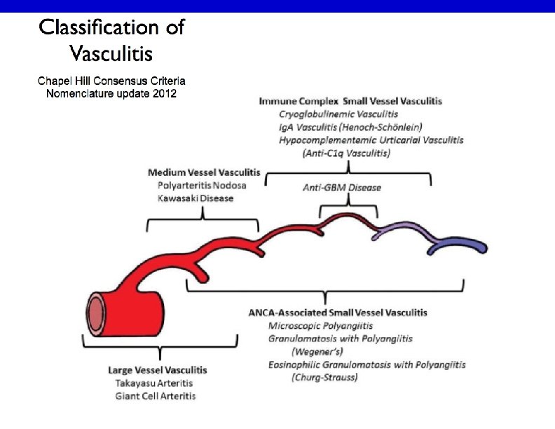
Classification -Large-vessel vasculitis Aorta and the great vessels (subclavian, carotid) Claudication, blindness, stroke -Medium-vessel vasculitis Arteries with muscular wall Mononeuritis multiplex (wrist/foot drop), mesenteric ischemia, cutaneous ulcers -Small-vessel vasculitis Capillaries, arterioles, venules Palpable purpura, glomerulonephritis, pulmonary hemorrhage
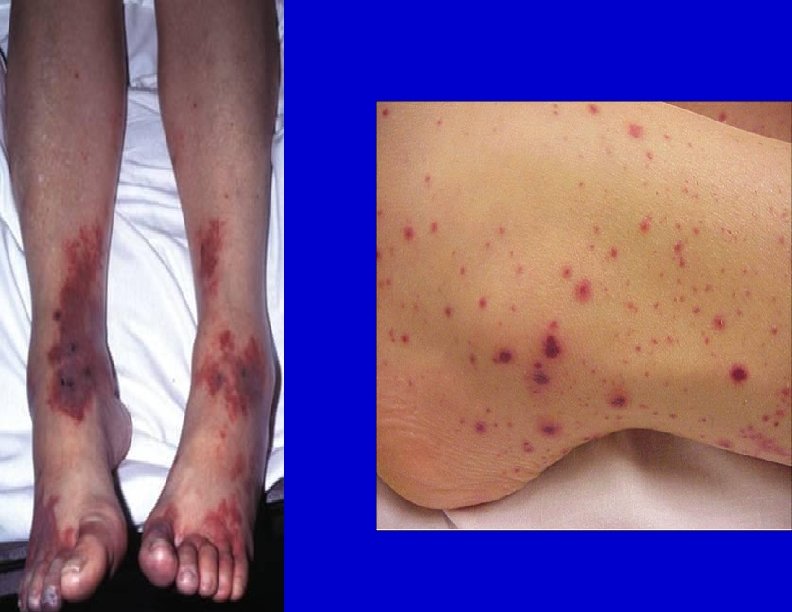
Cutaneous small-vessel vasculitis (CSVV) • Also called Leukocytoclastic vasculitis. • characterized by neutrophilic infiltration into the peripheral small dermal blood vessels. • Purpura, urticaria, erythema-multiforme-like erythema, papules, nodules, pustules, blistering, erosion and ulceration occur, mainly in the lower extremities. • An immune complex of an antigen (e. g. , bacterium, virus, drug) and the antibody against that antigen deposit on the arteriolovenular walls. These activate the immune system and cause vasculitis (type III allergic reaction). Penicillin, sulfa drugs, thiazides, NSAIDs and other drugs, chemical substances, hemolytic streptococcus bacteria, or viruses may be the foreign antigen. Collagen diseases and antibodies against malignant tumors can also be causes.
Cutaneous small-vessel vasculitis (CSVV)
Cutaneous small vessel vasculitis Pathogenesis: -Many forms of small-vessel vasculitis are felt to be caused by circulating immune complexes -These lodge in vessel walls and activate compliment
Cutaneous small vessel vasculitis
Pathology In the upper and middle dermal layer, fragments of nuclear debris and leakage of erythrocytes are found in the peripheral arteriola. Neutrophilic infiltration occurs in the arteriolovenous small blood vessels and capillaries. Thickening of the blood vessel walls and fibrinoid necrosis.
Investigations • • • CBC, urea, creatinine. ESR ( usually raised) Complements (decreased) Urinalysis ( for protein and hematuria if kidneys involved) Occult blood in stool ANA, ANCA Cryoglobulins , hepatitis B, C, HIV. CRP, ASO titer and throat swab culture for streptococcal infection Skin biopsy
Treatment -treatment of cause. -Symptomatic treatment (if skin is only involved): rest , NSAIDS , Antihistamine -severe visceral involvement may require high doses of corticosteroids with or without an immunosuppressive agent -Immunosuppressive agents for rapidly progressive course and severe systemic involvement
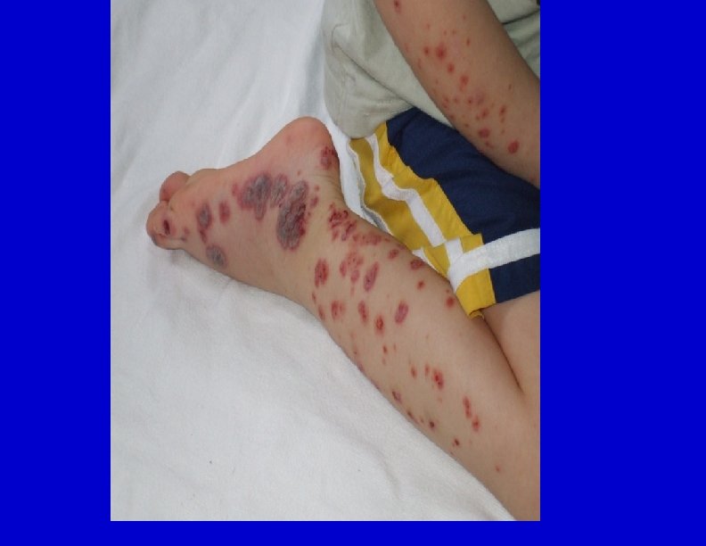
Henoch-Schönlein purpura (HSP) • In children, the onset is mostly after upper respiratory infection; association with hemolytic streptococcus has been pointed out. • Drugs (penicillin, aspirin) • These antigens combine with antibodies (mainly Ig. A) in the body, and the immunocomplex deposits on the vascular walls. Immunoreaction is induced to cause vasculitis and purpura. • Pathology shows leukocytoclastic vasculitis with Ig. A deposition is observed by direct immunofluorescence. • The histology of the kidney in HSP patients often shows crescentic glomerulonephritis.
Henoch-Schönlein purpura (HSP) • Treatment by bed rest, pain relief and antibiotics if strep throat infection is present. • Systemic steroids for severe cases with systemic involvement. • HSP generally has a good prognosis and resolves within several weeks in most cases; however, it may recur. • Serious complications may occur in other organs, such as nephritis with Ig. A deposition in the mesangium area, enterorrhagia, intussusception, intestinal perforation, or cerebral hemorrhage. • Adults may have prolonged kidney involvement.
Henoch-Schönlein purpura (HSP)
-The long-term prognosis in children with gross hematuria is very good; however, progressive glomerular disease and renal failure may develop in a small percentage -Ig. A, C 3 and fibrin depositions have been demonstrated in biopsies of both involved and uninvolved skin by immunofluorescence techniques
Urticarial vasculitis • Presents with urticarial weals that lasts more than 24 hours unlike urticaria. And usually leave hyperpigmentation after resolution. • Could be associated with fever, arthralgia, abdominal pain and angioedema especially in hypocomplementaemic urticarial vasculitis that is associated with SLE. • Causes include connective tissue diseases, viral and bacterial infections, drugs like ACE inhibitors, penicillin, sulfonamides, fluoxetine and thiazides. Also leukemia could cause it. • Most cases are idiopathic and improve spontaneously after few months. • Treatment is to remove the offending agents if present, treat the underlying diseases. Symptomatic therapy with antihistamines and NSAIDs. • Dapsone, colchicine, hydroxycholorquine, steroids, azathioprine, and cyclosporine.
Urticarial vasculitis
Cutaneous polyarteritis nodosa • a rare form of vasculitis which involves small and medium sized arteries of dermis and subcutaneous tissue. • tender subcutaneous nodules and livedo reticularis that may ulcerate on legs and feet. • Pathogenesis is unknown. • Systemic involvement is uncommon except for peripheral neuropathy, and progression to classical polyarteritis nodosa is an exception. • may have neuromuscular involvement in the form of peripheral neuropathy that presents with tingling, numbness, sensory disturbances, weakness, and absent reflexes.
Cutaneous polyarteritis nodosa • In cutaneous PAN, histopathological examination shows features of nodular arteritis with polymorphonuclear infiltrates involving medium sized arteries in deep reticular dermis. There is extensive fibrinoid necrosis. This is in contrast to classical PAN which rarely shows nodular arteritis and the picture is of small vessel leukocytoclastic. • Cutaneous PAN runs a chronic course lasting months to years, and has a waxing and waning phenomenon. Patients are generally treated with non-steroidal anti-inflammatory drugs and oral steroids. Immunosuppressive drugs can also be used in low doses in more severe kinds of cutaneous PAN and as steroid-sparing drugs.
Cutaneous polyarteritis nodosa
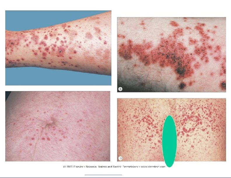
pigmented purpuric dermatoses • a group of chronic diseases of mostly unknown etiology characterized by extravasation of erythrocytes in the skin with marked hemosiderin deposition. • Mostly affect males. • Venous hypertension, exercise, and gravitational dependency are important cofactors that appear to influence disease presentation. • Histologically, a perivascular T-cell lymphocytic infiltrate is centered on the superficial small blood vessels of the skin, which show signs of endothelial cell swelling and narrowing of the lumen. Extravasation of red blood cells with marked hemosiderin deposition in macrophages is also found but no vasculitis. • The lesions are chronic and persist for years. With time, many of the lesions tend to extend and may become darker brown in color. • No effective treatment available. It is a cosmetic problem mostly.
pigmented purpuric dermatoses