
Cutaneous manifestation of SLE and other CTD Diseases












- Slides: 66
Cutaneous manifestation of SLE and other CTD Diseases By: Dr. Eman Almukhadeb Consultant dermatologist, Assistant professor
Objectives At the conclusion of this lectures the student should be able to: • Differentiate between the various types of Lupus • Recognize how Lupus affects the various systems of the body • Identify all of the current treatment options available for Lupus
Lupus Erythematosus (LE) �LE is an autoimmune diseases associated with antibodies directed against components of cell nuclei. � Is a multisystem disorder that may affect any tissue, kidneys, CNS, lungs and others. It might affects only skin without systemic involvement �It is an immune complex disease that target collagen or ground substance.
Lupus Erythematosus( LE) �Classification : LE classified as 1. Cutaneous 2. Systemic
Classification of cutaneous lupus erythematous �Acute �Subacute �Chronic
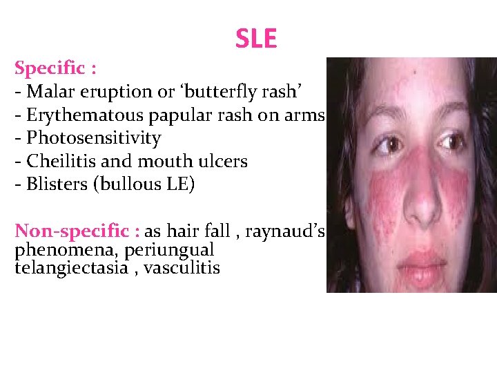
Cutaneous classification 1 - Acute LE : Specific : - Malar eruption or ‘butterfly rash’ - Erythematous papular rash on arms - Photosensitivity - Cheilitis and mouth ulcers - Blisters (bullous LE) Non-specific : as hair fall , raynaud’s phenomena, periungual telangiectasia , vasculitis
Cutaneous classification 2 - Subacute cutaneous LE ( SCLE ): �Annular �Papulosquamous �Associated with syndromes : - neonatal LE - complement deficiency syndrome - drug induced SCLE
Cutaneous classification 3 - chronic : � Discoid LE � Verrucous ( hypertrophic ) LE � Lupus erythematous –Lichen planus overlap � Chilblain LE � Tumid LE � Lupus panniculitis
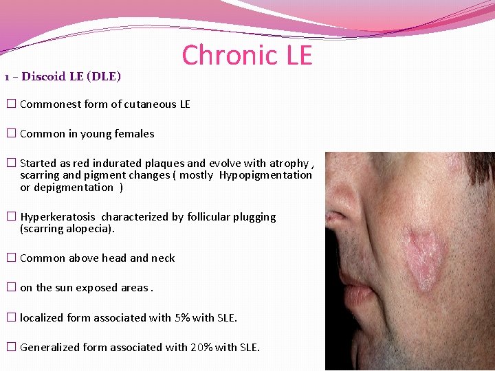
1 – Discoid LE (DLE) Chronic LE � Commonest form of cutaneous LE � Common in young females � Started as red indurated plaques and evolve with atrophy , scarring and pigment changes ( mostly Hypopigmentation or depigmentation ) � Hyperkeratosis characterized by follicular plugging (scarring alopecia). � Common above head and neck � on the sun exposed areas. � localized form associated with 5% with SLE. � Generalized form associated with 20% with SLE.
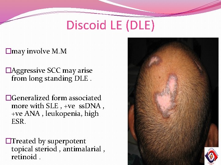
Discoid LE (DLE) �may involve M. M �Aggressive SCC may arise from long standing DLE. �Generalized form associated more with SLE , +ve ss. DNA , +ve ANA , leukopenia, high ESR. �Treated by superpotent topical steriod , antimalarial , retinoid.
Discoid Lupus Erythematosus
Discoid Lupus Erythematosus
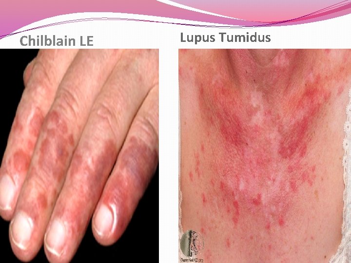
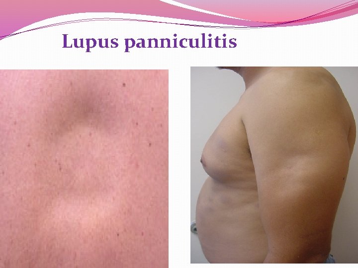
Other chronic cutaneous LE 2 - Hypertrophic LE (Verrucous ): commonly seen on the extremities. 3 - LE-LP : common on the extremities , palm and soles associated with oral ulcers and nail involvement. 4 - Chilblain LE ( pernio): presents as tender red or purple skin lesion that involve fingertips , rim of the ears, calves and heals , exacerbated by cold exposure 5 - Tumid LE : presented with edematous erythematous plaques on the trunk and respond to antimalarial. 6 - LE panniculitis : presented as non-tender subcutaneous nodules on face or proximal extremities that heals with atrophy
Chilblain LE Lupus Tumidus
Lupus panniculitis
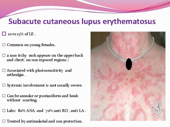
Subacute cutaneous lupus erythematosus � 10 to 15% of LE. � Common on young females. � a non-itchy rash appears on the upper back and chest( on sun exposed regions ) � Associated with photosensitivity and arthralgia. � Systemic involvement is not usually severe. � Can be annular or psoriasiform and heals without scarring. � Labs: 80% ANA and 70% anti-RO , anti-LA. � Treated by antimalarial and sun protection.
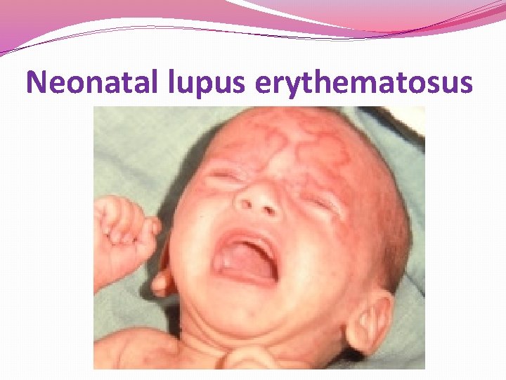
Neonatal lupus erythematosus �Newborn babies born to mothers with subacute LE may develop annular rash, known as neonatal LE that resolve spontaneously. � The neonates could be at risk of complete heart block, thrombocytopenia , increased liver enzymes
Neonatal lupus erythematosus
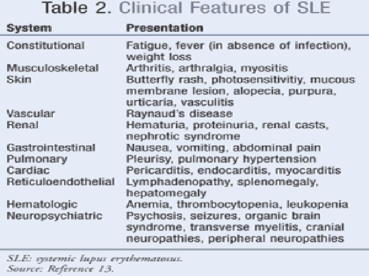
SLE �Only a few patients with cutaneous LE also have SLE. The most common presentation is with a malar eruption or butterfly. Other skin changes in SLE are photosensitivity, mouth ulcers, and diffuse hair loss. �SLE may also affect joints, kidneys, lungs, heart, liver, brain, blood vessels and blood cells
SLE �ACR criteria for the diagnosis : 4 out of 11 need for the diagnosis. 1. Malar rash. 2. DLE. 3. Photosensitivity. 4. Oral ulcers. 5. Arthritis 6. Serositis ( pleuritis , pericarditis )
SLE 7 - renal : proteinuria more than 0. 5 g/day 8 - CNS : Seizure or psychosis 9 - blood : hemolytic anemia, leukopenia, thrombocytopenia 10 - +VE ANA 11 - +VE anti-ds DNA and anti-smith and antiphospholipid antibodies.
�Drug induced Lupus : �associated with ANA and anti-histone antibodies. �does not usually affect the skin. �hydralazine, procainamide, sulfonamide, anticonvulsants, Minocycline.
SLE Specific : - Malar eruption or ‘butterfly rash’ - Erythematous papular rash on arms - Photosensitivity - Cheilitis and mouth ulcers - Blisters (bullous LE) Non-specific : as hair fall , raynaud’s phenomena, periungual telangiectasia , vasculitis
Lab findings � CBC : anemia , Leucopenia , thrombocytopenia � High ESR � Coombs test : hemolytic anemia � Urinalysis and 24 hr urine collection : for proteinuria � U &E. � Serology : ANA (+ve in 95% of SLE patients, screening test ) , anti-ds DNA (specific ), anti-ss DNA , anti smith (specific ) , anti-RO , anti-LA , lupus anticoagulant and anti cardiolipin , anti-n RNP. � low C 2 and C 4. � Skin biopsy may be diagnostic especially in DLE. Direct IF may show positive antibody deposition along the basement membrane (lupus band test).
Treatment of cutaneous lupus �The aim of treatment for cutaneous LE is to alleviate symptoms and to prevent scarring. Local therapy : �Sun protection. �Topical or intralesional steroid �Topical calcineurin inhibitor
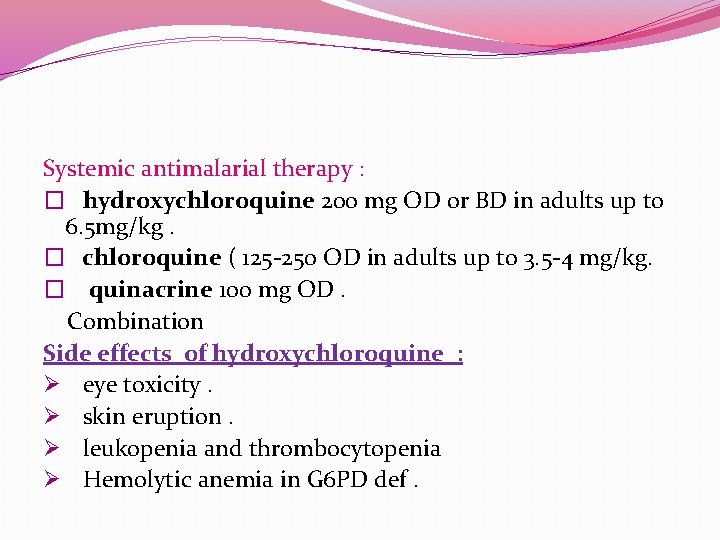
Systemic antimalarial therapy : � hydroxychloroquine 200 mg OD or BD in adults up to 6. 5 mg/kg. � chloroquine ( 125 -250 OD in adults up to 3. 5 -4 mg/kg. � quinacrine 100 mg OD. Combination Side effects of hydroxychloroquine : Ø eye toxicity. Ø skin eruption. Ø leukopenia and thrombocytopenia Ø Hemolytic anemia in G 6 PD def.
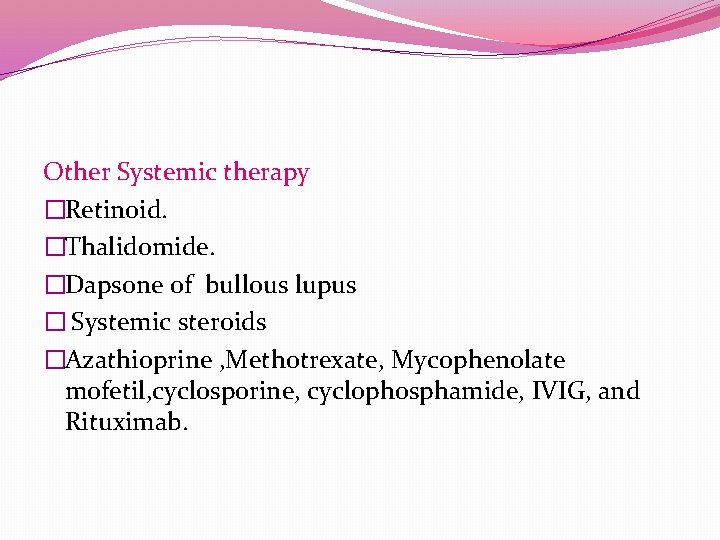
Other Systemic therapy �Retinoid. �Thalidomide. �Dapsone of bullous lupus � Systemic steroids �Azathioprine , Methotrexate, Mycophenolate mofetil, cyclosporine, cyclophosphamide, IVIG, and Rituximab.
Objectives • To learn how to diagnose , investigate and manage dermatomyositis. • To learn the presentation of morphea and systemic sclerosis and ways to manage them.
Dermatomyositis • An uncommon inflammatory disease affects adults between 40 -60 (females mainly) and children 5 -15. • Skin changes. • Muscle weakness
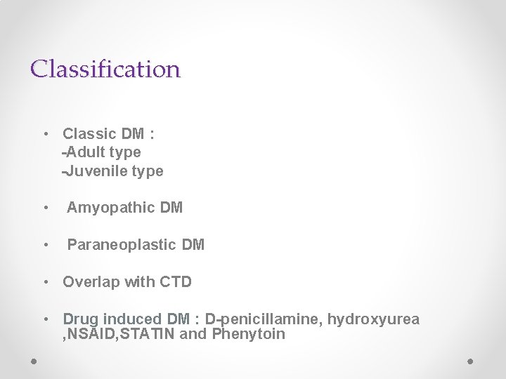
Classification • Classic DM : -Adult type -Juvenile type • Amyopathic DM • Paraneoplastic DM • Overlap with CTD • Drug induced DM : D-penicillamine, hydroxyurea , NSAID, STATIN and Phenytoin
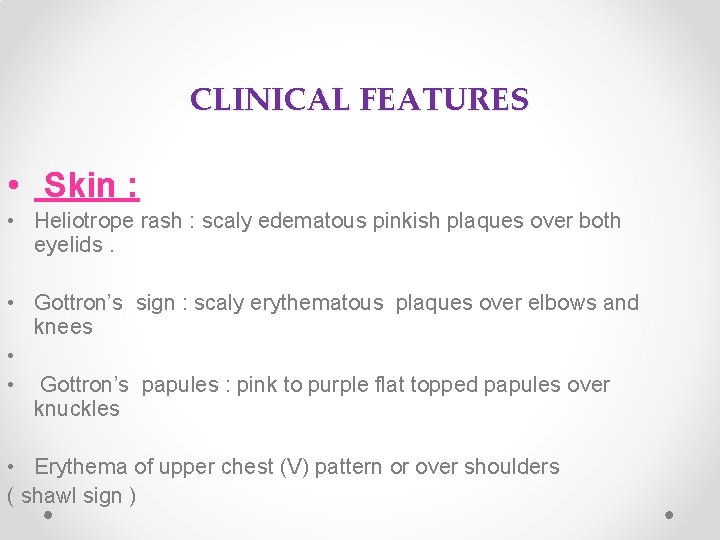
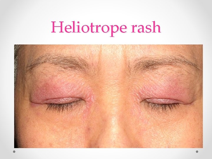
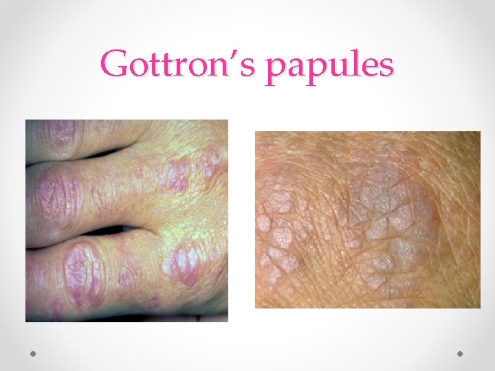
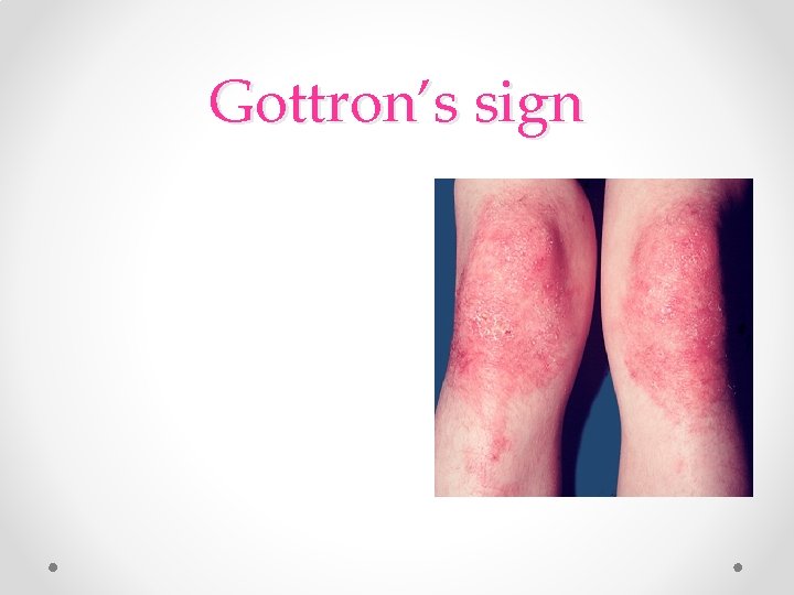
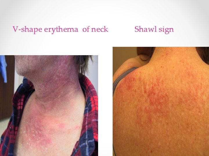
CLINICAL FEATURES • Skin : • Heliotrope rash : scaly edematous pinkish plaques over both eyelids. • Gottron’s sign : scaly erythematous plaques over elbows and knees • • Gottron’s papules : pink to purple flat topped papules over knuckles • Erythema of upper chest (V) pattern or over shoulders ( shawl sign )
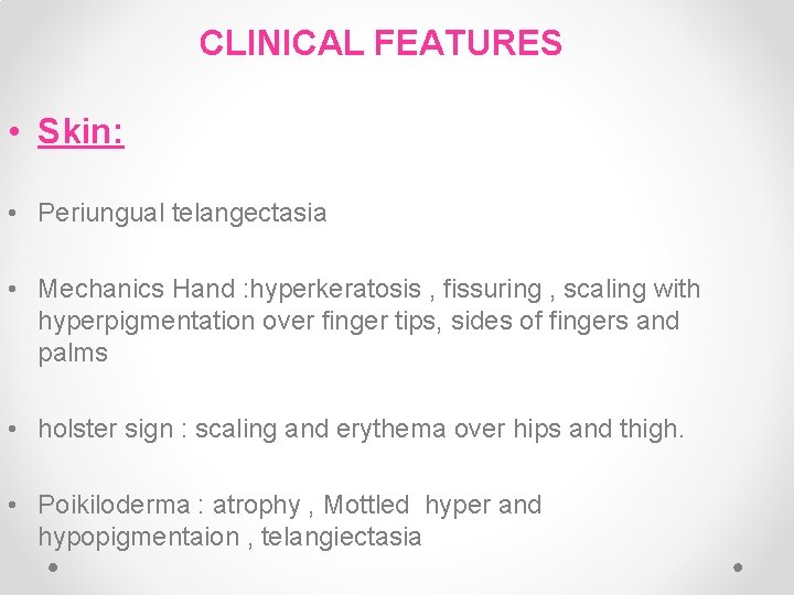
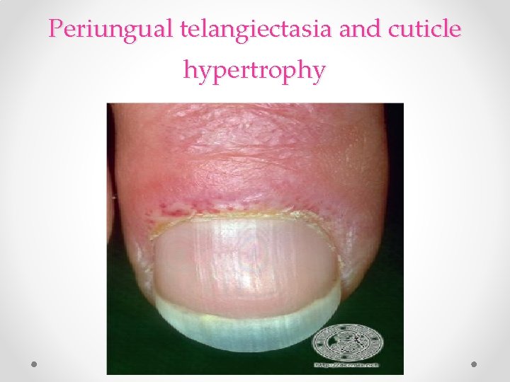
CLINICAL FEATURES • Skin: • Periungual telangectasia • Mechanics Hand : hyperkeratosis , fissuring , scaling with hyperpigmentation over finger tips, sides of fingers and palms • holster sign : scaling and erythema over hips and thigh. • Poikiloderma : atrophy , Mottled hyper and hypopigmentaion , telangiectasia
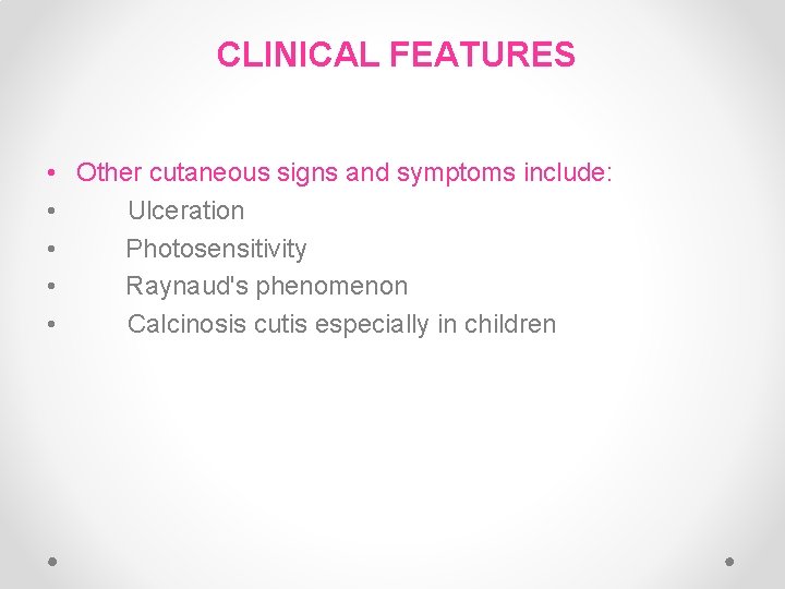
CLINICAL FEATURES • Other cutaneous signs and symptoms include: • Ulceration • Photosensitivity • Raynaud's phenomenon • Calcinosis cutis especially in children
Heliotrope rash
Gottron’s papules
Gottron’s papules
Gottron’s sign
V-shape erythema of neck Shawl sign
Periungual telangiectasia and cuticle hypertrophy
Clinical features : • Muscle Weakness, progressive proximal muscle weakness involves the hips, thighs, shoulders, upper arms and neck. The weakness is symmetrical and more in the extensor muscles. • Lungs : interstitial lung disease
Associated disease • It can be associated with: Other CTD: Such as lupus, rheumatoid arthritis, scleroderma and Sjogren's syndrome. Cancer: o Especially in older patients. o common : ovarian CA o Cancer of the cervix, lungs, pancreas, breasts, lymphoma and gastrointestinal tract. o Malignancy could precede, coincide or follow the diagnosis of DM.
Investigations and Diagnosis CRITERIA : 1 - Typical skin rash. 2 - Muscle weakness ( proximal upper or lower extremity) 3 - +ve ms enzymes ( CK and aldolase ) 4 - +ve EMG ( short duration , polyphasic) 5 - +ve muscle biopsy Definite : rash with 3 muscle findings. OTHER LABS: Ø Serology: Anti- Jo-1 ( histidyl- t. RNA synthetase): lung disease & mechanical hands. Mi-2 : specific for DM with skin findings Ø Skin Bx Ø Magnetic resonance imaging (MRI).
Treatment SKIN : • Sunscreen • Topical steroid , Topical Tacrolimus • Hydoxychloroquine • Oral steroids are the mainstay treatment. Systemic therapy: • Steroid sparing agents are: • Methotrexate, azathioprine, mycophenolate mofetil, cyclosporine, cyclophosphamide, IVIG, and Rituximab. • Physiotherapy to improve strength and flexibility of the muscles. • Surgical excision or Co 2 laser could be utilized to remove tender calcium deposits.
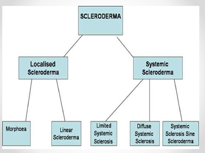
Systemic sclerosis(Scleroderma) • A group of diseases that involve the hardening and tightening of the skin and connective tissues • affects women more often than men. • most commonly occurs between the ages of 30 and 50.
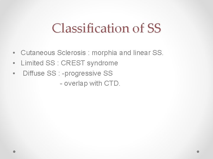
Classification of SS • Cutaneous Sclerosis : morphia and linear SS. • Limited SS : CREST syndrome • Diffuse SS : -progressive SS - overlap with CTD.
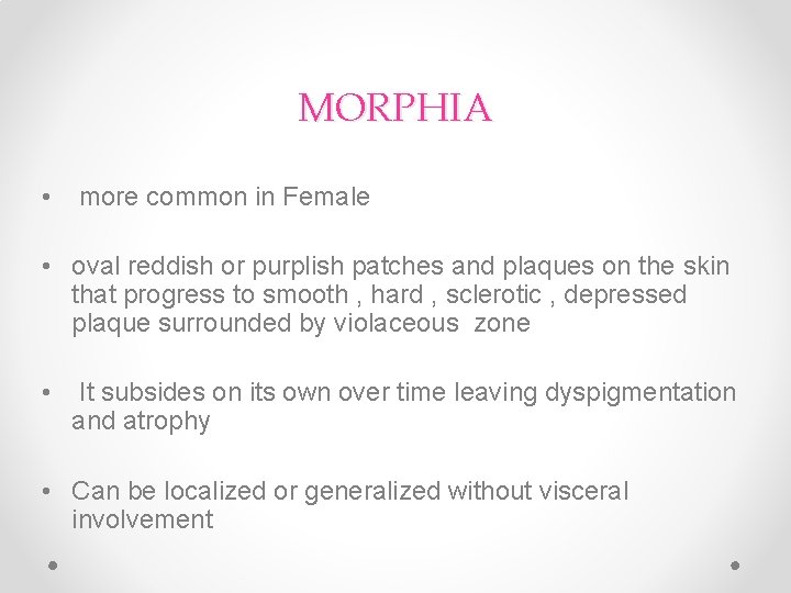
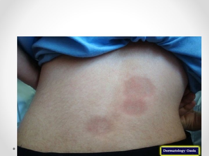
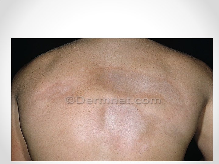
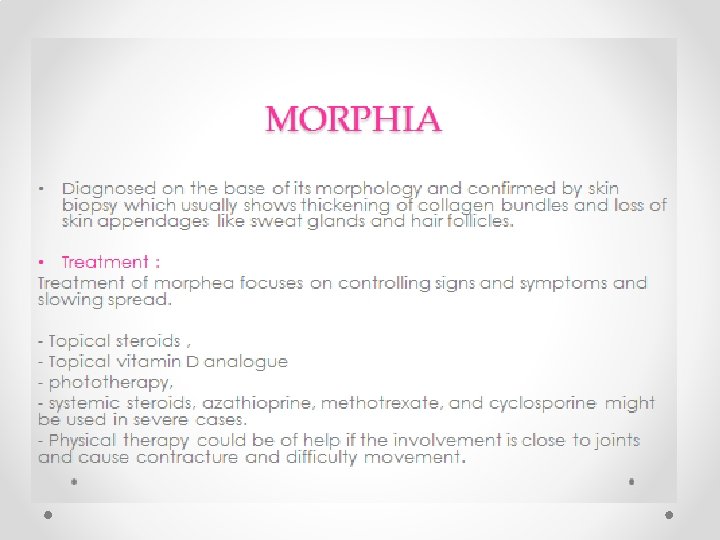
MORPHIA • more common in Female • oval reddish or purplish patches and plaques on the skin that progress to smooth , hard , sclerotic , depressed plaque surrounded by violaceous zone • It subsides on its own over time leaving dyspigmentation and atrophy • Can be localized or generalized without visceral involvement
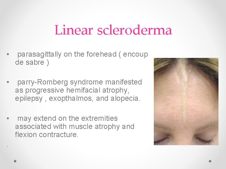
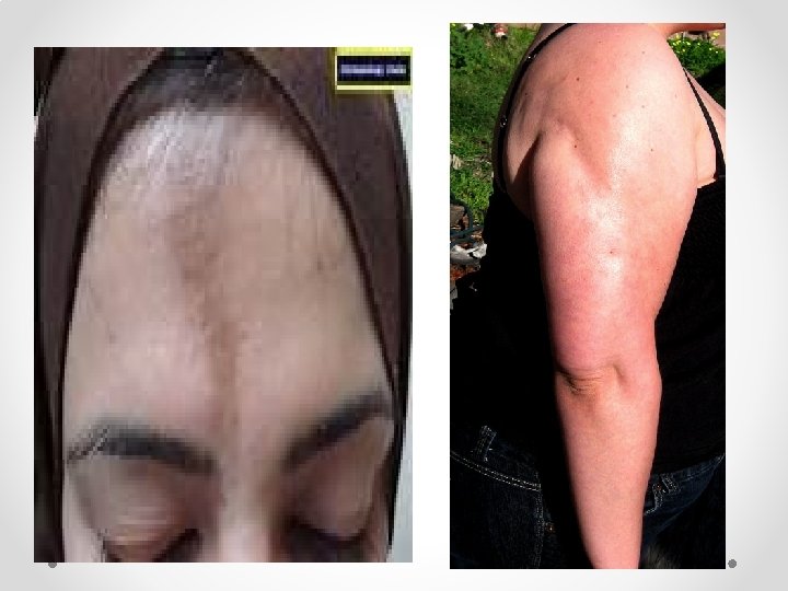
Linear scleroderma • parasagittally on the forehead ( encoup de sabre ) • parry-Romberg syndrome manifested as progressive hemifacial atrophy, epilepsy , exopthalmos, and alopecia. • may extend on the extremities associated with muscle atrophy and flexion contracture. .
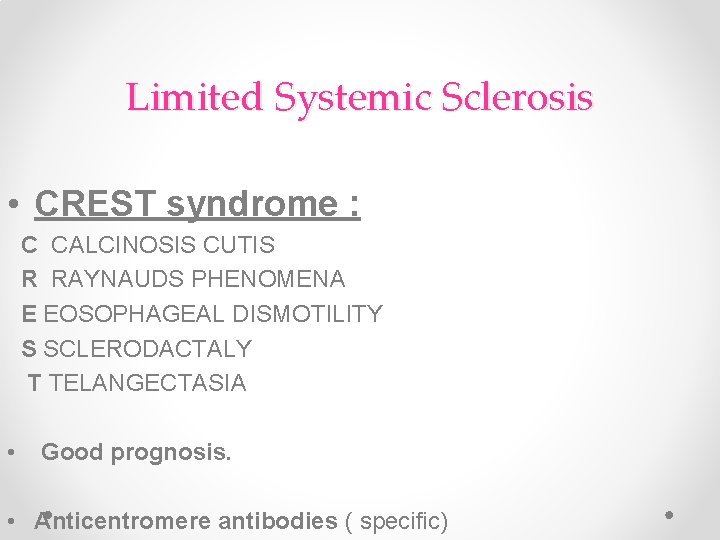
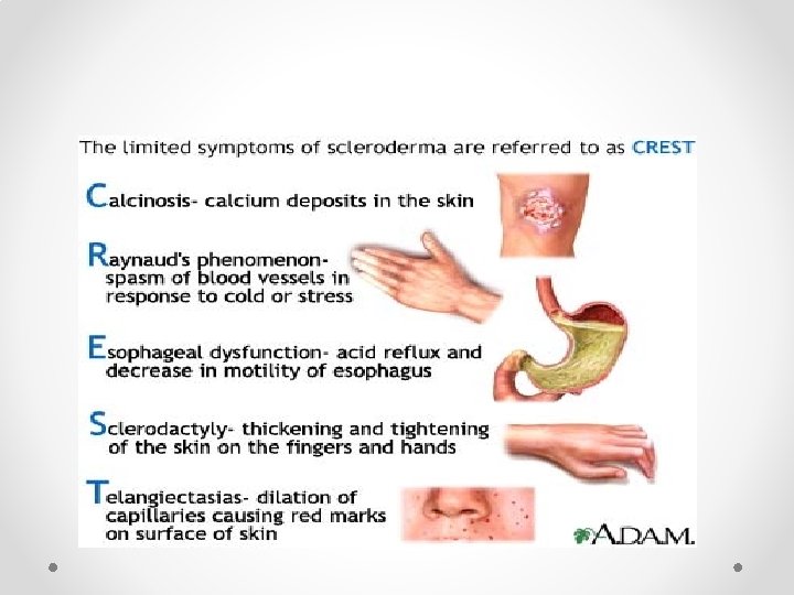
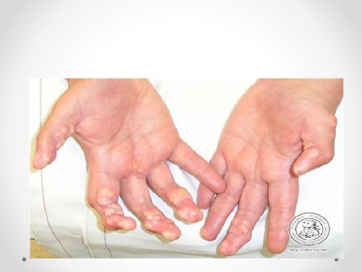
Limited Systemic Sclerosis • CREST syndrome : C CALCINOSIS CUTIS R RAYNAUDS PHENOMENA E EOSOPHAGEAL DISMOTILITY S SCLERODACTALY T TELANGECTASIA • Good prognosis. • Anticentromere antibodies ( specific)
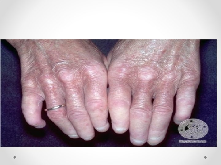
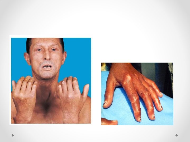
Systemic Sclerosis (Diffuse SS ) • An autoimmune multisystem disease that results in fibrosis and vascular abnormalities in association with autoimmune changes. • Usually starts between 30 -40 years. More in women • Pathophysiology may involve some injury to the endothelial cells and this results in excessive activation of the dermal connective tissue cells, the fibroblasts. • Usually presents with Raynaud's phenomena, Thickening of the skin of the fingers, then atrophy and sclerosis. The fingers become spindle-shaped (sclerodactyly) from resorption of the fingertips
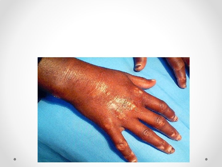
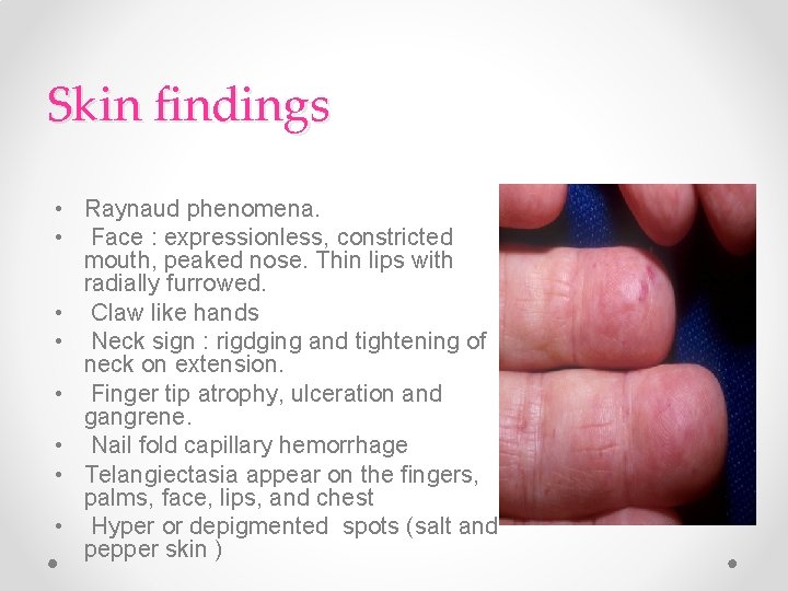
Skin findings • Raynaud phenomena. • Face : expressionless, constricted mouth, peaked nose. Thin lips with radially furrowed. • Claw like hands • Neck sign : rigdging and tightening of neck on extension. • Finger tip atrophy, ulceration and gangrene. • Nail fold capillary hemorrhage • Telangiectasia appear on the fingers, palms, face, lips, and chest • Hyper or depigmented spots (salt and pepper skin )
Internal involvement • • • Esophageal dysmotility( distal ) Esophageal reflux and dysphagia: Pulmonary fibrosis Pericarditis , HTN Retinopathy polyarthritis Progressive kidney disease resulting in proteinuria, high blood pressure and eventually renal failure
Investigations and Diagnosis • Diagnosis is made based on clinical features and presentation. • ANA is usually positive. Anti topoisomerase I (Scl 70) is characteristic for it especially in severe cases. • High ESR , thrombocytopenia, hemolytic anemia • High CK with muscle involvement • CXR : diffuse ground glass and honeycomb lung pattern • CT : lung fibrosis • ECG : arrythemia • Skin biopsy will show skin atrophy with thickened collagen bundles
Treatment of SS • Is symptomatic. • Raynaud's phenomena: Stop smoking, keep hands warm and decrease trauma. calcium channel blockers, aspirin and vasodilating drugs including nifedipine and iloprost infusions. • Calcinosis cutis: nifedipine, surgical or laser excision. • Skin sclerosis: physiotherapy, phototherapy. • GI: proton pump inhibitor, surgery for strictures. • Kidney: ACE inhibitors. • In severe cases: immunosuppressant , D-Penicillamine might be used
Thank you