
CUP patient pathway with CUP team MDT NICE
- Slides: 24
CUP patient pathway with CUP team & MDT • NICE guidelines mandated CUP networks across hospitals • CUP team in each hospital with cancer care • Link with acute oncology • CUP specialist nurse or keyworker • Major role in coordinating patient's care • CUP network multidisciplinary team (MDT) including pathologist set up to review treatment and care of patients with confirmed CUP • Provide clinical management & support like type- and site-specific cancer MDTs • Avoid “MDT tennis”
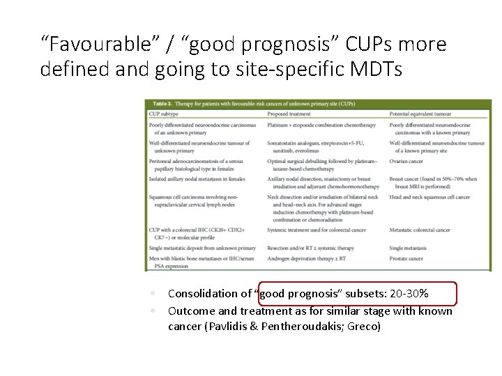
“Favourable” / “good prognosis” CUPs more defined and going to site-specific MDTs • Consolidation of “good prognosis” subsets: 20 -30% • Outcome and treatment as for similar stage with known cancer (Pavlidis & Pentheroudakis; Greco) ESMO (European Society Medical Oncology), Fizazi et al 2015
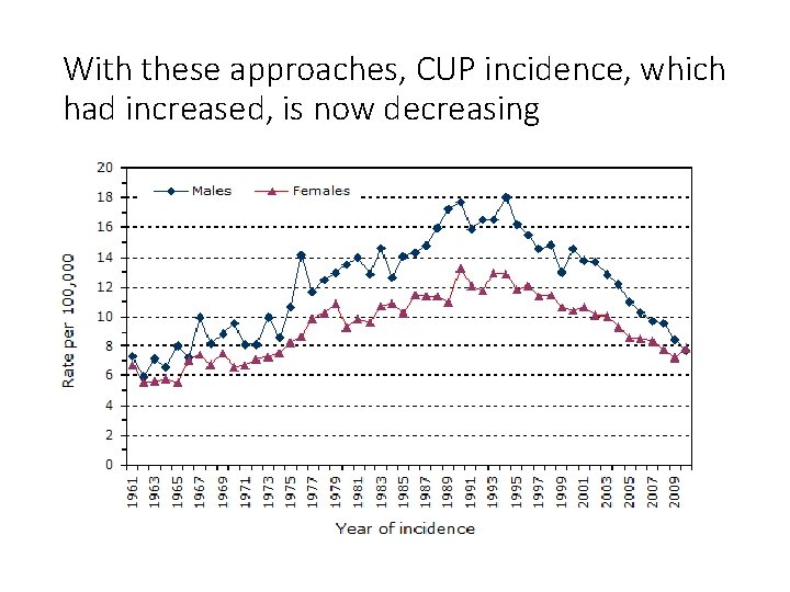
With these approaches, CUP incidence, which had increased, is now decreasing Brewster (2014). Cancer Epidemiology.
Outline • Cancer of Unknown Primary • Pathology classification for cancer in general and CUP in particular including immunohistochemistry (IHC) (morphomolecular part 1) • Performance & practice • RCPath dataset • CUP management through MDT approach • Morphomolecular approaches beyond IHC (part 2) • What is CUP today - MUO through to provisional CUP, confirmed CUP “residual CUP”? • Main challenges in pathology esp. “residual CUP” and therefore unmet needs • Approaches, implementation and next steps
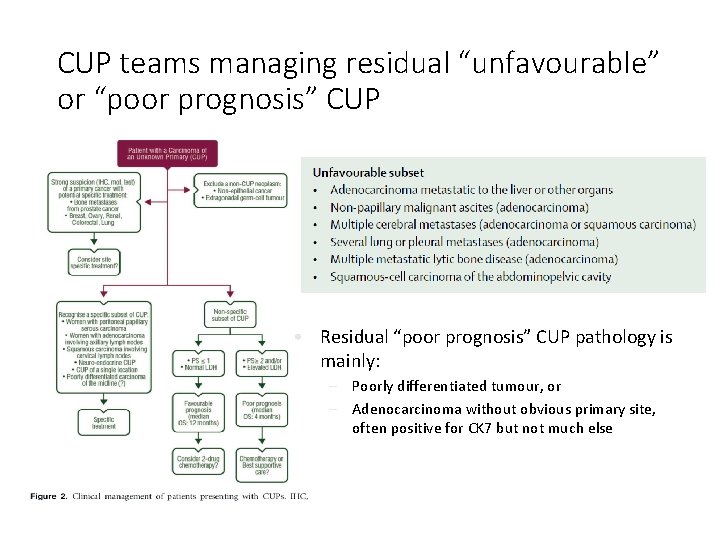
CUP teams managing residual “unfavourable” or “poor prognosis” CUP • Residual “poor prognosis” CUP pathology is mainly: – Poorly differentiated tumour, or – Adenocarcinoma without obvious primary site, often positive for CK 7 but not much else
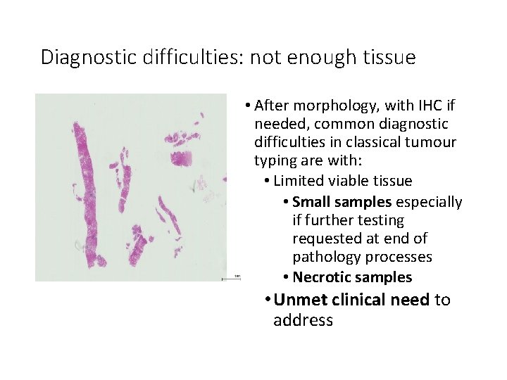
Diagnostic difficulties: not enough tissue • After morphology, with IHC if needed, common diagnostic difficulties in classical tumour typing are with: • Limited viable tissue • Small samples especially if further testing requested at end of pathology processes • Necrotic samples • Unmet clinical need to address
Diagnostic difficulties: difficult morphology • Common diagnostic difficulties in classical tumour typing for CUP are with: • “Very poorly differentiated or undifferentiated tumours • Adenocarcinoma, even well-differentiated, without obvious primary site • Pancreatico-biliary including cholangiocarcinoma • Gastro-oesophageal • Ovarian mucinous • Atypical tumours from lung, breast etc • Unmet clinical need to address
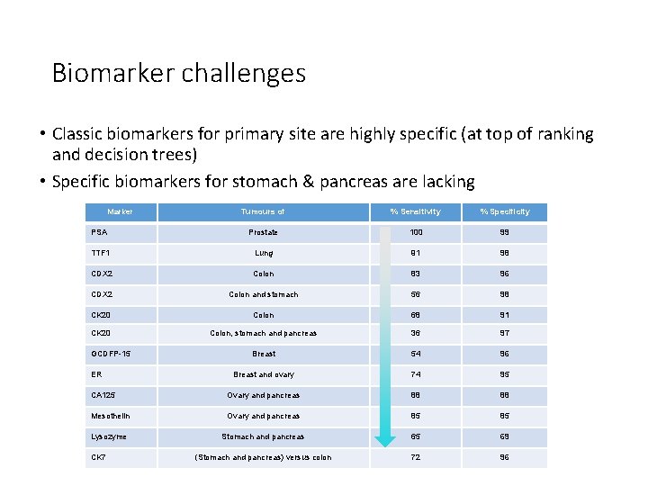
Biomarker challenges • Classic biomarkers for primary site are highly specific (at top of ranking and decision trees) • Specific biomarkers for stomach & pancreas are lacking Marker Tumours of % Sensitivity % Specificity PSA Prostate 100 99 TTF 1 Lung 91 98 CDX 2 Colon 83 96 CDX 2 Colon and stomach 56 98 CK 20 Colon 68 91 CK 20 Colon, stomach and pancreas 36 97 Breast 54 96 Breast and ovary 74 95 CA 125 Ovary and pancreas 88 88 Mesothelin Ovary and pancreas 85 85 Lysozyme Stomach and pancreas 65 69 (Stomach and pancreas) versus colon 72 96 GCDFP-15 ER CK 7 Based on Dennis, lin Cancer Res 2005; 11(10): 3766 -3772.
Diagnostic difficulties: difficult immunohistochemistry • IHC is subjective: technical performance and microscopic interpretation varies (as does actual tissue expression), so IHC biomarkers used in panels • IHC is selective: often limited tissue & time so only few biomarkers can be tested (study showed 7 -8 was usual in CUP) • In CUP, a barrier to correct tumour classification is not using the most appropriate markers Image from Wellcome Images; Weiss et al 2012, Schroeder et al 2012
Can we quantify performance of current pathology in CUP classification? • 5 -6 studies identified in meta-analysis • (Difficult to study by definition) • Sensitivity of IHC panels for primary site was consistent, around 82% in mixed primary and metastatic tumours and 6670% in metastases alone • Confirms metastases are harder to classify than primary tumours by IHC • Sets baseline for comparisons with molecular tests Anderson et al 2010, Weiss et al 2012, Sanden et al 2012
Molecular profiling in CUP • Large-scale profiling for CUP has been achieved at m. RNA, mi. RNA, DNA and epigenetic levels • Molecular profiling for primary site: esp. m. RNA/mi. RNA; also epigenetic • Adds to existing classifications? • Commercial tests • Molecular analysis for mutations especially “actionable targets” • May suggest primary site • But mainly for therapeutic prediction • Adds to or new classification/taxonomy Image from Royal College of Pathologists; Pillai et al 2011, Stancel et al 2012, Erlander et al 2011, Meiri et al 2012; reviewed in Oien 2012 Ann Oncol; Moran 2016 Lancet Oncology
Molecular profiling for primary site: rationale • “Different tissue types have distinct RNA profiles” (…or epigenetic, or protein…etc) • Yield tumour type, site and/or subtype i. e. classic taxonomy • IHC and m. RNA molecular profiling use similar tissuespecific genes: • Molecular profiling tests more genes and may be less subjective Greco 2014, Pillai et al 2011, Stancel et al 2012, Erlander et al 2011, Meiri et al 2012, Oien 2012
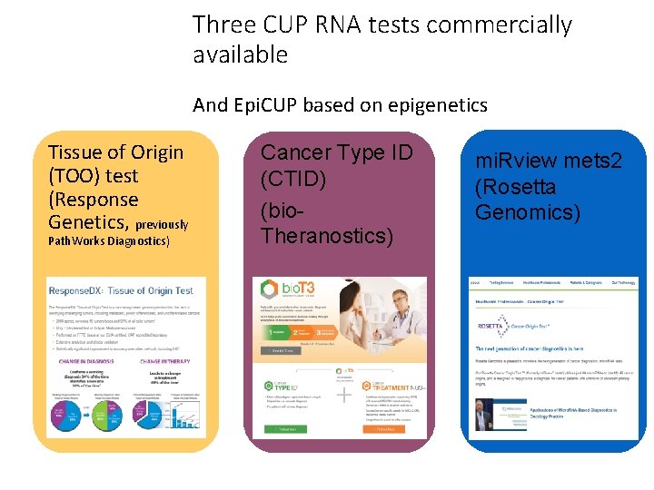
Three CUP RNA tests commercially available And Epi. CUP based on epigenetics Tissue of Origin (TOO) test (Response Genetics, previously Path. Works Diagnostics) Cancer Type ID (CTID) (bio. Theranostics) mi. Rview mets 2 (Rosetta Genomics) http: //rosettagenomics. com/ http: //www. responsegenetics. com/products-services/tissue-of-origin-testing/
Molecular profiling for primary site: results • “Clinical accuracy of all three tests is similar: 85 -88%. Test accuracy in CUP cases not easily determined, because origin not identified in most cases. ” • 10% of RNA tests yield no result • Tumours difficult for morphology and IHC often also difficult for molecular profiling • In poorly differentiated tumours, molecular profiling may: • Yield likely final diagnosis in 78 -92% • Compared with 68 -70% for IHC • Thus out-perform IHC by 10 -20% Greco 2015 Mol Diagn Ther, Weiss et al 2012, Sanden et al 2012
Guidelines and health economics in CUP profiling tests NCCN Recommendations 2014 NCCN Recommendations 2015 Elsewhere: is testing costeffective? • US NCCN Clinical Practice Guidelines on Occult Primary: Although GEP (Gene Expression Profiling) has a diagnostic benefit, clinical benefit has not been demonstrated. The panel recommends against GEP as standard management… • US NCCN Clinical Practice Guidelines on Occult Primary: Until more robust outcomes and comparative effectiveness data are available, pathologists & oncologists must collaborate on the judicious use of these modalities (IHC and GEP) on a case-by-case basis, with the best possible individualized patient outcome in mind. • In models: yes if influences survival. • For healthcare: may save work-up costs but may bring in additional oncology costs • Molecular profiling not currently recommended in NICE or ESMO guidelines • FDA approved and used in USA https: //www. nccn. org/professionals/physician_gls/f_guidelines. asp http: //www. ncbi. nlm. nih. gov/pubmedhealth/PMH 0073073/pdf/TOC. pdf/ Hannouf 2016 Pharmacogenomics Journal, Prasad 2015 Eur J Cancer
Where from, where going and how getting there? Where from? RNA etc for tissue of origin
Where from, where going and how getting there? Where from? RNA etc for tissue of origin Where going to? “Actionable” mutations
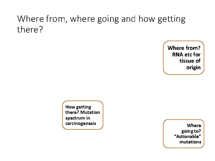
Where from, where going and how getting there? Where from? RNA etc for tissue of origin How getting there? Mutation spectrum in carcinogenesis Where going to? “Actionable” mutations
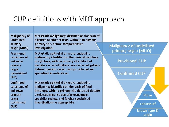
CUP definitions with MDT approach Malignancy of undefined primary origin (MUO) Metastatic malignancy identified on the basis of a limited number of tests, without an obvious primary site, before comprehensive investigation. Provisional carcinoma of unknown primary origin (provisional CUP) Metastatic epithelial or neuro-endocrine malignancy identified on the basis of histology or cytology, with no primary site detected despite a selected initial screen of investigations, before specialist review and possible further specialised investigations. Confirmed carcinoma of unknown primary origin (confirmed CUP) Metastatic epithelial or neuro-endocrine malignancy identified on the basis of final histology, with no primary site detected despite a selected initial screen of investigations, specialist review, and further specialised investigations as appropriate. Malignancy of undefined primary origin (MUO) Provisional CUP Confirmed CUP More cancers of known type & origin NICE guidance/guideline on CUP, 2010 https: //www. nice. org. uk/guidance/cg 104/resources/metastatic-malignant-disease-of-unknown-primary-origin-diagnosis-andmanagement-of-metastatic-malignant-disease-of-unknown-primary-origin-35109328970437
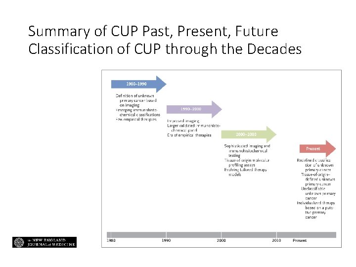
Summary of CUP Past, Present, Future Classification of CUP through the Decades
Conclusions: Pathology incl. IHC in CUP • Pathology, with IHC if needed, remains “gold standard” in tumour classification, especially where: • • Tumour has obvious likely primary and/or Tumour is at least moderately differentiated and/or IHC results are classical and/or The clinical context is appropriate • Optimal pathology enabled by: • Standardised approach incl. IHC • MDT approach to enable full clinical context and knowledge of potential treatments
Conclusions: CUP pathology unmet needs and morpho-molecular approaches • Adenocarcinomas without obvious primary & poorly differentiated cancers, esp. if: • Current pathology equivocal for management or conflicting with clinical context • Diagnosis either truly unknown or includes multiple possible differentials • Limited tissue or time? • Better prediction of treatment benefit • Molecular profiling and screening for actionable mutations provide additional information, often potentially clinically significant incl. treatment guidance, but not yet included in most guidelines • How best to use? And how to evidence towards funding and implementation of: • Molecular classifiers for site/type • Molecular analysis for actionable mutations • Complementary & in clinicopathological context
Acknowledgements Personal thanks to: CUP Heroes • Dr Jayne Dennis • Dr Harpreet Wasan &Jayson Wang • Prof David Bowtell and colleagues • Colleagues in Bio. Theranostics • Prof Nicol Keith • Prof Jeff Evans • Profs Andrew Biankin & Sean Grimmond and colleagues • John Symons & Jo’s Friends • Profs John Schofield, Paul Barrett and colleagues Personal thanks to Cancer Research UK and University of Glasgow for funding; but this presentation is my personal opinion Pathology Heroes