
Cumulative Exam Review PPT Statistics Digestion and Absorption
Cumulative Exam Review PPT Statistics, Digestion and Absorption, The Blood System, and Ventillation
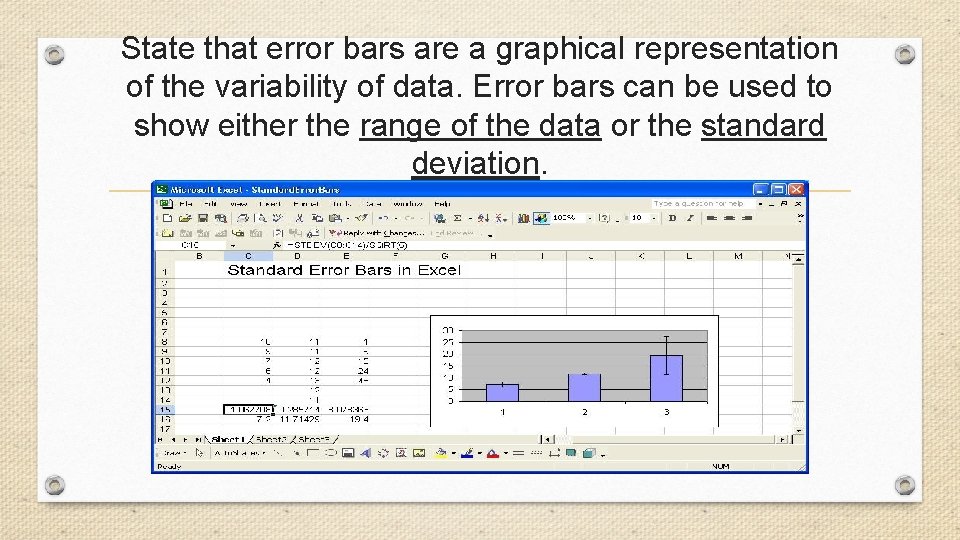
State that error bars are a graphical representation of the variability of data. Error bars can be used to show either the range of the data or the standard deviation.
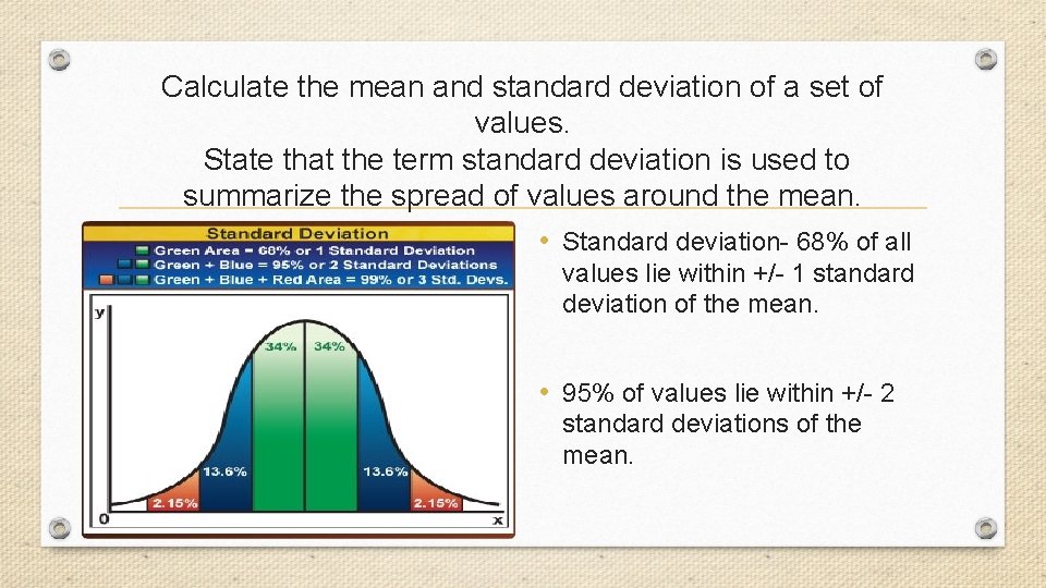
Calculate the mean and standard deviation of a set of values. State that the term standard deviation is used to summarize the spread of values around the mean. • Standard deviation- 68% of all values lie within +/- 1 standard deviation of the mean. • 95% of values lie within +/- 2 standard deviations of the mean.
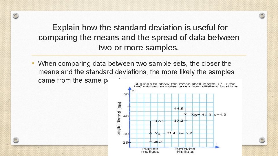
Explain how the standard deviation is useful for comparing the means and the spread of data between two or more samples. • When comparing data between two sample sets, the closer the means and the standard deviations, the more likely the samples came from the same population
Deduce the significance of the difference between two sets of data using calculated values for t. • Criteria for t -test : • Normal distribution • Sample size of at least 10 • The t -test can be used to compare two sets of data and measure the amount of overlap. (two-tailed, unpaired t test). • If the value of p is < the critical p value at. 05, then there is a significant difference between the two sets of data, and the null hypothesis should be rejected.
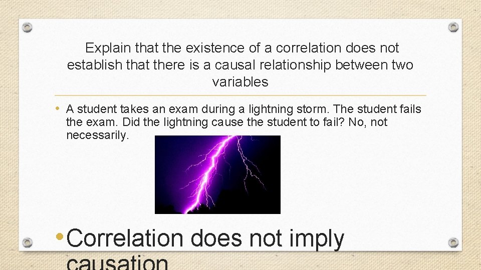
Explain that the existence of a correlation does not establish that there is a causal relationship between two variables • A student takes an exam during a lightning storm. The student fails the exam. Did the lightning cause the student to fail? No, not necessarily. • Correlation does not imply
Digestion and Absorption
Understandings
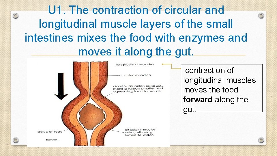
U 1. The contraction of circular and longitudinal muscle layers of the small intestines mixes the food with enzymes and moves it along the gut. contraction of longitudinal muscles moves the food forward along the gut.
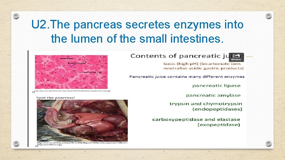
U 2. The pancreas secretes enzymes into the lumen of the small intestines.
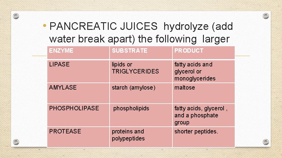
• PANCREATIC JUICES hydrolyze (add water break apart) the following larger ENZYME PRODUCT molecules: SUBSTRATE LIPASE lipids or TRIGLYCERIDES fatty acids and glycerol or monoglycerides AMYLASE starch (amylose) maltose PHOSPHOLIPASE phospholipids fatty acids, glycerol , and a phosphate group PROTEASE proteins and polypeptides shorter peptides.
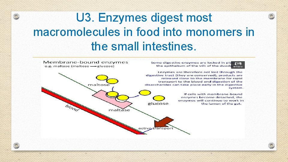
U 3. Enzymes digest most macromolecules in food into monomers in the small intestines.
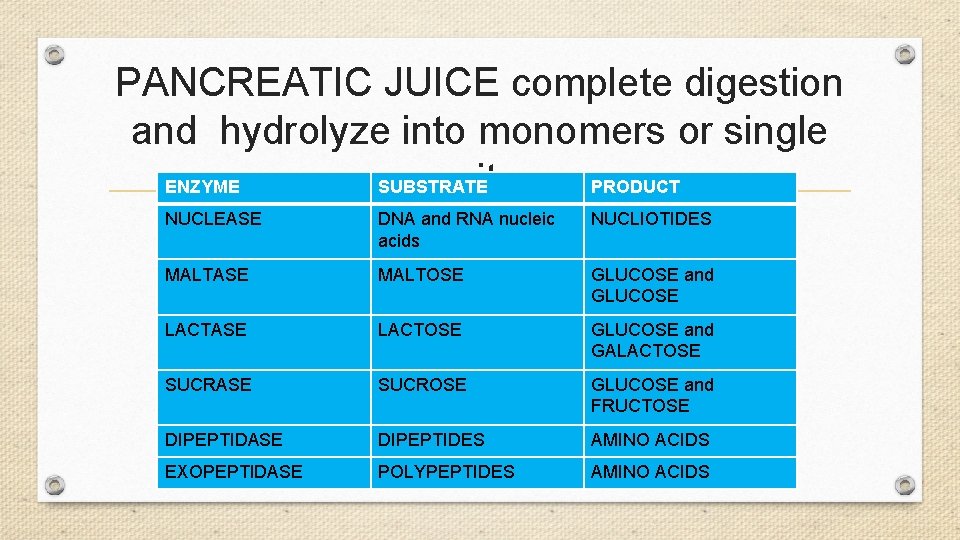
PANCREATIC JUICE complete digestion and hydrolyze into monomers or single units. ENZYME SUBSTRATE PRODUCT NUCLEASE DNA and RNA nucleic acids NUCLIOTIDES MALTASE MALTOSE GLUCOSE and GLUCOSE LACTASE LACTOSE GLUCOSE and GALACTOSE SUCRASE SUCROSE GLUCOSE and FRUCTOSE DIPEPTIDASE DIPEPTIDES AMINO ACIDS EXOPEPTIDASE POLYPEPTIDES AMINO ACIDS
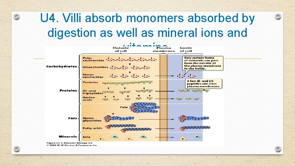
U 4. Villi absorb monomers absorbed by digestion as well as mineral ions and vitamins.
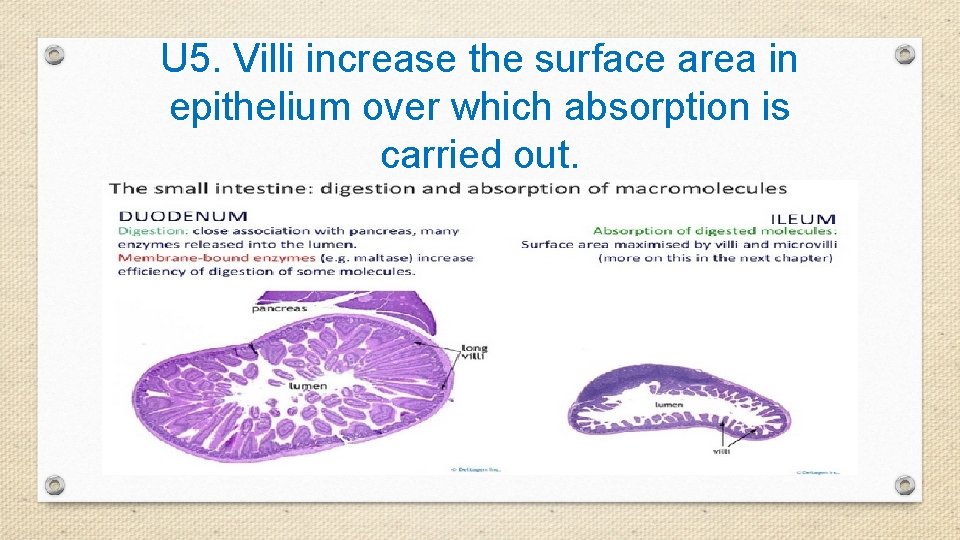
U 5. Villi increase the surface area in epithelium over which absorption is carried out.
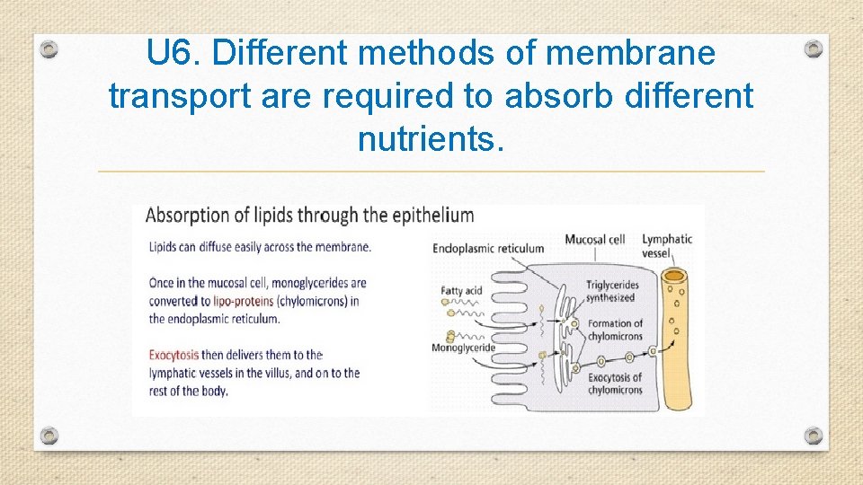
U 6. Different methods of membrane transport are required to absorb different nutrients.
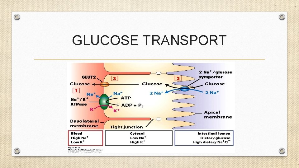
GLUCOSE TRANSPORT
Applications
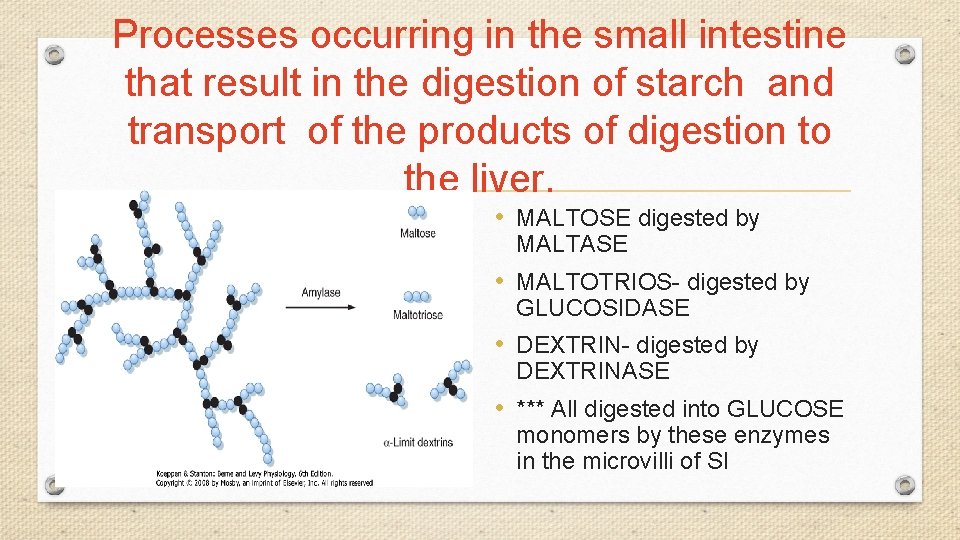
Processes occurring in the small intestine that result in the digestion of starch and transport of the products of digestion to the liver. • MALTOSE digested by MALTASE • MALTOTRIOS- digested by GLUCOSIDASE • DEXTRIN- digested by DEXTRINASE • *** All digested into GLUCOSE monomers by these enzymes in the microvilli of SI
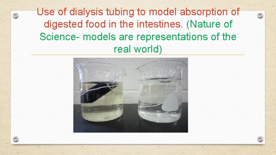
Use of dialysis tubing to model absorption of digested food in the intestines. (Nature of Science- models are representations of the real world)
Skills
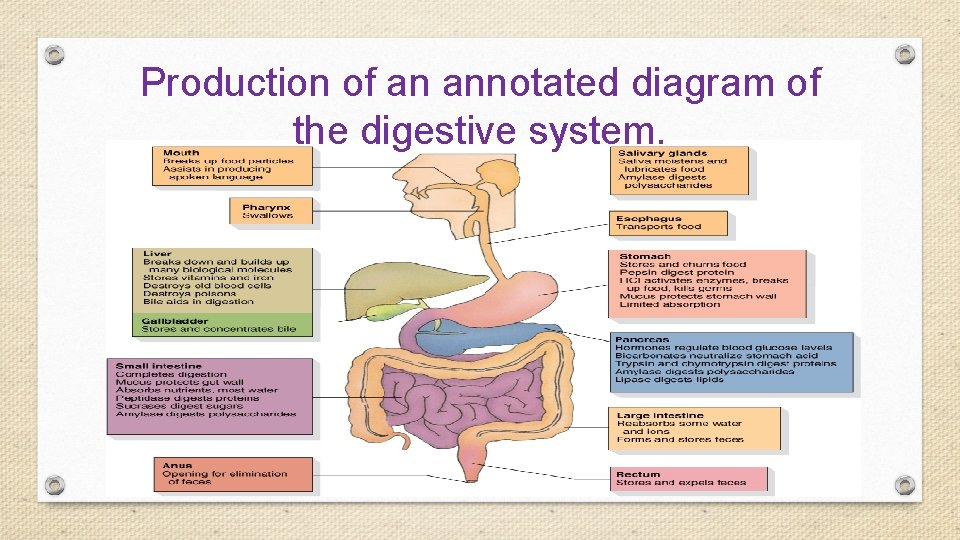
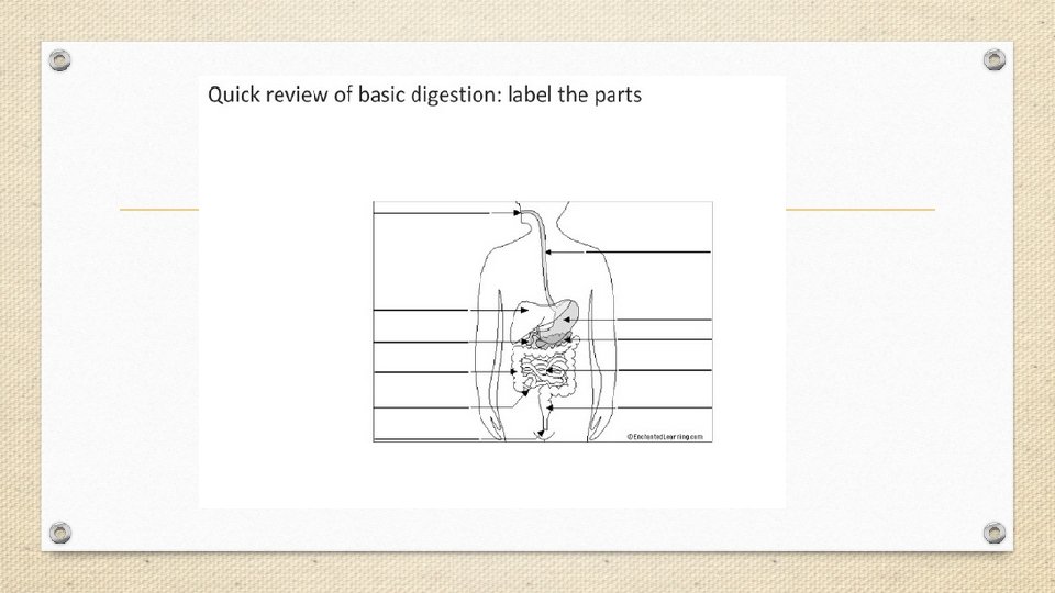
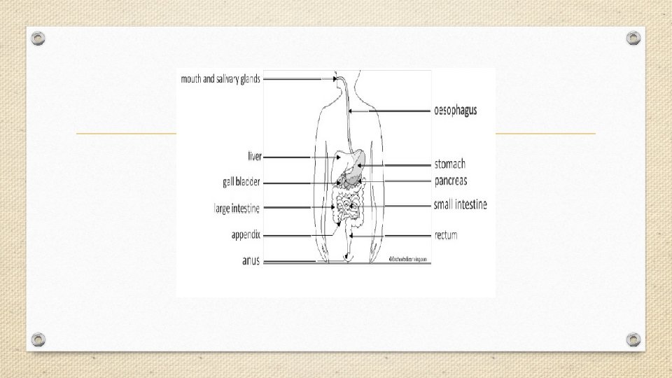
Production of an annotated diagram of the digestive system.
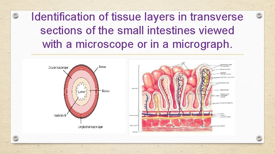
Identification of tissue layers in transverse sections of the small intestines viewed with a microscope or in a micrograph.
The Blood System
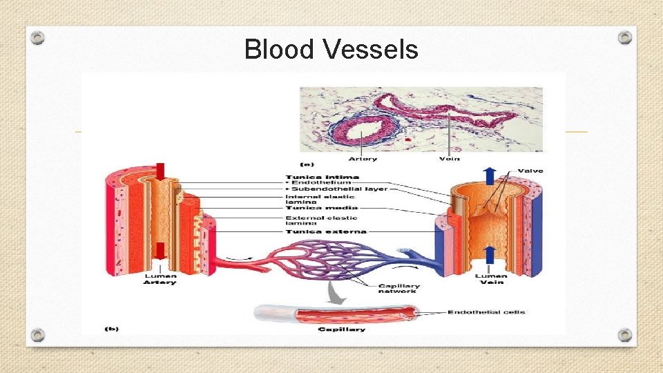
Blood Vessels
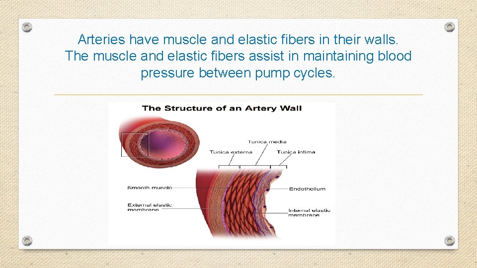
Arteries have muscle and elastic fibers in their walls. The muscle and elastic fibers assist in maintaining blood pressure between pump cycles.
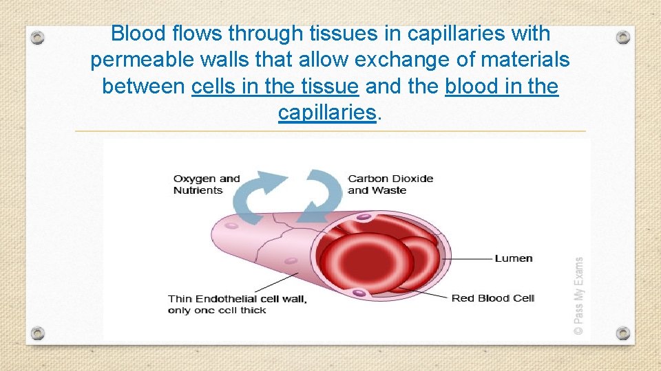
Blood flows through tissues in capillaries with permeable walls that allow exchange of materials between cells in the tissue and the blood in the capillaries.
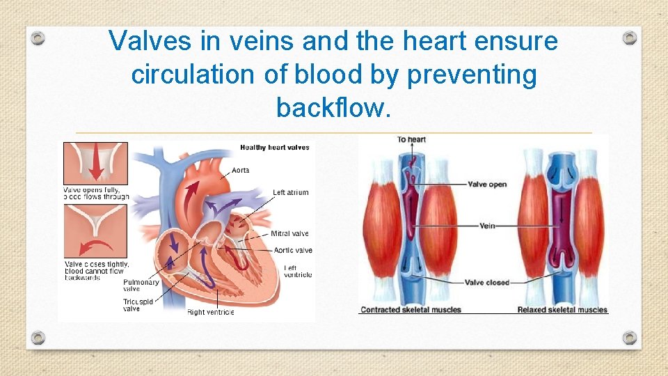
Valves in veins and the heart ensure circulation of blood by preventing backflow.
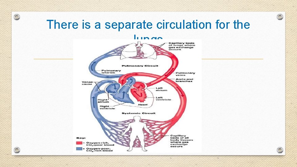
There is a separate circulation for the lungs
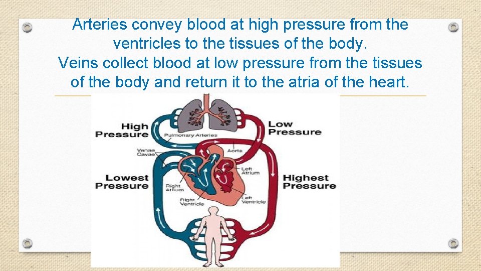
Arteries convey blood at high pressure from the ventricles to the tissues of the body. Veins collect blood at low pressure from the tissues of the body and return it to the atria of the heart.
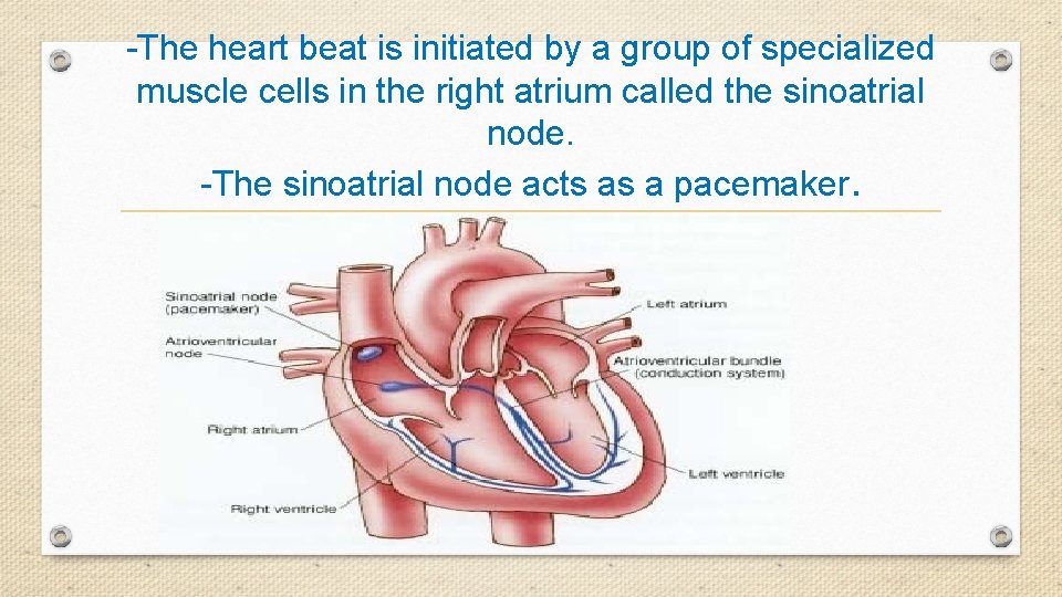
-The heart beat is initiated by a group of specialized muscle cells in the right atrium called the sinoatrial node. -The sinoatrial node acts as a pacemaker.
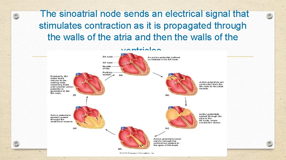
The sinoatrial node sends an electrical signal that stimulates contraction as it is propagated through the walls of the atria and then the walls of the ventricles.
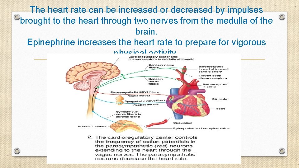
The heart rate can be increased or decreased by impulses brought to the heart through two nerves from the medulla of the brain. Epinephrine increases the heart rate to prepare for vigorous physical activity.
Skills, Applications, and Nature of Science
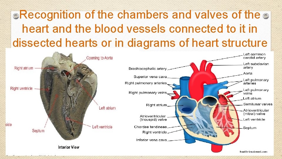
Recognition of the chambers and valves of the heart and the blood vessels connected to it in dissected hearts or in diagrams of heart structure
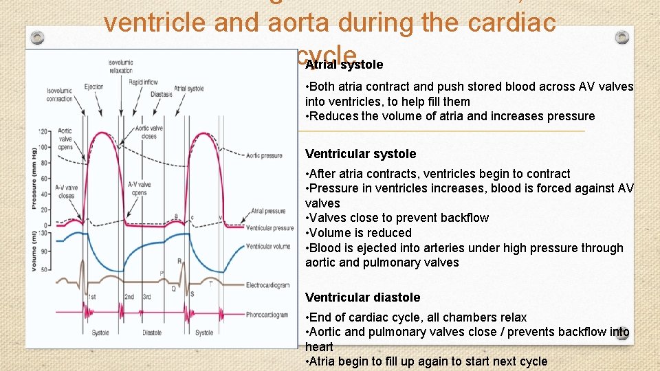
ventricle and aorta during the cardiac cycle. Atrial systole • Both atria contract and push stored blood across AV valves into ventricles, to help fill them • Reduces the volume of atria and increases pressure Ventricular systole • After atria contracts, ventricles begin to contract • Pressure in ventricles increases, blood is forced against AV valves • Valves close to prevent backflow • Volume is reduced • Blood is ejected into arteries under high pressure through aortic and pulmonary valves Ventricular diastole • End of cardiac cycle, all chambers relax • Aortic and pulmonary valves close / prevents backflow into heart • Atria begin to fill up again to start next cycle
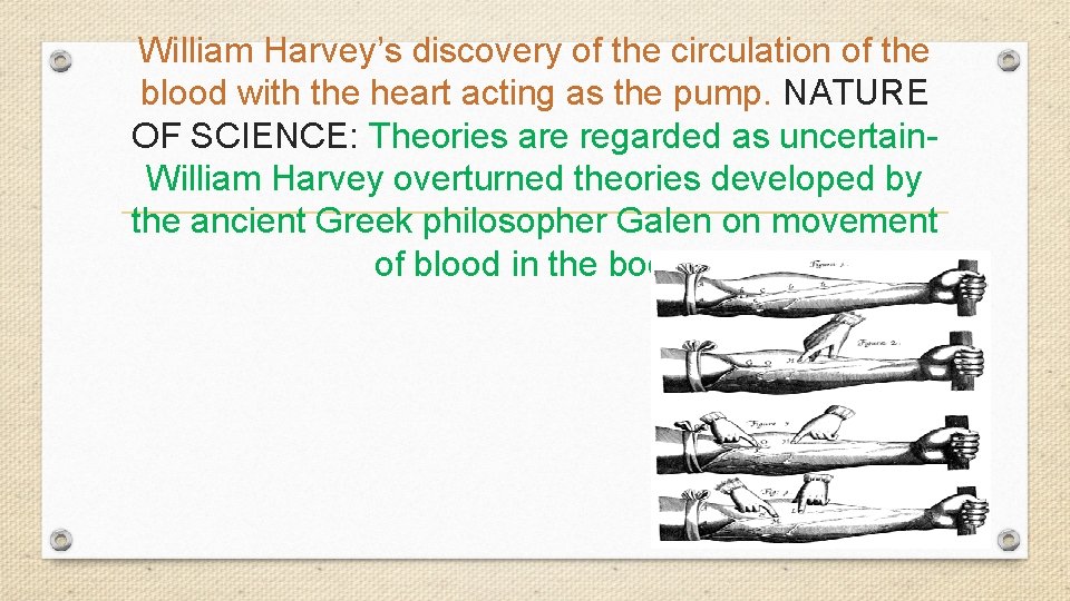
William Harvey’s discovery of the circulation of the blood with the heart acting as the pump. NATURE OF SCIENCE: Theories are regarded as uncertain. William Harvey overturned theories developed by the ancient Greek philosopher Galen on movement of blood in the body.
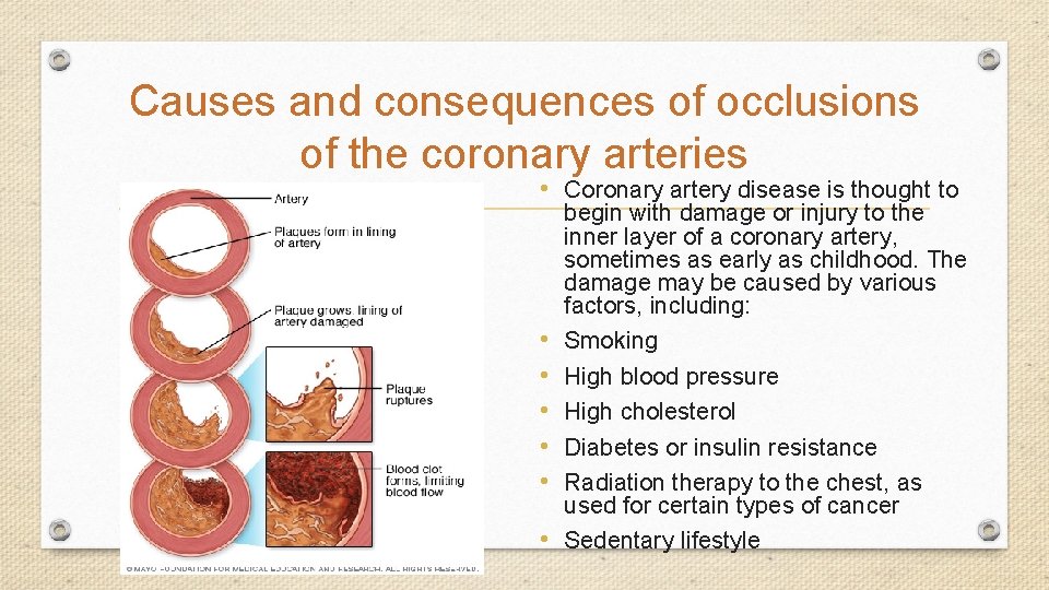
Causes and consequences of occlusions of the coronary arteries • Coronary artery disease is thought to • • • begin with damage or injury to the inner layer of a coronary artery, sometimes as early as childhood. The damage may be caused by various factors, including: Smoking High blood pressure High cholesterol Diabetes or insulin resistance Radiation therapy to the chest, as used for certain types of cancer Sedentary lifestyle
VENTILATION
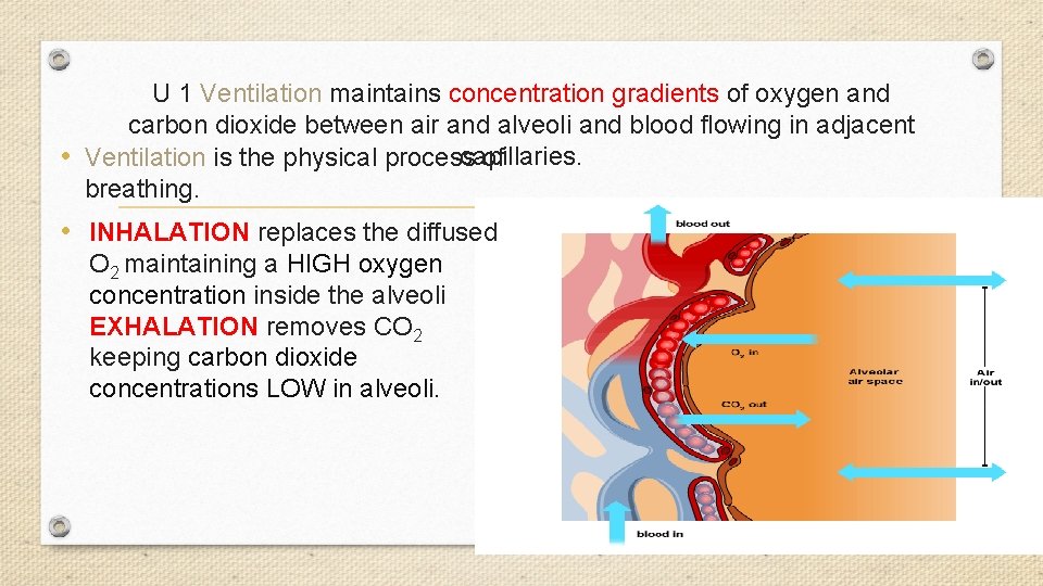
U 1 Ventilation maintains concentration gradients of oxygen and carbon dioxide between air and alveoli and blood flowing in adjacent capillaries. • Ventilation is the physical process of breathing. • INHALATION replaces the diffused O 2 maintaining a HIGH oxygen concentration inside the alveoli EXHALATION removes CO 2 keeping carbon dioxide concentrations LOW in alveoli.
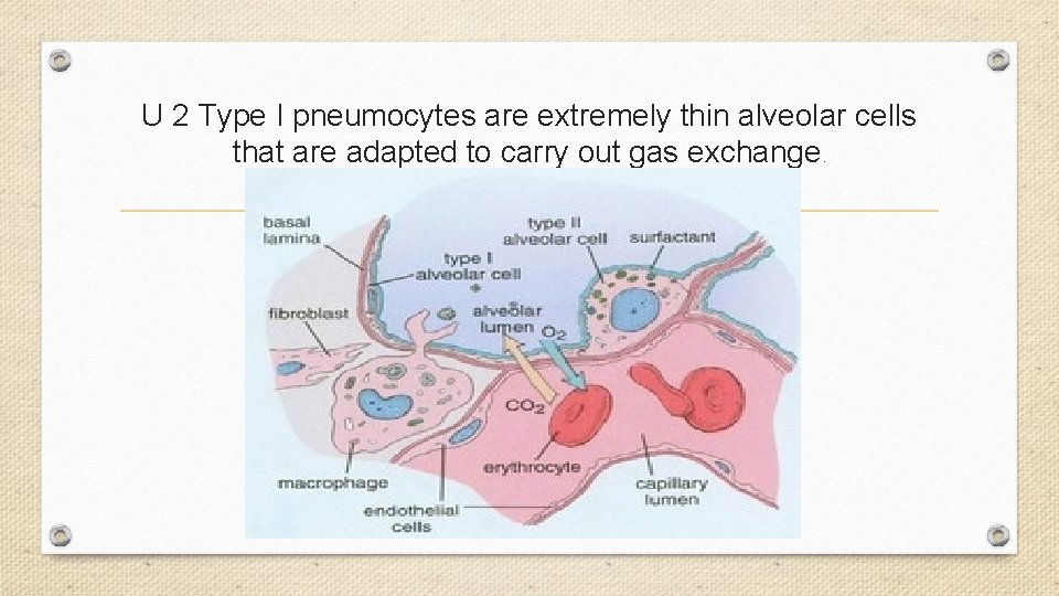
U 2 Type I pneumocytes are extremely thin alveolar cells that are adapted to carry out gas exchange.
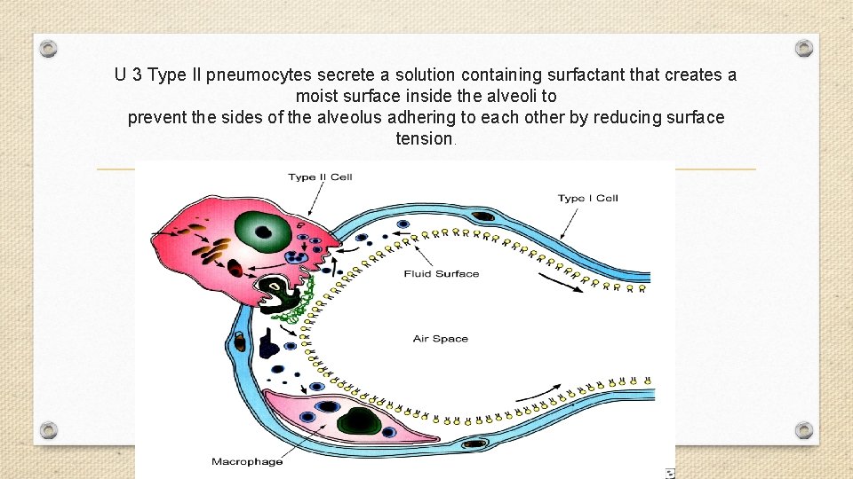
U 3 Type II pneumocytes secrete a solution containing surfactant that creates a moist surface inside the alveoli to prevent the sides of the alveolus adhering to each other by reducing surface tension.
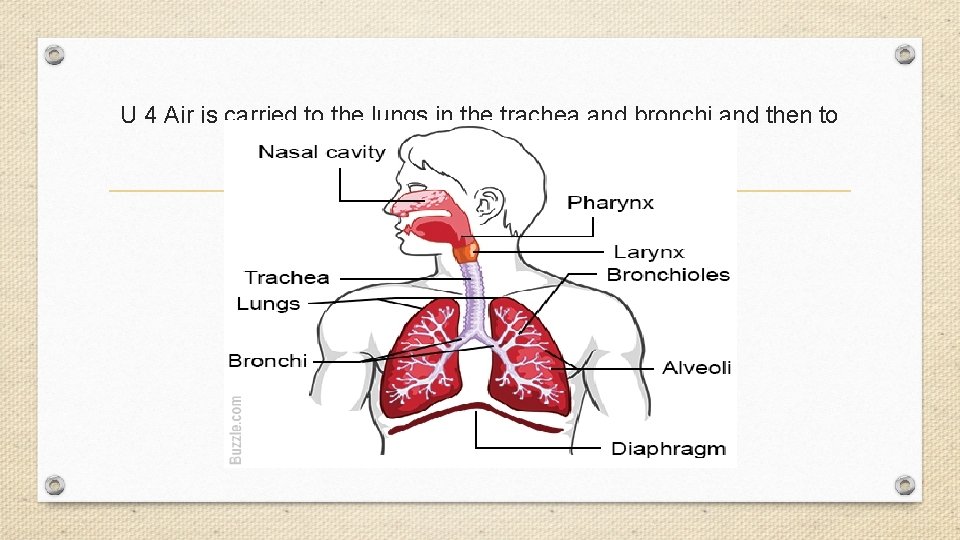
U 4 Air is carried to the lungs in the trachea and bronchi and then to the alveoli in bronchioles
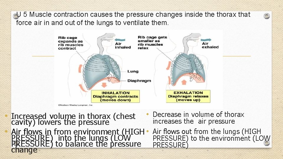
U 5 Muscle contraction causes the pressure changes inside thorax that force air in and out of the lungs to ventilate them. • Increased volume in thorax (chest • Decrease in volume of thorax cavity) lowers the pressure • Air flows in from environment (HIGH • PRESSURE) into the lungs (LOW PRESSURE) to balance the pressure change increases the air pressure Air flows out from the lungs (HIGH PRESSURE) to the environment (LOW PRESSURE)
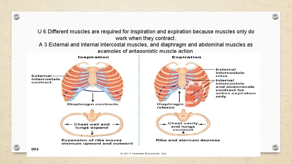
U 6 Different muscles are required for inspiration and expiration because muscles only do work when they contract. A 3 External and internal intercostal muscles, and diaphragm and abdominal muscles as examples of antagonistic muscle action.
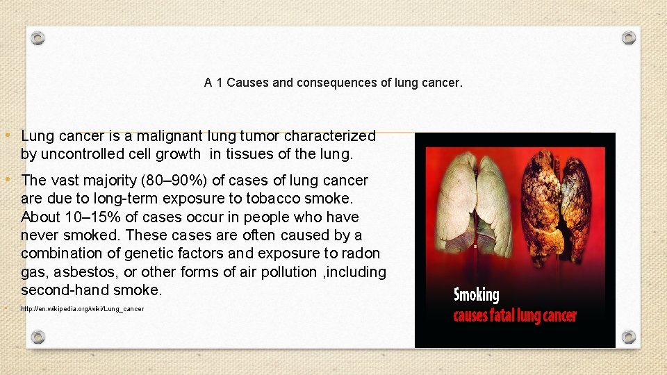
A 1 Causes and consequences of lung cancer. • Lung cancer is a malignant lung tumor characterized by uncontrolled cell growth in tissues of the lung. • The vast majority (80– 90%) of cases of lung cancer are due to long-term exposure to tobacco smoke. About 10– 15% of cases occur in people who have never smoked. These cases are often caused by a combination of genetic factors and exposure to radon gas, asbestos, or other forms of air pollution , including second-hand smoke. • http: //en. wikipedia. org/wiki/Lung_cancer
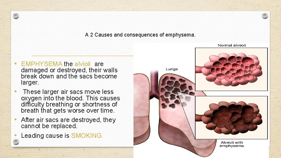
A 2 Causes and consequences of emphysema. • EMPHYSEMA the alvioli are damaged or destroyed, their walls break down and the sacs become larger. • These larger air sacs move less oxygen into the blood. This causes difficulty breathing or shortness of breath that gets worse over time. • After air sacs are destroyed, they cannot be replaced. • Leading cause is SMOKING.
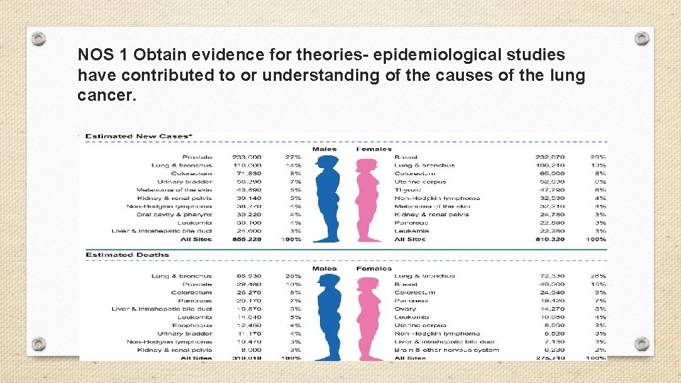
NOS 1 Obtain evidence for theories- epidemiological studies have contributed to or understanding of the causes of the lung cancer.
- Slides: 50