CT 18 FFDG PET Luso della PET nella
![CT [18 F]FDG PET L'uso della PET nella diagnosi precoce e la valutazione del CT [18 F]FDG PET L'uso della PET nella diagnosi precoce e la valutazione del](https://slidetodoc.com/presentation_image_h2/d41129d0f6ea79360a83e85c307dfcff/image-1.jpg)
![[18 F]FDG-PET [18 F]FDG-PET](https://slidetodoc.com/presentation_image_h2/d41129d0f6ea79360a83e85c307dfcff/image-2.jpg)
![Why [18 F]FDG-PET in tumors? Why [18 F]FDG-PET in tumors?](https://slidetodoc.com/presentation_image_h2/d41129d0f6ea79360a83e85c307dfcff/image-3.jpg)
![Why [18 F]FDG-PET in tumors? Elevated glucose metabolism in tumor Why [18 F]FDG-PET in tumors? Elevated glucose metabolism in tumor](https://slidetodoc.com/presentation_image_h2/d41129d0f6ea79360a83e85c307dfcff/image-4.jpg)
![Why [18 F]FDG-PET in tumors? Elevated glucose metabolism in tumor [18 F]FDG is a Why [18 F]FDG-PET in tumors? Elevated glucose metabolism in tumor [18 F]FDG is a](https://slidetodoc.com/presentation_image_h2/d41129d0f6ea79360a83e85c307dfcff/image-5.jpg)
![Why [18 F]FDG-PET in tumors? Elevated glucose metabolism in tumor [18 F]FDG is a Why [18 F]FDG-PET in tumors? Elevated glucose metabolism in tumor [18 F]FDG is a](https://slidetodoc.com/presentation_image_h2/d41129d0f6ea79360a83e85c307dfcff/image-6.jpg)
![[18 F]FDG PET: APPLICATION IN LUNG CANCER Differentiating benign from malignant lesions (SPN) Staging [18 F]FDG PET: APPLICATION IN LUNG CANCER Differentiating benign from malignant lesions (SPN) Staging](https://slidetodoc.com/presentation_image_h2/d41129d0f6ea79360a83e85c307dfcff/image-7.jpg)

![[18 F]FDG PET Measure of metabolic activity of SPN Visual Analysis Quantitative analysis CT [18 F]FDG PET Measure of metabolic activity of SPN Visual Analysis Quantitative analysis CT](https://slidetodoc.com/presentation_image_h2/d41129d0f6ea79360a83e85c307dfcff/image-9.jpg)
![[18 F]FDG PET Measure of metabolic activity of SPN Visual Analysis: nodule activity vs [18 F]FDG PET Measure of metabolic activity of SPN Visual Analysis: nodule activity vs](https://slidetodoc.com/presentation_image_h2/d41129d0f6ea79360a83e85c307dfcff/image-10.jpg)
![[18 F]FDG PET Measure of metabolic activity of SPN Visual Analysis: nodule activity vs [18 F]FDG PET Measure of metabolic activity of SPN Visual Analysis: nodule activity vs](https://slidetodoc.com/presentation_image_h2/d41129d0f6ea79360a83e85c307dfcff/image-11.jpg)
![[18 F]FDG PET Measure of metabolic activity of SPN Visual Analysis Quantitative analysis: SUV [18 F]FDG PET Measure of metabolic activity of SPN Visual Analysis Quantitative analysis: SUV](https://slidetodoc.com/presentation_image_h2/d41129d0f6ea79360a83e85c307dfcff/image-12.jpg)

![[18 F]FDG PET Measure of metabolic activity of SPN Visual Analysis Quantitative analysis: SUV [18 F]FDG PET Measure of metabolic activity of SPN Visual Analysis Quantitative analysis: SUV](https://slidetodoc.com/presentation_image_h2/d41129d0f6ea79360a83e85c307dfcff/image-14.jpg)
![[18 F]FDG PET Measure of metabolic activity of SPN Visual Analysis Quantitative analysis: SUV [18 F]FDG PET Measure of metabolic activity of SPN Visual Analysis Quantitative analysis: SUV](https://slidetodoc.com/presentation_image_h2/d41129d0f6ea79360a83e85c307dfcff/image-15.jpg)
![[18 F]FDG PET Lung cancer is hypermetabolic A SPN with SUV more than 2. [18 F]FDG PET Lung cancer is hypermetabolic A SPN with SUV more than 2.](https://slidetodoc.com/presentation_image_h2/d41129d0f6ea79360a83e85c307dfcff/image-16.jpg)
![CT [18 F]FDG PET Fused SPN: 1. 2 cm in diameter HSR - Milano CT [18 F]FDG PET Fused SPN: 1. 2 cm in diameter HSR - Milano](https://slidetodoc.com/presentation_image_h2/d41129d0f6ea79360a83e85c307dfcff/image-17.jpg)
![CT [18 F]FDG PET CT- PET SPN: 1. 4 cm in diameter HSR - CT [18 F]FDG PET CT- PET SPN: 1. 4 cm in diameter HSR -](https://slidetodoc.com/presentation_image_h2/d41129d0f6ea79360a83e85c307dfcff/image-18.jpg)
![Accuracy of PET with [18 F]FDG in SPN or Pulmonary Opacity Authors Sensitivity Duhaylongsod Accuracy of PET with [18 F]FDG in SPN or Pulmonary Opacity Authors Sensitivity Duhaylongsod](https://slidetodoc.com/presentation_image_h2/d41129d0f6ea79360a83e85c307dfcff/image-19.jpg)


![CT [18 F]FDG PET ? Fused R. F. 53 aa Lung nodule 4 mm CT [18 F]FDG PET ? Fused R. F. 53 aa Lung nodule 4 mm](https://slidetodoc.com/presentation_image_h2/d41129d0f6ea79360a83e85c307dfcff/image-22.jpg)
![CT [18 F]FDG PET CT- PET Z. A. 41 aa SPN: 4 mm in CT [18 F]FDG PET CT- PET Z. A. 41 aa SPN: 4 mm in](https://slidetodoc.com/presentation_image_h2/d41129d0f6ea79360a83e85c307dfcff/image-23.jpg)

![CT [18 F]FDG PET CARCINOID HSR - Milano CT [18 F]FDG PET CARCINOID HSR - Milano](https://slidetodoc.com/presentation_image_h2/d41129d0f6ea79360a83e85c307dfcff/image-25.jpg)
![CT [18 F]FDG PET/CT P. G. 68 aa SPN: 10 mm in diameter Well CT [18 F]FDG PET/CT P. G. 68 aa SPN: 10 mm in diameter Well](https://slidetodoc.com/presentation_image_h2/d41129d0f6ea79360a83e85c307dfcff/image-26.jpg)
![[18 F]FDG PET IN LUNG CANCER SUV and histological type Mean SUV BAC (12) [18 F]FDG PET IN LUNG CANCER SUV and histological type Mean SUV BAC (12)](https://slidetodoc.com/presentation_image_h2/d41129d0f6ea79360a83e85c307dfcff/image-27.jpg)
![[18 F]FDG PET IN LUNG CANCER FDG uptake in BAC No [18 F]FDG uptake [18 F]FDG PET IN LUNG CANCER FDG uptake in BAC No [18 F]FDG uptake](https://slidetodoc.com/presentation_image_h2/d41129d0f6ea79360a83e85c307dfcff/image-28.jpg)
![Pure Bronchioloalveolar Carcinoma CT 18 F]FDG PET [11[C]Choline PET SUV = 1. 73 B. Pure Bronchioloalveolar Carcinoma CT 18 F]FDG PET [11[C]Choline PET SUV = 1. 73 B.](https://slidetodoc.com/presentation_image_h2/d41129d0f6ea79360a83e85c307dfcff/image-29.jpg)

![VALUTAZIONE DELLA RISPOSTA ALLA TERAPIA con PET e [18 F]FDG NEL TUMORE POLMONARE 113 VALUTAZIONE DELLA RISPOSTA ALLA TERAPIA con PET e [18 F]FDG NEL TUMORE POLMONARE 113](https://slidetodoc.com/presentation_image_h2/d41129d0f6ea79360a83e85c307dfcff/image-31.jpg)


- Slides: 33
![CT 18 FFDG PET Luso della PET nella diagnosi precoce e la valutazione del CT [18 F]FDG PET L'uso della PET nella diagnosi precoce e la valutazione del](https://slidetodoc.com/presentation_image_h2/d41129d0f6ea79360a83e85c307dfcff/image-1.jpg)
CT [18 F]FDG PET L'uso della PET nella diagnosi precoce e la valutazione del SUV come indicatore di prognosi Gino Pepe Medicina Nucleare – PET H. San. Raffaele IRCCS - Milano
![18 FFDGPET [18 F]FDG-PET](https://slidetodoc.com/presentation_image_h2/d41129d0f6ea79360a83e85c307dfcff/image-2.jpg)
[18 F]FDG-PET
![Why 18 FFDGPET in tumors Why [18 F]FDG-PET in tumors?](https://slidetodoc.com/presentation_image_h2/d41129d0f6ea79360a83e85c307dfcff/image-3.jpg)
Why [18 F]FDG-PET in tumors?
![Why 18 FFDGPET in tumors Elevated glucose metabolism in tumor Why [18 F]FDG-PET in tumors? Elevated glucose metabolism in tumor](https://slidetodoc.com/presentation_image_h2/d41129d0f6ea79360a83e85c307dfcff/image-4.jpg)
Why [18 F]FDG-PET in tumors? Elevated glucose metabolism in tumor
![Why 18 FFDGPET in tumors Elevated glucose metabolism in tumor 18 FFDG is a Why [18 F]FDG-PET in tumors? Elevated glucose metabolism in tumor [18 F]FDG is a](https://slidetodoc.com/presentation_image_h2/d41129d0f6ea79360a83e85c307dfcff/image-5.jpg)
Why [18 F]FDG-PET in tumors? Elevated glucose metabolism in tumor [18 F]FDG is a glucose analog
![Why 18 FFDGPET in tumors Elevated glucose metabolism in tumor 18 FFDG is a Why [18 F]FDG-PET in tumors? Elevated glucose metabolism in tumor [18 F]FDG is a](https://slidetodoc.com/presentation_image_h2/d41129d0f6ea79360a83e85c307dfcff/image-6.jpg)
Why [18 F]FDG-PET in tumors? Elevated glucose metabolism in tumor [18 F]FDG is a glucose analog [18 F]FDG uptake into viable neoplastic cells
![18 FFDG PET APPLICATION IN LUNG CANCER Differentiating benign from malignant lesions SPN Staging [18 F]FDG PET: APPLICATION IN LUNG CANCER Differentiating benign from malignant lesions (SPN) Staging](https://slidetodoc.com/presentation_image_h2/d41129d0f6ea79360a83e85c307dfcff/image-7.jpg)
[18 F]FDG PET: APPLICATION IN LUNG CANCER Differentiating benign from malignant lesions (SPN) Staging N and M and re-staging for therapy planning Predicting and monitoring response to therapy

Solitary Pulmonary Nodule (SPN) Solitary lung lesion < 3 cm in diameter 20% - 40% of SPN are malignant Initial presentation in 20% - 30% of lung cancer Coleman et al. J Nuc Med 1999
![18 FFDG PET Measure of metabolic activity of SPN Visual Analysis Quantitative analysis CT [18 F]FDG PET Measure of metabolic activity of SPN Visual Analysis Quantitative analysis CT](https://slidetodoc.com/presentation_image_h2/d41129d0f6ea79360a83e85c307dfcff/image-9.jpg)
[18 F]FDG PET Measure of metabolic activity of SPN Visual Analysis Quantitative analysis CT [18 F]FDG PET SUV = 7. 3
![18 FFDG PET Measure of metabolic activity of SPN Visual Analysis nodule activity vs [18 F]FDG PET Measure of metabolic activity of SPN Visual Analysis: nodule activity vs](https://slidetodoc.com/presentation_image_h2/d41129d0f6ea79360a83e85c307dfcff/image-10.jpg)
[18 F]FDG PET Measure of metabolic activity of SPN Visual Analysis: nodule activity vs mediastinal blood pool activity Quantitative analysis CT [18 F]FDG PET SUV = 7. 3
![18 FFDG PET Measure of metabolic activity of SPN Visual Analysis nodule activity vs [18 F]FDG PET Measure of metabolic activity of SPN Visual Analysis: nodule activity vs](https://slidetodoc.com/presentation_image_h2/d41129d0f6ea79360a83e85c307dfcff/image-11.jpg)
[18 F]FDG PET Measure of metabolic activity of SPN Visual Analysis: nodule activity vs mediastinal blood pool activity Quantitative analysis CT [18 F]FDG PET SUV = 7. 3
![18 FFDG PET Measure of metabolic activity of SPN Visual Analysis Quantitative analysis SUV [18 F]FDG PET Measure of metabolic activity of SPN Visual Analysis Quantitative analysis: SUV](https://slidetodoc.com/presentation_image_h2/d41129d0f6ea79360a83e85c307dfcff/image-12.jpg)
[18 F]FDG PET Measure of metabolic activity of SPN Visual Analysis Quantitative analysis: SUV (standardized uptake value) CT [18 F]FDG PET SUV = 7. 3

Standardized Uptake Value = SUV – SUVBW = Ct/(ID/BW) = (k. Bq/ml) / (MBq/Kg) – SUVLBM = Ct/(ID/LBM) = (k. Bq/ml) / (MBq/Kg) • LBM uomini =1. 1 * W - 128 *(W/h)2 • LBM donne = 1. 07 * W - 148 *(W/h)2 – SUVBSA = Ct/(ID/BSA) = (k. Bq/ml) / (MBq/m 2) • BSA = 0. 007184 * W 0. 425 * h 0. 725
![18 FFDG PET Measure of metabolic activity of SPN Visual Analysis Quantitative analysis SUV [18 F]FDG PET Measure of metabolic activity of SPN Visual Analysis Quantitative analysis: SUV](https://slidetodoc.com/presentation_image_h2/d41129d0f6ea79360a83e85c307dfcff/image-14.jpg)
[18 F]FDG PET Measure of metabolic activity of SPN Visual Analysis Quantitative analysis: SUV (standardized uptake value) CT [18 F]FDG PET SUV = 7. 3
![18 FFDG PET Measure of metabolic activity of SPN Visual Analysis Quantitative analysis SUV [18 F]FDG PET Measure of metabolic activity of SPN Visual Analysis Quantitative analysis: SUV](https://slidetodoc.com/presentation_image_h2/d41129d0f6ea79360a83e85c307dfcff/image-15.jpg)
[18 F]FDG PET Measure of metabolic activity of SPN Visual Analysis Quantitative analysis: SUV (standardized uptake value) CT [18 F]FDG PET SUV = 7. 9 7. 3
![18 FFDG PET Lung cancer is hypermetabolic A SPN with SUV more than 2 [18 F]FDG PET Lung cancer is hypermetabolic A SPN with SUV more than 2.](https://slidetodoc.com/presentation_image_h2/d41129d0f6ea79360a83e85c307dfcff/image-16.jpg)
[18 F]FDG PET Lung cancer is hypermetabolic A SPN with SUV more than 2. 5 is considered to be malignant No difference between visual vs. quantitative analysis Visual SUV Sens Spec PPV NPV Acc 100 69 90 100 92 96 69 90 85 89 Hain SF et al. Eur J Nucl Med 2001
![CT 18 FFDG PET Fused SPN 1 2 cm in diameter HSR Milano CT [18 F]FDG PET Fused SPN: 1. 2 cm in diameter HSR - Milano](https://slidetodoc.com/presentation_image_h2/d41129d0f6ea79360a83e85c307dfcff/image-17.jpg)
CT [18 F]FDG PET Fused SPN: 1. 2 cm in diameter HSR - Milano
![CT 18 FFDG PET CT PET SPN 1 4 cm in diameter HSR CT [18 F]FDG PET CT- PET SPN: 1. 4 cm in diameter HSR -](https://slidetodoc.com/presentation_image_h2/d41129d0f6ea79360a83e85c307dfcff/image-18.jpg)
CT [18 F]FDG PET CT- PET SPN: 1. 4 cm in diameter HSR - Milano
![Accuracy of PET with 18 FFDG in SPN or Pulmonary Opacity Authors Sensitivity Duhaylongsod Accuracy of PET with [18 F]FDG in SPN or Pulmonary Opacity Authors Sensitivity Duhaylongsod](https://slidetodoc.com/presentation_image_h2/d41129d0f6ea79360a83e85c307dfcff/image-19.jpg)
Accuracy of PET with [18 F]FDG in SPN or Pulmonary Opacity Authors Sensitivity Duhaylongsod (1995) Specificity Accuracy 97% 82% 92% Bury (1996) 100% 88% 96% Lowe (1998) 98% 69% 89% Prauer (1998) 90% 83% 87% Graeber (1999) 97% 89% 92% Hung (2001) 95% 50% 86% Hellwig (2001) (meta-analysis) 96% 80% - Gould (2001) (meta-analysis) 97% 78% - Hickeson (2002) (using PET-CT) 97% 82% 92%

Limitations of FDG-PET for Lung Nodule Characterization: False-positive results Inflammatory lesions (mainly granulomas) Tubercolosis Histoplasmosis Sarcoidosis Cryptococcosis Aspergillosis

Limitations of FDG-PET for Lung Nodule Characterization: False-negative results Lesion Dimension Histological Type Hyperglycemia Small lesion < 5 -6 mm % of viable neoplastic cells in SPN
![CT 18 FFDG PET Fused R F 53 aa Lung nodule 4 mm CT [18 F]FDG PET ? Fused R. F. 53 aa Lung nodule 4 mm](https://slidetodoc.com/presentation_image_h2/d41129d0f6ea79360a83e85c307dfcff/image-22.jpg)
CT [18 F]FDG PET ? Fused R. F. 53 aa Lung nodule 4 mm in diameter HSR - Milano
![CT 18 FFDG PET CT PET Z A 41 aa SPN 4 mm in CT [18 F]FDG PET CT- PET Z. A. 41 aa SPN: 4 mm in](https://slidetodoc.com/presentation_image_h2/d41129d0f6ea79360a83e85c307dfcff/image-23.jpg)
CT [18 F]FDG PET CT- PET Z. A. 41 aa SPN: 4 mm in diameter HSR - Milano

Limitations of FDG-PET for Lung Nodule Characterization: False-negative results Lesion Dimension Carcinoid Histological Type Pure Bronchioloalveolar Carcinoma (BAC), mucinous ca, neuroendocrine tumor Hyperglycemia Well differentiated type
![CT 18 FFDG PET CARCINOID HSR Milano CT [18 F]FDG PET CARCINOID HSR - Milano](https://slidetodoc.com/presentation_image_h2/d41129d0f6ea79360a83e85c307dfcff/image-25.jpg)
CT [18 F]FDG PET CARCINOID HSR - Milano
![CT 18 FFDG PETCT P G 68 aa SPN 10 mm in diameter Well CT [18 F]FDG PET/CT P. G. 68 aa SPN: 10 mm in diameter Well](https://slidetodoc.com/presentation_image_h2/d41129d0f6ea79360a83e85c307dfcff/image-26.jpg)
CT [18 F]FDG PET/CT P. G. 68 aa SPN: 10 mm in diameter Well differentiated tumor HSR - Milano
![18 FFDG PET IN LUNG CANCER SUV and histological type Mean SUV BAC 12 [18 F]FDG PET IN LUNG CANCER SUV and histological type Mean SUV BAC (12)](https://slidetodoc.com/presentation_image_h2/d41129d0f6ea79360a83e85c307dfcff/image-27.jpg)
[18 F]FDG PET IN LUNG CANCER SUV and histological type Mean SUV BAC (12) 1. 3 Well Differentiated (10) 3. 1 Moderately differentiated (12) 4. 3 Poorly differentiated (4) 5. 6 Higashi et al. Nucl Med Comm 2000
![18 FFDG PET IN LUNG CANCER FDG uptake in BAC No 18 FFDG uptake [18 F]FDG PET IN LUNG CANCER FDG uptake in BAC No [18 F]FDG uptake](https://slidetodoc.com/presentation_image_h2/d41129d0f6ea79360a83e85c307dfcff/image-28.jpg)
[18 F]FDG PET IN LUNG CANCER FDG uptake in BAC No [18 F]FDG uptake in more than 50% of patients with BAC 85, 7% of BAC are negative for Glut-1 (glucose transporter) expression (Higashi 2000) [18 F]FDG-PET FN A different PET tracer may solve the problem
![Pure Bronchioloalveolar Carcinoma CT 18 FFDG PET 11CCholine PET SUV 1 73 B Pure Bronchioloalveolar Carcinoma CT 18 F]FDG PET [11[C]Choline PET SUV = 1. 73 B.](https://slidetodoc.com/presentation_image_h2/d41129d0f6ea79360a83e85c307dfcff/image-29.jpg)
Pure Bronchioloalveolar Carcinoma CT 18 F]FDG PET [11[C]Choline PET SUV = 1. 73 B. A. 65 aa HSR Milano

Limitations of FDG-PET for Lung Nodule Characterization: False-negative results Lesion Dimension Histological Type Hyperglycemia > 200 mg/dl = PET not performed
![VALUTAZIONE DELLA RISPOSTA ALLA TERAPIA con PET e 18 FFDG NEL TUMORE POLMONARE 113 VALUTAZIONE DELLA RISPOSTA ALLA TERAPIA con PET e [18 F]FDG NEL TUMORE POLMONARE 113](https://slidetodoc.com/presentation_image_h2/d41129d0f6ea79360a83e85c307dfcff/image-31.jpg)
VALUTAZIONE DELLA RISPOSTA ALLA TERAPIA con PET e [18 F]FDG NEL TUMORE POLMONARE 113 pazienti con neoplasia polmonare NSC trattati con chemio o radiochemioterapia Differenza significativa nella sopravvivenza tra pazienti con PET positiva e pazienti con PET negativa Mediana di sopravvivenza 12. 1 mesi nei pz con PET + dopo terapia 34. 2 mesi nell’ 85% dei pz con PET - dopo terapia Patz et al AJR, 2000

FDG-PET: VALUTAZIONE PRECOCE DELLA RISPOSTA ALLA CHEMIOTERAPIA 57 pazienti in stadio IIIB o IV studiati con PET prima e dopo I° ciclo di chemio PET Responder ( SUV – 61%) PR in 20/28 PET responders (71%) SD o PD in 26/27 PET non responders (96%) Weber et al. , J Clin Oncol 2003; 21: 2651

PET in SPN • Who: – Patient with indetermined lung nodule • When: – After a CT study • Why: – Metabolic characterization of indetermined lung nodule (benign vs malignant)
 Amore e follia nella letteratura
 Tema della diversità nella letteratura italiana
 Sono esenti dall'applicazione della cmr i traslochi
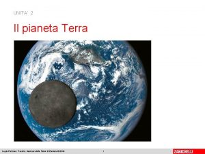
 Prove della sfericità della terra
 Soluzioni il racconto della chimica e della terra
 Soluzioni chimica capitolo 13
 Serie di bowen
 I tre principi dell'io di fichte
 Prove della sfericità della terra
 Seta origini
 Attestato di credito formativo compilato
 F92 disturbi misti della condotta e della sfera emozionale
 Il ritratto della mia bambina
 Culture e formati della televisione e della radio
 Soluzioni il racconto delle scienze naturali
 Soluzioni il racconto della chimica e della terra
 La coccinella in cerca della felicità pdf
 Elena bettinelli
 Il racconto della chimica e della terra soluzioni
 Moti millenari della terra zanichelli
 Fuoco nella sera klee
 Cosa avviene nella metafase
 Metafore nella locandiera
 O sbagliato tante volte
 Scoperte scientifiche della belle epoque
 Arco di distorsione nella comunicazione
 La strada entra nella casa
 Vincolo manicotto
 La luna geme sui fondali del mare
 La luce nella bibbia
 Le donne nella divina commedia
 Lepica
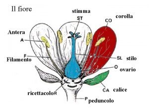
 Filamento fiore
 Non sputare nella carrozza


