Crystalline lens diseases by Hatem G Ammar Assistant


Crystalline lens diseases by Hatem G Ammar Assistant Professor of Ophthalmology Sohag University

Anatomy

Biconvex, avascular, transparent structure enclosed by a capsule. The capsule is the basement membrane secreted by the lens epithelium. The capsule is thickest at the equatorial zone and thinnest at the posterior pole.

The lens epithelium is one layer that lines the ant capsule and the equatorial region(mitotic activity). The cells elongate and lose organelles to form the new lens fibres.

Anatomy
. while the surrounding lens matter")
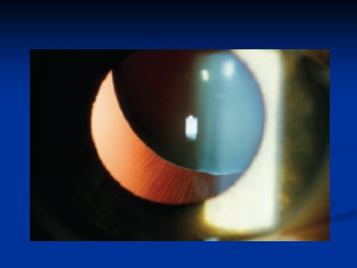
The central compact core is termed the nucleus, (hard). while the surrounding lens matter is the cortex, (soft)

Diameter: 9 -10 mm Thickness: 4 mm Weight: 60 mg-250 mg Refractive index: 1. 42 Dioptric power: 18 (inside), 70 D (outside? ? ? )

physiology Lens transparency? ? ? Lens functions.

Cataract = lens opacity
 n Site of")
Classifications Aetiology n Age (soft ≤ 25 ys, hard >25 ys) n Site of opacity (capular and lenticular) n maturity n
 Congenital 2) Acquired 1) Senile 2) Traumatic 3) Complicated (2 ry)")
Aetiologic Classification 1) Congenital 2) Acquired 1) Senile 2) Traumatic 3) Complicated (2 ry) 4) Drug induced 5) Radiation(uv, xray, infrared) 6) Deficiency (vit C&D) 7) Occupational
 cataract Morphological classification: 1) Subcapsular: Anterior Posterior 2) Nuclear 3) Cortical")
Senile (age related) cataract Morphological classification: 1) Subcapsular: Anterior Posterior 2) Nuclear 3) Cortical

Anterior subcapsular

Posterior subcapsular

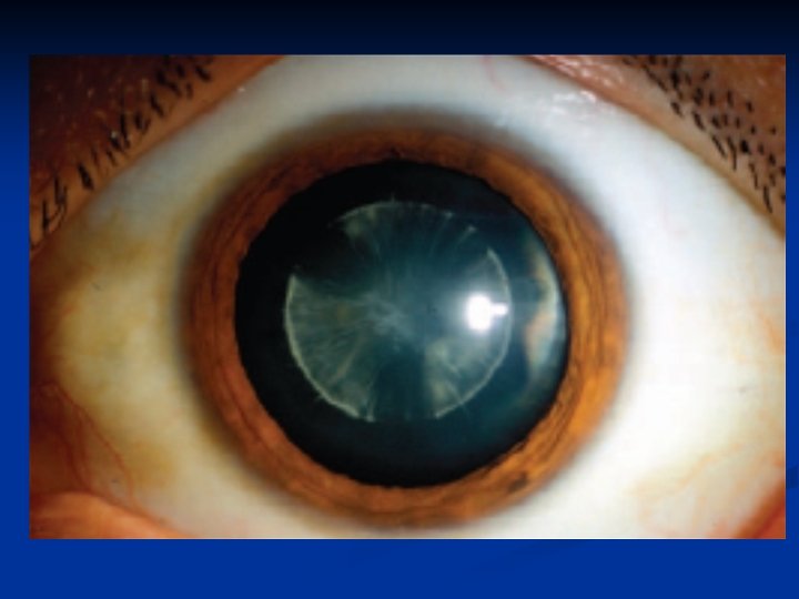
Nuclear Cataract

Cortical Cataract
 Immature. 2) Mature 3) Hypermature & Morgagnian")
Classification according to maturity: 1) Immature. 2) Mature 3) Hypermature & Morgagnian

Mature cataract

Mature cataract

Hypermature cataract

Morgagnian cataract
 Non-surgical ? ? ? 2) Surgical: Indications: 1) Visual")
Treatment of Senile Cataract 1) Non-surgical ? ? ? 2) Surgical: Indications: 1) Visual 2) Medical 3) Cosmetic ? ? ?

Surgical treatment # Preoperative evaluation. # Biometry: IOL power calculation # Intraocular Lenses: * Positioning: - PC IOL - AC IOL * Types: - Rigid - Foldable

Intraocular Lenses
 General 2) Local : Retrobulbar Peribulbar Parabulbar (sub-Tenon) Topical Intracameral")
# Anaesthesia: 1) General 2) Local : Retrobulbar Peribulbar Parabulbar (sub-Tenon) Topical Intracameral
 Extracapsular cataract extraction. 2) Intra capsular cataract extraction. 3)")
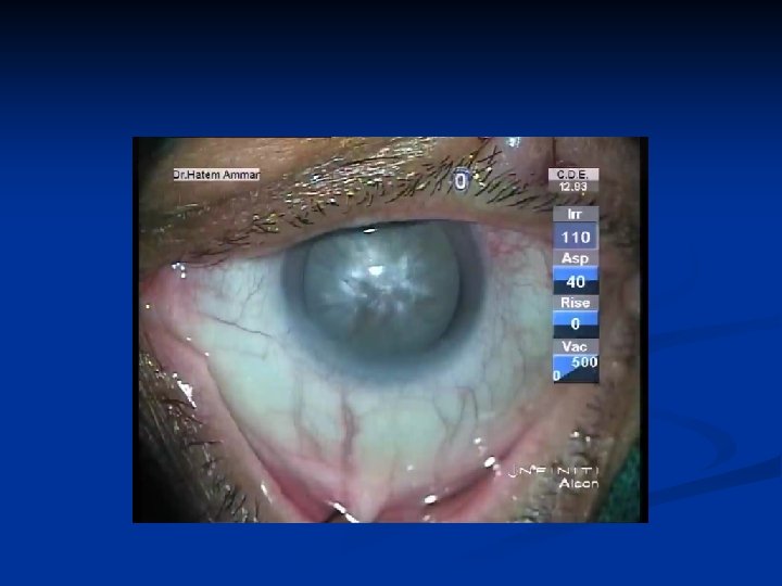
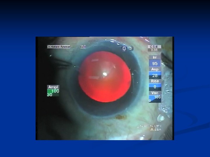
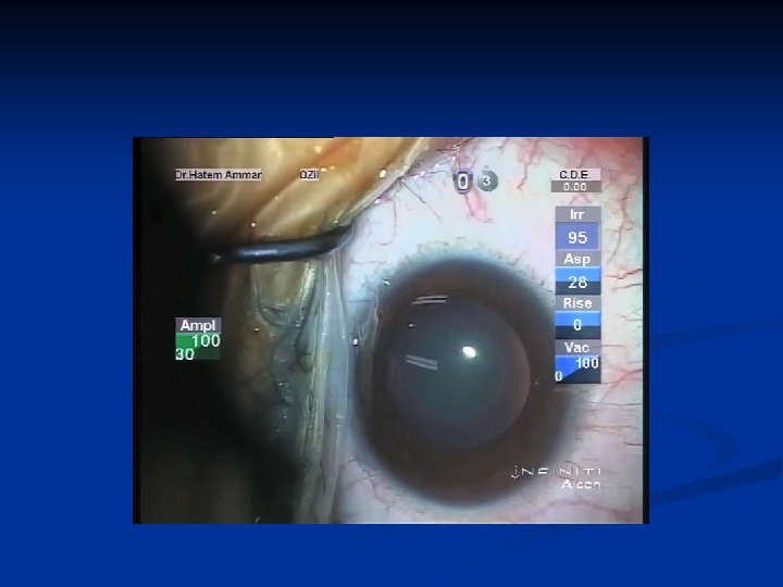
# Types of surgery: 1) Extracapsular cataract extraction. 2) Intra capsular cataract extraction. 3) Phacoemulsification.

Extracapsular cataract extraction.
")
Extracapsular cataract extraction. (with ACIOL)

Phacoemulsification.
 Complications of anesthesia 2) Rupture of posterior capsule 3) Posterior")
# Operative complications: 1) Complications of anesthesia 2) Rupture of posterior capsule 3) Posterior loss of lens fragments. 4) Posterior dislocation of IOL 5) Suprachoroidal hemorrhage.
 Endophthalmitis. 2) Aftercataract 3) Corneal edema. 4) Iris prolapse 5)")
# Postoperative complications: 1) Endophthalmitis. 2) Aftercataract 3) Corneal edema. 4) Iris prolapse 5) IOL malposition 6) RD 7) Cystoid macular edema

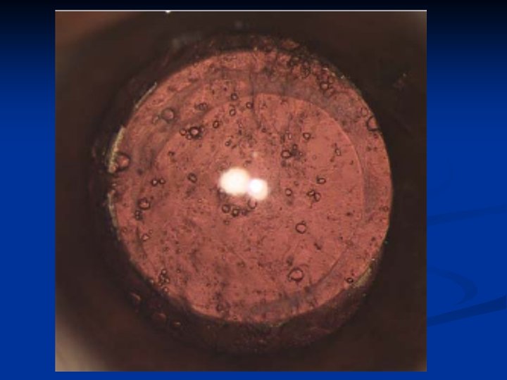
After Cataract
 Cortical material 2) Inflammatory debris 3) Elsching pearls 4) Sommering 5)")
Definition: Etiology: 1) Cortical material 2) Inflammatory debris 3) Elsching pearls 4) Sommering 5) Capsular fibrosis

Elsching pearls

Capsular fibrosis
 Surgical 2) YAG laser")
Treatment 1) Surgical 2) YAG laser
 Cataract without systemic association 2) Cataract with systemic association")
Congenital Cataract 1) Cataract without systemic association 2) Cataract with systemic association
 Cataract without systemic association 1) Zonular: affecting Discrete zone – Nuclear")
Congenital Cataract 1) Cataract without systemic association 1) Zonular: affecting Discrete zone – Nuclear - Lamellar - Capsular - Sutural 2) Polar : anterior OR posterior 3) Others: Coronary – Blue dot – Total Membranous

Congenital Nuclear cataract

Congenital Lamellar cataract

Anterior Polar

Congenital Sutural cataract

Congenital Coronary cataract

Congenital Blue dot cataract
 Cataract with systemic association 1) Metabolic: Galactosaemia 2) Prenatal infections: Congenital rubella 3)")
2) Cataract with systemic association 1) Metabolic: Galactosaemia 2) Prenatal infections: Congenital rubella 3) Chromosomal abnormalities: Down syndrome
")
Galactosaemia (oil droplet appearance)
 Management of the eye (Ocular examination) 2) Management of")
Management of congenital cataract 1) Management of the eye (Ocular examination) 2) Management of the patient (Systemic investigations) 3) Management of the parents (Genetic analysis and serological tests)
 Management of the eye (Ocular examination) 1) Density: the impact on vision -")
1) Management of the eye (Ocular examination) 1) Density: the impact on vision - Very dense - Less dense - Visually insignificant 2) Morphology: a guide for the etiology 3) Associated ocular pathology: (EUGA) - Anterior segment - Posterior segment 4) Other indicators of visual impairment: Nystagmus – Squint 5) Special tests: VEP – Preferential looking
 Management of the patient (Systemic investigations) 1) Examination by pediatrician 2) Serological tests")
2) Management of the patient (Systemic investigations) 1) Examination by pediatrician 2) Serological tests (TORSH) 3) Urine analysis: Galactosemia
 Management of the parents - Reassurance - Informations about the disease - Genetic")
3) Management of the parents - Reassurance - Informations about the disease - Genetic analysis and serological tests
 Bilateral dense 2) Unilateral dense")
Timing for surgery According to laterality and density 1) Bilateral dense 2) Unilateral dense 3) Bilateral partially dense 4) Unilateral partially dense
 Lensectomy. 2) I/A. Management of the posterior capsule")
Surgical techniques: 1) Lensectomy. 2) I/A. Management of the posterior capsule

Lensectomy
 1) Glasses 2) Contact lenses 3) IOLs 4) Occlusion")
Visual rehabilitation: (Correction of aphakia) 1) Glasses 2) Contact lenses 3) IOLs 4) Occlusion (management of amblyopia)
 Cataract")
Complicated (2 ry) Cataract
 Cataract Causes: secondary to 1) Systemic diseases 2) Local (ocular) diseases")
Complicated (2 ry) Cataract Causes: secondary to 1) Systemic diseases 2) Local (ocular) diseases
 Complicated Cataract 2 ry to Systemic diseases: A) D. M: 1) Classical diabetic")
1) Complicated Cataract 2 ry to Systemic diseases: A) D. M: 1) Classical diabetic cataract 2) Age related cataract (earlier in D. M) B) Myotonic dystrophy Stellate posterior subcapsular cataract C) Atopic dermatitis D) Neurofibromatosis type 2 E) Hypocalcaemia F) Ankylostoma infestation
 Complicated Cataract 2 ry to Ocular diseases: 1) Chronic uveitis 2) Glaucoma 3)")
1) Complicated Cataract 2 ry to Ocular diseases: 1) Chronic uveitis 2) Glaucoma 3) High Myopia 4) Hereditary Fundus dystrophies e. g Retinitis pigmentosa

Complicated Cataract 2 ry to Chronic uveitis

Complicated Cataract 2 ry to Chronic uveitis

Drug induced cataract
 Steroids - Systemic & local - Children more susceptible -")
Drug induced cataract: 1) Steroids - Systemic & local - Children more susceptible - Individual (Genetic) variation 2) Miotics 3) Iron & Gold 4) Chlorpromazine, Amiodaron & Allopurinol

Traumatic cataract
 Mechanical Trauma: - Penetrating trauma to the lens - Concussion")
Traumatic cataract Causes: 1) Mechanical Trauma: - Penetrating trauma to the lens - Concussion (blunt) trauma: 1) Vossius ring 2) Rosette cataract 2) Electric shock 3) Ionizing radiation: in ocular tumors 4) Infra-red radiation: true exfoliation (What is pseudoexfoliation syndrome ? )

Vossius ring

Rosette cataract

Lens Subluxation & Dislocation

Definitions Lens Subluxation: partially displaced lens & still remaining in the pupillary area. Dislocation: completely dislocated rendering the pupil aphakic

Lens Subluxation

Posterior Dislocation
 Hereditary: A) Without systemic associations: - Familial ectopia lentis B) With systemic")
Etiology 1) Hereditary: A) Without systemic associations: - Familial ectopia lentis B) With systemic associations: - Marfan syndrome - Homocystinuria - Stickler syndrome 2) Acquired: - Trauma - Large eye: Myopia & Buphthalmos - Anterior uveal tumors - Hypermature cataract
 Observation 2) Surgical")
Evaluation: Ocular & Systemic Management: 1) Observation 2) Surgical

Thank you
- Slides: 80