CRRT Its Not Just for Renal Failure Anymore


 heart failure is commonly")


 relate to distension and fluid")


 Can be triggered by infectious and non-infectious events Infectious causes bacteria")


 MIDDLE MOLECULES- 500 -5000 daltons")


 Continuous Veno-Venous Hemofiltration Effluent")


- Slides: 70
CRRT: It’s Not Just for Renal Failure Anymore Presented by: Sue Fallone, MS, RN, CNN Clinical Nurse Specialist Adult and Pediatric Dialysis Albany Medical Center
Objectives Define Heart Failure Define Sepsis Discuss medical management of heart failure and sepsis Describe indications for CRRT for these disorders Case Study
Clinical syndrome that can result from any structural or functional cardiac disorder that impairs the ability of the ventricle to fill with or eject blood
Incidence of Heart Failure More deaths from heart failure than from all forms of cancer Nearly 1 millions people are admitted to the hospital with CHF and 30%-60% are readmitted Contributed to 53, 000 deaths in the U. S. each year About 550, 000 new cases per year Affects men and women equally Related to the aging population, lower death rate from MI, and improved treatment for heart disease http: //health. usnews. com/health-conditions/heart-health/congestive-heart
Main causes Ischemic heart disease, Cardiomyopathy, Hypertension, Diabetes Other causes: Valvular heart disease, Congenital heart disease, Alcohol and drugs, Hyperdynamic circulation (anemia, thyrotoxicosis, hemochromatosis, Paget's disease), Right heart failure (RV infarct, pulmonary hypertension, pulmonary embolism, cor pulmonale (COPD)), Arrhythmia and Pericardial disease.
Impaired cardiac contractility as in myocardial infarction and cardiomyopathy Ventricular outflow obstruction (pressure overload) as in hypertension and aortic stenosis Impaired ventricular fillings as in mitral stenosis and constrictive pericarditis Volume overload as in mitral regurgitation
Infections Arrhythmias Physical, Dietary, Fluid, Environmental, and Emotional Excesses. Myocardial infarction Pulmonary embolism Anemia Thyrotoxicosis and pregnancy Aggravation of hypertension Rheumatic, Viral, and Other Forms of Myocarditis Infective endocarditis Diabetes
Cardiac remodeling: 1. Hypertrophy & Dilatation C. O. P 2. Sympathetic activity: • H. R. • V. C • E. D. V Angiotensin � Aldosterone
TYPES OF HEART FAILURE Left- sided or left ventricular (LV) heart failure is commonly caused by ischemic heart disease but can also occur with valvular heart disease and hypertension. 2 types of (LV) heart failure diastolic failure is a syndrome consisting of symptoms and signs of heart failure with preserved left ventricular ejection fraction above 45– 50% and abnormal left ventricular relaxation assessed by echocardiography systolic failure is when the left ventricle loses it’s ability to contract normally, can pump enough blood into the systemic circulation Right-sided or right ventricular (RV)heart failure may be secondary to chronic( LV ) heart failure but can occur with primary and secondary pulmonary hypertension, right ventricular infarction.
TYPES of HEART FAILURE Congestive Heart Failure Blood flow out of the heart slows, blood returning to the heart through the veins backs up and congestion in the body’s tissues Will see edema, SOB, can affect kidney function
Symptoms & Signs OF Heart Failure Left heart failure Symptoms are predominantly fatigue, exertional dyspnea, orthopnea and PND Physical signs: Cardiomegaly, gallop functional mitral regurgitation and crackles a the lung bases.
Right Heart Failure Symptoms (fatigue, breathlessness, anorexia and nausea) relate to distension and fluid accumulation in areas drained by the systemic veins. Physical signs are usually more prominent than the symptoms, with: jugular venous distension tender smooth hepatic enlargement dependent pitting edema development of free abdominal fluid (ascites) Pleural effusion (commonly right-sided). Dilatation of the right ventricle produces cardiomegaly and may give rise to functional tricuspid regurgitation. Tachycardia and a right ventricular third heart sound are usual.
Major symptoms & signs of heart failure
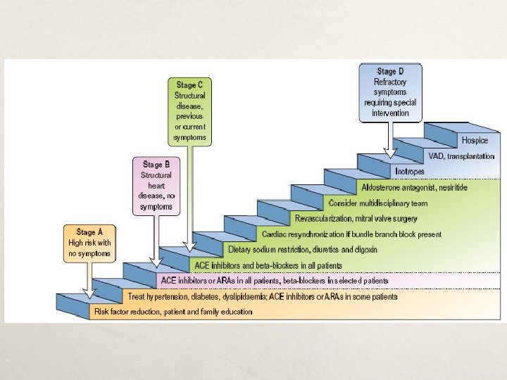
Classification of Heart Failure Functional Capacity Class I – patients with cardiac disease and no limitation of physical activity Class II- patients with cardiac disease slight limitation of physical activity results in fatigue, palpitation, dyspnea or angina Class III-patients with cardiac disease marked limitation of physical activity comfortable at rest Class IV-patients with cardiac disease inability to carry on any physical activity, symptoms of heart failure at rest http: //www. heartorg/HEARTORG/Conditions/Heart
Treatment of heart failure C. O. P 1. Hypertrophy & Dilatation Positive Inotropics 2. Sympathetic activity: • H. R. • V. C • E. D. V vasodilators Angiotensin Diuretics � Aldosterone ACE inhibitors
If Resistant to Diuretics MAY NEED Ultrafiltration
UNLOAD STUDY The UNLOAD study was a randomized, multicenter study of 200 patients involving 28 hospitals and medical centers across the United States. UNLOAD compared the short and long-term safety and efficacy of an advanced form of ultrafiltration therapy(Aquapheresis) to the use of conventional diuretic drug therapy in fluid overloaded heart failure patients. The UNLOAD study was published in the February 13, 2007 issue of Journal of American College of Cardiology. (Costanzo MR et al. JACC 2007; 49(6): 675 -683).
UNLOAD Study Results 28% with greater fluid loss with UF 43% reduction in patients being re-hospitalization for HF 63% fewer hospital days for HF
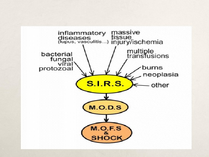
What is SIRS? The systemic inflammatory response syndrome is systemic level of acute inflammation, that may or may not be due to infection, and is generally manifested as a combination of vital sign abnormalities including fever or hypothermia, tachycardia, and tachypnea.
Definitions Severe SIRS – SIRS in which at least 1 major organ system has failed. Sepsis – SIRS which is secondary to infection. Severe Sepsis – Severe SIRS which is secondary to infection. Septic Shock – Severe sepsis resulting in hypotensive cardiovascular failure.
Systemic Inflammatory Reponse(SIRS) Can be triggered by infectious and non-infectious events Infectious causes bacteria or fungi Non infectious causes are prancreatitis, burns, trauma SIRS is the term used for noninfectious causes
Criteria for SIRS Requires 2 of the following 4 features to be present: Temp >38. 3° or <36. 0° C Tachypnea (RR>20 or MV>10 L) Tachycardia (HR>90, in the absence of intrinsic heart disease) WBC > 10, 000/mm 3 or <4, 000/mm 3 or >10% band forms on differential
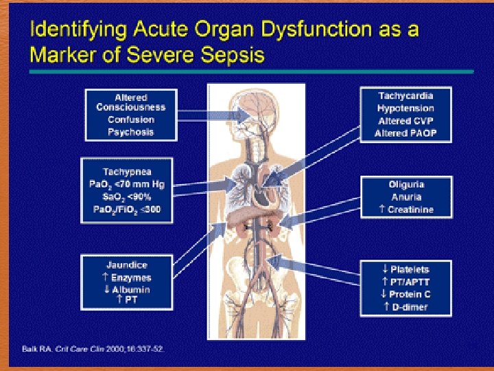
Criteria for Severe SIRS Must meet criteria for SIRS, plus 1 of the following: Altered mental status SBP<90 mm. Hg or fall of >40 mm. Hg from baseline Impaired gas exchange Metabolic acidosis (p. H<7. 30 & lactate > 1. 5 x upper limit of normal) Oliguria (<0. 5 m. L/kg/hr) or renal failure Hyperbilirubinemia Coagulopathy (platelets < 80, 000 -100, 000/mm 3, INR >2. 0, PTT >1. 5 x control, or elevated fibrin degredation products)
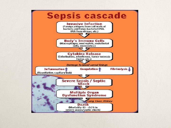
Pathophysiology of Sepsis Overwhelming inflammatory response Increased production of proimflamatory cytokines and decreased production of cytokines( which inhibit inflammation) Clotting cascade activated Peripheral Vasodilatation systemic vascular resistance
Pathophysiology of Sepsis continued C/O decreases Intravascular fluid loss Decreased pre load-hypotension ATN-renal hypoperfusion and ischemic injury MODS MOF
Relationship Between SIRS and Sepsis Adapted from: Marini JJ, et al. Critical Care Medicine, 2 nd ed. 1997.
Risk Factors for SIRS/Sepsis Age Indwelling lines/catheters Immunocompromised states Malnutrition Alcoholism Malignancy Diabetes Cirrhosis Male sex Genetic predisposition?
Prognosis Overall mortality from SIRS/sepsis in the U. S. is approximately 20%. Mortality is roughly linearly related to the number of organ failures, with each additional organ failure raising the mortality rate by 15%. Hypothermia is one of the worst prognostic signs. Patients presenting with SIRS and hypothermia have an overall mortality of ~80%.
Treatment Fluid Resuscitation Vasopressors Antibiotics Eradication of infection Ventilatory support, activated protein C, steroids, glycemic control, nutrition CRRT
CONTIN UOUS RENAL REPLACMENT THERAPY
CRRT Definition CRRT = Continuous Renal Replacement Therapy Defined as “Any extracorporeal blood purification therapy intended to substitute for impaired renal function over an extended period of time and applied for or aimed at being applied for 24 hours /day. ” * * Bellomo R. , Ronco C. , Mehta R, Nomenclature for Continuous Renal Replacement Therapies, AJKD, Vol 28, No. 5, Suppl 3, November 1996
Introduction to CRRT Why continuous therapies? Continuous therapies closely mimic the native kidney in treating ARF and fluid overload Slow & gentle Remove fluid and waste products over time Tolerated well by the hemodynamically unstable patient
Three Compartment Model Intracellular Space Extracellular Space Circulating Blood Volume Toxins Fluid 23 L 17 L 40 Liters Toxins Fluid 5 Liters Dialyzer Intra 2 Vascular Space Toxins Fluid
Indications for Therapy Acute kidney injury- preferred in the critically ill patient Fluid overload- can removed large amounts of fluid slowly Hemodynamically unstable- continuous therapy allow for slow hourly fluid removal which allows the intravascular spaces to refill
Indications continued Highly catabolic patients who need increased clearance rates Patients needing large molecular weight substances removed Sepsis
Molecular Weight SMALL MOLECULES- 0 -500 daltons (urea, creatinine) MIDDLE MOLECULES- 500 -5000 daltons ( vitamin B 12) LARGE MOLECULES- 5000 -50, 000 daltons ( heparin, Beta 2 drugs)
CRRT Modalities SCUF OR ULTRAFILTRATION - Slow Continuous Ultra. Filtration CVVHD - Continuous Veno-Venous Hemo. Dialysis CVVH - Continuous Veno-Venous Hemofiltration CVVHDF – Continuous Veno-Venous Hemodiafiltration
SCUF/Ultrafiltration Primary therapeutic goal: Safe management of fluid removal Patient UF rate ranges up to 2 L/Hr No dialysate; No replacement fluids No molecule removal Large fluid removal via ultrafiltration Blood Flow rates = 100 -200 ml/min
Access SCUF/ULTRAFILTRATION Return Slow Continuous Ultra. Filtration Effluent
Ultrafiltration Particles move through a semi-permeable membrane by use of HYDROSTATIC pressure. The separation of particles from a suspension by passage through a filter. The separation is accomplished by convective transport. 45
Convection – Step 1 Filter Action Na Red Cell Na H 2 O U K Red Cell U Red Cell Na K U U H 2 O K Red Cell U Na K Na H 2 O U Na K K H 2 O Na Red Cell Na Na Na U H 2 O Na H 2 O K U Na Na H 2 O Na Na Na U H 2 O Na K K
Solute Removal by Convection: The movement of solutes with a water-flow, “solvent drag”, e. g. . . the movement of membrane-permeable solutes with water across the semipermeable membrane
Access CVVH Return Replacement (pre or post dilution) Continuous Veno-Venous Hemofiltration Effluent
Molecular Transport Mechanisms Convection - The movement of solutes with a water-flow, “solvent drag”, the movement of membranepermeable solutes with water across the semipermeable membrane
Convection – Step 1 Filter Action Na Red Cell Na H 2 O U K Red Cell U Red Cell Na K U U H 2 O K Red Cell U Na K Na H 2 O U Na K K H 2 O Na Red Cell Na Na Na U H 2 O Na H 2 O K U Na Na H 2 O Na Na Na U H 2 O Na K K
Solute Removal by Convection: The movement of solutes with a water-flow, “solvent drag”, e. g. . . the movement of membrane-permeable solutes with water across the semipermeable membrane
CVVHD - Continuous VV Hemodialysis Primary therapeutic goal: Solute removal by diffusion Safe fluid volume management by ultrafiltration Requires Dialysate solution Patient UF rate ranges 2 -7 L/24 hours (~300 ml/hr) Dialysate Flow rate = 15 -45 ml/min (~2 L/hr) Blood Flow rate = 100 -200 ml/min No replacement solution Solute removal determined by Dialysate Flow rate.
Diffusion – Filter Action Na Na K Mg Mg Na H 2 O Na U Na K H 2 O Mg U H 2 O Na K H 2 O Na Na U H 2 O Na K H 2 O Mg K U H 2 O U Na Mg U U Na K Na U U K Na Na Na K
Vascular Access Depending on the device used lumen size matters If using Aqua. Dex Flex. Flow Fluid Removal System midline catheters can be used If using CRRT devices hemodialysis type catheters need to be placed.
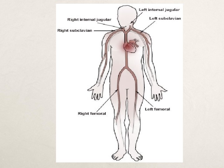
Catheter Size q Adults q q q 12. 5 to 14 french Length will vary 16, 19, 24, cm Femoral placement least preferred q Children (weight based) q 5 french single catheter 7 fr dual lumen 8 fr dual lumen 10 fr dual lumen 11 fr dual lumen q Length 9 cm, 10 cm, 12 cm, 15 cm
Case Study #1 Mr. G is a 60 year old man with CAD s/p MI and PTCA to LAD in 1997, dyslipidemia, and tobacco use who called 911 for severe chest pain on 11/01/10. This pain was similar in nature to his previous MI.
ECG in the ambulance
History In the ambulance en route to the emergency room, the patient developed two episodes of ventricular fibrillation which both successfully responded to DC cardioversion. After arrival to the cath lab, the patient developed cardiogenic shock and recurrent ventricular fibrillation requiring multiple shocks (he was shocked 11 times in the cath lab prior to intervention) and intubation with mechanical ventilation.
Cath Lab Course Coronary angiography showed: Totally occluded mid LAD with thrombus Mild diffuse atherosclerosis of left circumflex and right coronary arteries Soon after the first injection there was proximal propagation of the LAD thrombus which occluded the left main coronary artery A wire was passed to the distal LAD and an Angio. Jet thrombectomy device was used which re-established flow
Cath Lab Course After the Impella device was placed, the patient had no further episodes of ventricular fibrillation
Immediately Post Cath Patient admitted to the CCU on IV Epinephrine, Dobutamine, and Dopamine continuous infusions Echocardiogram the next day showed severe anterior wall hypokinesis with EF 25% The patient was placed on CVVH then on SCUF to remove excess fluid
Hospital Course Hospital day 3: Impella device was removed Hospital Day 6: Repeat echocardiogram, EF 50 -55% Hospital Day 8: Extubated, neurologically intact Hospital Day 16: Discharged to home
Case Study #2 Alan is a 20 year old admitted to a cardiology unit with CHF and Situs Inversus. He had SOB , anascara, arrythmias. His blood pressure was 110/60 mm Hg. He has a serum creatinine of 1. 5 mg/dl. He is in need of a pacemaker but first needs 10 liters of fluid removed before placement of a pacemaker. He is started on furosemide 80 mg every 8 hours and metolazone 10 mg/d for 2 days. On day three he is given mannitol 25 g every eight hours. He is putting out 3 L of urine a day but has only decreased his net fluid loss by 3 L due to lack of adherance to his fluid restriciton
Case Study Continued Because of his need for a pacemaker, the decision was made to place the patient on SCUF. After three days of therapy the patient was at his dry weight and stable and was able to receive his pacemaker Consideration has to be given related to rate of fluid removal and his overall renal function Patient was discharged to home with a follow up to a nephrologist
Case Study #3 Mrs. D was admitted to MICU for sepsis. She had been hypotensive that required vasopressors. During the course of her stay in MICU, she developed AKI. To manage her fluid and electrolytes, she was started on CRRT. She seemed to tolerate CRRT well. On her 5 th day of therapy, her Serum Creatinine was down to 1. 2 from 6. 9 and her electrolytes were stable, her BP was borderline with MAP > 60 mm. Hg and < 70 mm. Hg. CRRT was discontinued and only to be restarted after 2 days when the patient became hypotensive again that regular HD was not possible given her hemodynamic parameters. Patient was started on phenylephrine at 200 mcg/min and norepinephrine at 10 mcg/min. On the 3 rd day of the 2 nd therapy, the patient had the following data:
Patient Data Time BP CVP Vasopressor I and O Balance 2/15 2200 125/60 14 Off -100 2/16 0600 110/65 12 Off -250 2/16 1900 73/85 6 ON -1100 2/17 2300 108/55 10 Off -500
Questions What happened in this scenario? What should have been considered in setting the net fluid removal rate? How would we assess for the intravascular vs extra-vascular fluid status? When will be the right time to advocate for discontinuance of CRRT?
Conclusion CRRT therapies can be applied to many clinical situations The patient goals/outcomes can be enhanced with early initiation of this therapy CRRT IS NOT JUST FOR RENAL FAILURE
THANK YOU