
Critical Care of the Stroke Patient Ryan Keiler
- Slides: 39
Critical Care of the Stroke Patient Ryan Keiler BSN, RN, CCRN, CNRN Presented to: Northern Illinois Chapter AANN Stroke Symposium – June 4 th, 2016
Conflicts and Disclosures This speaker has no financial conflicts or disclosures.
Objective Describe key concepts in the management of critical care stroke patients in the first 24 hours Identify key complications that can arise in the critical care stroke patient and discuss management of these complications Identify patient care management to improve functional outcomes and decrease complications Describe the nurse’s role in incorporating the patient and family into care
Critical Care of the Stroke Patient What are some reasons that stroke patients end up in the ICU? Need for intensive neurological assessment and monitoring Need for aggressive hemodynamic management Need for interventions that require frequent monitoring, i. e. EVD, post cerebral angiography with intervention Need for intubation due to neurological status Other comorbidities
Re-Perfusing the Penumbra For the acute stroke patient in the critical care setting, the overarching goal of care is to re-perfuse the penumbra. How can you as a Neurocritical care nurse achieve this?
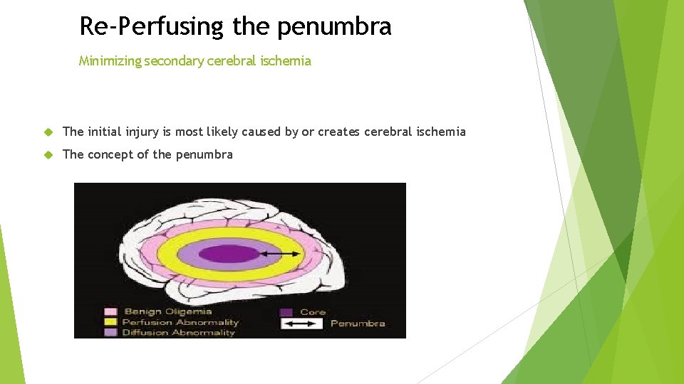
Re-Perfusing the penumbra Minimizing secondary cerebral ischemia The initial injury is most likely caused by or creates cerebral ischemia The concept of the penumbra
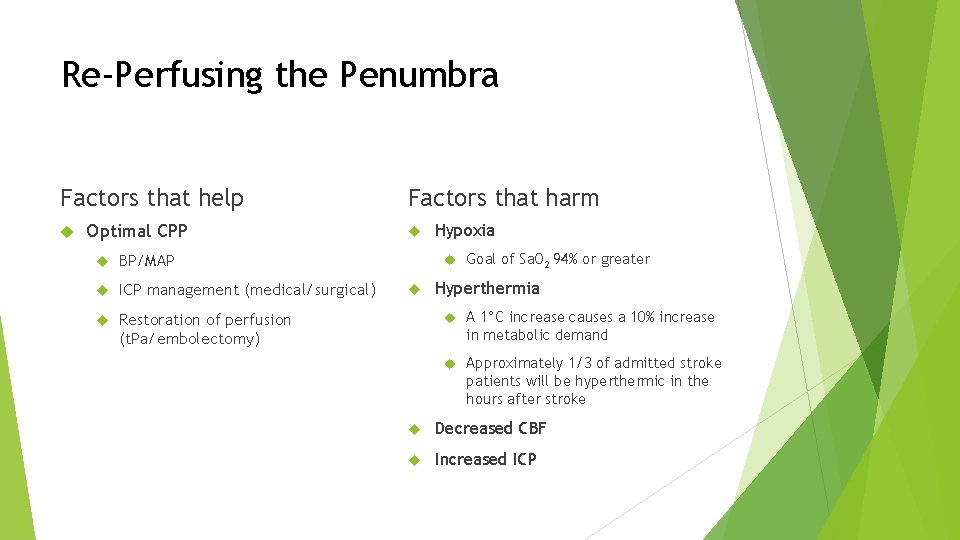
Re-Perfusing the Penumbra Factors that help Optimal CPP BP/MAP ICP management (medical/surgical) Restoration of perfusion (t. Pa/embolectomy) Factors that harm Hypoxia Goal of Sa. O 2 94% or greater Hyperthermia A 1°C increase causes a 10% increase in metabolic demand Approximately 1/3 of admitted stroke patients will be hyperthermic in the hours after stroke Decreased CBF Increased ICP
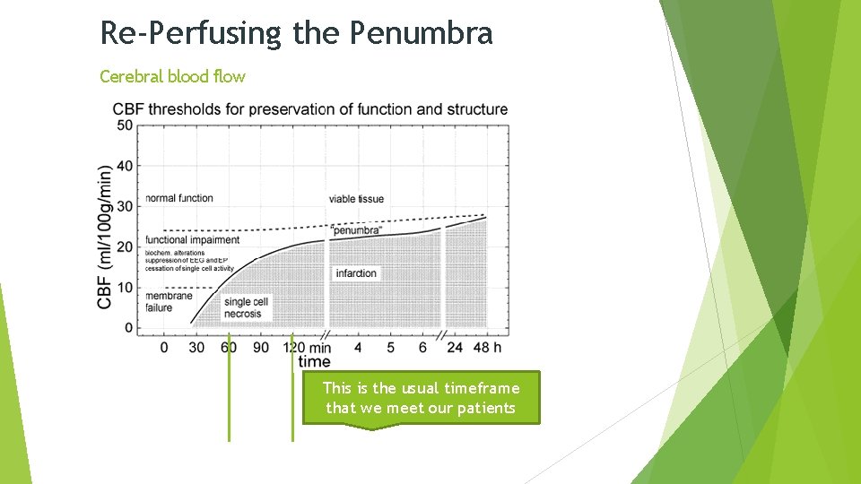
Re-Perfusing the Penumbra Cerebral blood flow This is the usual timeframe that we meet our patients
Critical Care of Stroke – 1 st 24 Hours Managing secondary cerebral ischemia Time is brain The role of the Neuro ICU nurse is also to manage the patient’s hemodynamics in such a manner that optimal cerebral perfusion can be maintained. Hemodynamics in the critical care stroke patient Ventilation Perfusion Circulation Pressure management
Critical Care of Stroke – 1 st 24 Hours ABC When caring for the stroke patient in the critical care setting there are many different aspects of care that differentiate this patient population from others, however there is one aspect of critical care that always remains the same: You must always maintain your patient’s ABC’s before you can take care of their neurological issues As with any critical care patient, the most important assessment begins with A and works its way to D Airway Breathing Circulation Disease
Critical Care of Stroke – 1 st 24 Hours Airway & Breathing Ventilation strategies are similar to those in other patient populations The airway is secured if the neurological injury is such that the patient’s ability to protect their airway is at risk Early intubation is a key strategy Proactive is much better and safer than reactive Ventilator strategies with Neuro ICU patients must also take into account the entire patient picture, still working with a low tidal volume goal (5 -7 m. L/kg) to minimize barotrauma What about minimizing PEEP for patients with an increased ICP?
Critical Care of Stroke – 1 st 24 Hours Ventilators and ICP Mechanical ventilation is a life-saving technology, however are the physiologic changes associated with positive pressure ventilation deleterious to the stroke patient? PEEP does not effect ICP There also some benefits to mechanical ventilation that may aid the nurse in managing the acutely-ill Neuro patient The patient can be better sedated to lower ICP The Pa. CO 2 can be more directly managed when ICP is an issue The Pa. O 2 must also be maximized in order to prevent hypoxia
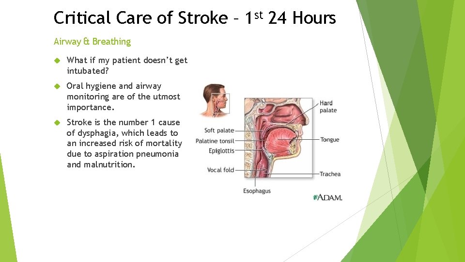
Critical Care of Stroke – 1 st 24 Hours Airway & Breathing What if my patient doesn’t get intubated? Oral hygiene and airway monitoring are of the utmost importance. Stroke is the number 1 cause of dysphagia, which leads to an increased risk of mortality due to aspiration pneumonia and malnutrition.
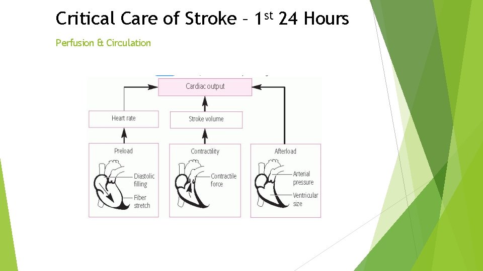
Critical Care of Stroke – 1 st 24 Hours Perfusion & Circulation What are the key physiologic mechanisms that control perfusion to the brain? Vasculature This is the key concept behind stroke, TBI, and many other neurological diseases Blockage in the arterial supply to the brain or increased pressure and mass effect from the disease process itself directly or indirectly alters the blood supply that the injured area Cardiac output At rest, CBF accounts for 15 -20% of the body’s total cardiac output
Critical Care of Stroke – 1 st 24 Hours Perfusion & Circulation
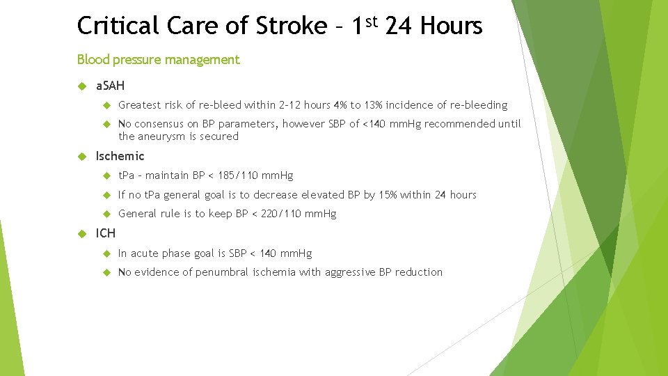
Critical Care of Stroke – 1 st 24 Hours Blood pressure management a. SAH Greatest risk of re-bleed within 2 -12 hours 4% to 13% incidence of re-bleeding No consensus on BP parameters, however SBP of <140 mm. Hg recommended until the aneurysm is secured Ischemic t. Pa – maintain BP < 185/110 mm. Hg If no t. Pa general goal is to decrease elevated BP by 15% within 24 hours General rule is to keep BP < 220/110 mm. Hg ICH In acute phase goal is SBP < 140 mm. Hg No evidence of penumbral ischemia with aggressive BP reduction
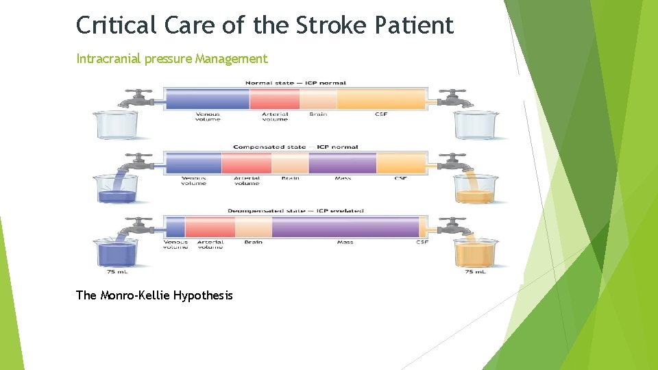
Critical Care of the Stroke Patient Intracranial pressure Management The Monro-Kellie Hypothesis
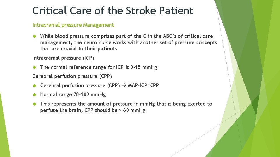
Critical Care of the Stroke Patient Intracranial pressure Management While blood pressure comprises part of the C in the ABC’s of critical care management, the neuro nurse works with another set of pressure concepts that are crucial to their patients Intracranial pressure (ICP) The normal reference range for ICP is 0 -15 mm. Hg Cerebral perfusion pressure (CPP) MAP-ICP=CPP Normal range 70 -100 mm. Hg This represents the amount of pressure in mm. Hg that is being exerted to perfuse the brain, CPP should be ≥ 60 mm. Hg
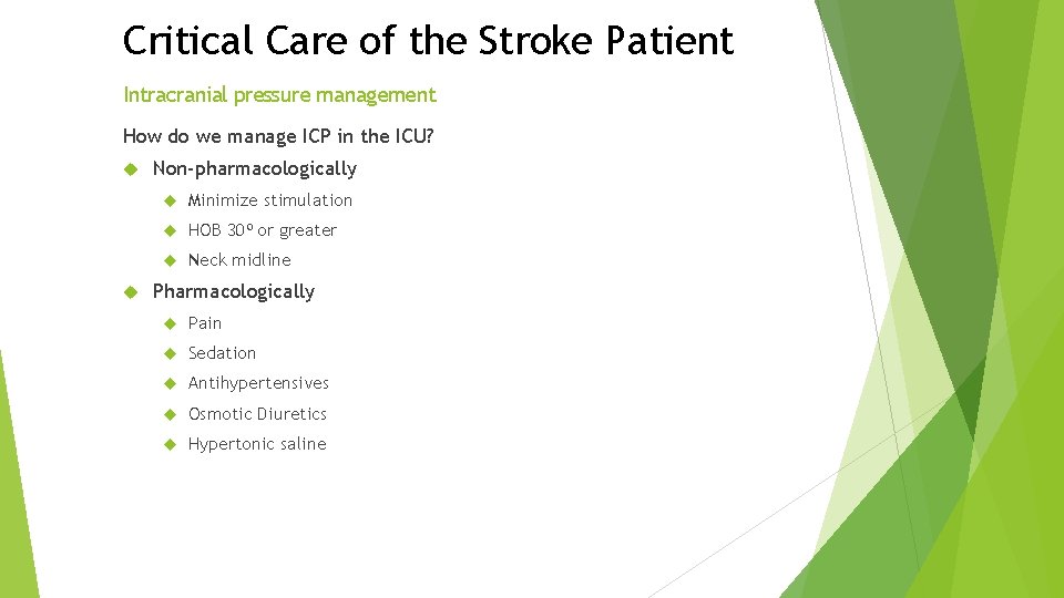
Critical Care of the Stroke Patient Intracranial pressure management How do we manage ICP in the ICU? Non-pharmacologically Minimize stimulation HOB 30⁰ or greater Neck midline Pharmacologically Pain Sedation Antihypertensives Osmotic Diuretics Hypertonic saline
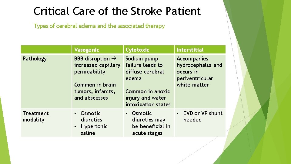
Critical Care of the Stroke Patient Types of cerebral edema and the associated therapy Vasogenic Cytotoxic Interstitial Pathology BBB disruption Sodium pump increased capillary failure leads to permeability diffuse cerebral edema Common in brain tumors, infarcts, Common in anoxic and abscesses injury and water intoxication states Accompanies hydrocephalus and occurs in periventricular white matter Treatment modality • Osmotic diuretics • Hypertonic saline • EVD or VP shunt needed • Osmotic diuretics may be beneficial in acute stages
Critical Care of the Stroke Patient Intracranial pressure management Mechanically – surgical interventions Extraventricular drain (EVD) Indications for EVD placement: Obstructive hydrocephalus SAH resulting in acute hydrocephalus SAH Hunt & Hess grade ≥ 3
Critical Care of the Stroke Patient Intracranial pressure management Cerebral edema Surgical mass lesions Why is an EVD useful? Utilization of an EVD is “the only reliable method to monitor cerebral perfusion pressure (CPP) and assess for cerebral hypo-perfusion and intracranial hypertension” (AANN, EVD, p. 6) An EVD allows the nurse to directly monitor the patient’s intracranial pressure (ICP) and manage that pressure through therapeutic drainage of CSF An EVD can also allow the clinician to instill antibiotics directly into the CSF (MD only)
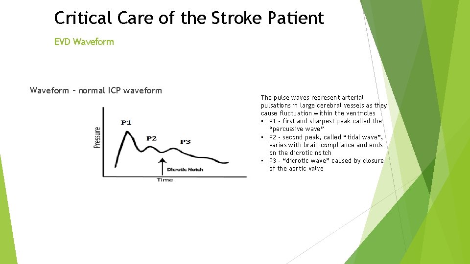
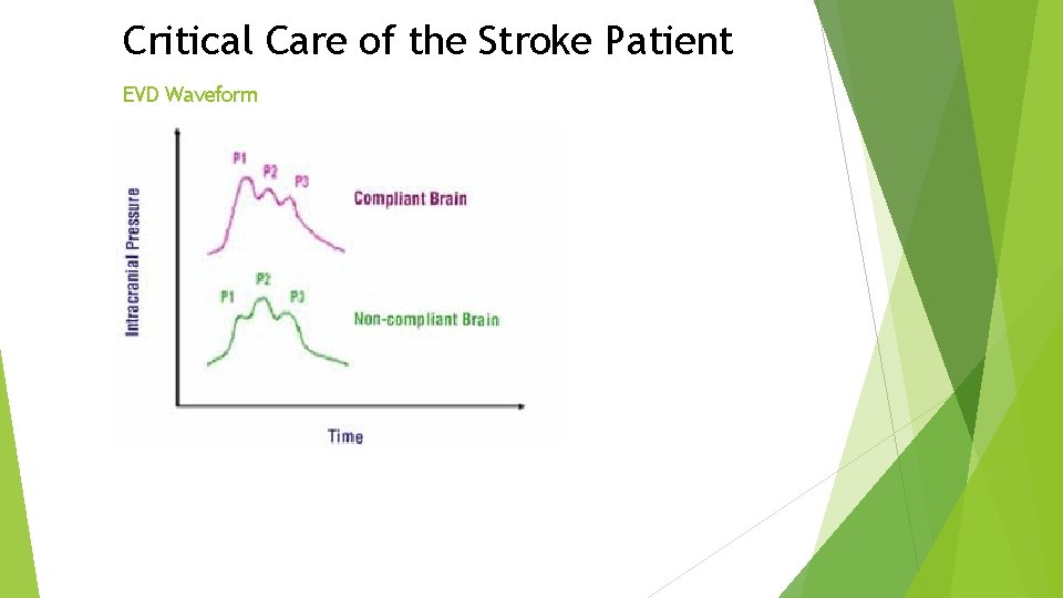
Critical Care of the Stroke Patient EVD Waveform – normal ICP waveform The pulse waves represent arterial pulsations in large cerebral vessels as they cause fluctuation within the ventricles • P 1 – first and sharpest peak called the “percussive wave” • P 2 – second peak, called “tidal wave”, varies with brain compliance and ends on the dicrotic notch • P 3 – “dicrotic wave” caused by closure of the aortic valve
Critical Care of the Stroke Patient EVD Waveform
Critical Care of the Stroke Patient Surgical Management Burr holes Often utilized to drain subdural or epidural hematomas Small holes are drilled in the skull depending of the area that needs to be evacuated Drains may be left in place Hemi-craniectomy One half of the skull is removed Indications are many Decompressive hemicrani Utilized in emergent situations when there is malignant cerebral edema or for refractory ICP management While this procedure may help decrease ICP and allow more room for swelling, it has not been shown to decrease overall mortality Sub-occipital craniectomy
Critical Care of the Stroke Patient Mobility and nutrition Mobility Mobilizing stroke patients has been shown to increase functional capacity at 3 months. Literature has shown that with stroke patients, there is no statistically significant improvement in functional status between 2 months post-stroke and 5 years post-stroke. Nutrition Plays a key role in healing and rehabilitation. Large bore NG tubes can initially be used for intubated patients, however small bowel feeding tubes better for long term and pose less risk of aspiration.
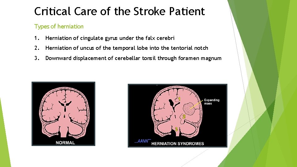
Critical Care of the Stroke Patient Herniation and brain death Prolonged elevation ICP result cerebral herniation Folds in dura mater divide the intracranial cavity into several compartments Cerebral herniation is distortion & displacement of brain from 1 compartment to another Herniation is a life-threatening event
Critical Care of the Stroke Patient Types of herniation 1. Herniation of cingulate gyrus under the falx cerebri 2. Herniation of uncus of the temporal lobe into the tentorial notch 3. Downward displacement of cerebellar tonsil through foramen magnum
Critical Care of the Stroke Patient Classic signs of herniation Decreased LOC Pupillary abnormalities – fixed & dilated pupil(s) Motor dysfunction (hemiplegia, decortication, or decerebration) Impaired brainstem reflexes – cranial nerves (corneal, gag, swallowing) “Cushing’s triad” of alterations in vital signs Bradycardia Hypertension Respiratory irregularity Vital to identify changes in patient early
Critical Care of the Stroke Patient Brain death – coma with known cause The first part of completing brain death testing is that the patient must have sustained an IRREVERSIBLE, CATASTROPHIC neurological injury. This injury would thus cause a coma and does not necessarily need to be neurological in origin (i. e. cardiac arrest). There are many metabolic and pharmacologic abnormalities that need to be accounted for first: Electrolyte abnormalities such as hyper or hyponatremia Severe acid-base imbalances, severe hyperammonemia Hypothermia (patient must have a core temp >96. 8⁰F or 36 ⁰C Drug intoxication, poisoning, sedatives, or neuromuscular blocking agents Pa. CO 2 must be normalized and SBP ≥ 100 Once these mitigating factors can be addressed, the care team can now move to step 2 in the process.
Critical Care of the Stroke Patient Brain death - absence of brain stem reflexes The coma with a known cause demonstrates that there is no cerebrally-mediated interaction with the environment, now we must work our way down to the brainstem. There are 5 key reflexes originating from the brainstem that must be tested for: 1. Pupillary response to light – CN II, III, midbrain 2. Absence of ocular movements (occulocephalic) – CN III, IV, VIII, pons, midbrain 3. Absence of corneal reflex – CN III, V, VII, pons 4. Cold calorics (occoulovestibular)– CN III, IV, VII, pons, midbrain 5. Absence of pharyngeal and tracheal reflexes CN IX, X, medulla
Critical Care of the Stroke Patient Brain death - apnea testing The apnea test is the final component of the brain death examination process. The apnea test, if positive proves that the patient no longer has the ability to initiate a breath when the Pa. CO 2 in arterial blood begins to rise. Apnea testing process: Prerequisites Normotension SBP ≥ 100 with or without pressors Normothermia core temp ≥ 96. 8⁰F or 36 ⁰C Pa. CO 2 35 -45 mm. Hg and no known history of CO 2 i. e. COPD Absence of hypoxia
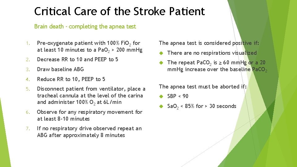
Critical Care of the Stroke Patient Brain death - completing the apnea test 1. Pre-oxygenate patient with 100% Fi. O 2 for at least 10 minutes to a Pa. O 2 > 200 mm. Hg 2. Decrease RR to 10 and PEEP to 5 3. Draw baseline ABG 4. Reduce RR to 10, PEEP to 5 5. Disconnect patient from ventilator, place a tracheal cannula at the level of the carina and administer 100% O 2 at 6 L/min 6. Observe for any respiratory movement for at least 8 -10 minutes 7. If no respiratory drive observed repeat an ABG after approximately 8 minutes The apnea test is considered positive if: There are no respirations visualized The repeat Pa. CO 2 is ≥ 60 mm. Hg or a 20 mm. Hg increase over the baseline Pa. CO 2 The apnea test must be aborted if: SBP < 90 Sa. O 2 < 85% for > 30 seconds
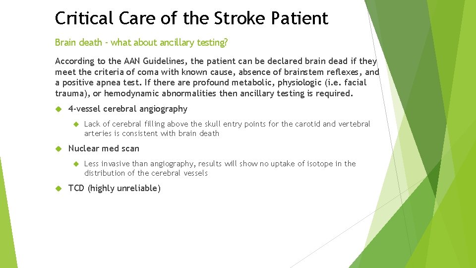
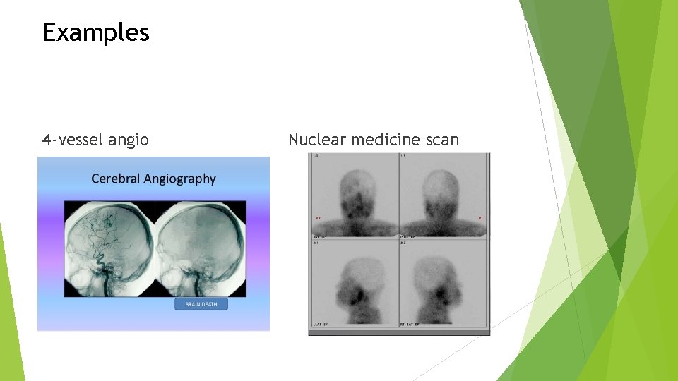
Critical Care of the Stroke Patient Brain death - what about ancillary testing? According to the AAN Guidelines, the patient can be declared brain dead if they meet the criteria of coma with known cause, absence of brainstem reflexes, and a positive apnea test. If there are profound metabolic, physiologic (i. e. facial trauma), or hemodynamic abnormalities then ancillary testing is required. 4 -vessel cerebral angiography Nuclear med scan Lack of cerebral filling above the skull entry points for the carotid and vertebral arteries is consistent with brain death Less invasive than angiography, results will show no uptake of isotope in the distribution of the cerebral vessels TCD (highly unreliable)
Examples 4 -vessel angio Nuclear medicine scan
Critical Care of the Stroke Patient Brain death - pitfalls Automatisms and spinally-mediated reflexes Cardiac-mediated ventilator triggering
Patient-centered care Putting it all together From the second we meet the stroke patient in the critical care setting until the second that they leave we must always keep the patient and their family at the center of the care we give Communication Compassion Integration
References American Association of Neuroscience Nurses (AANN) Clinical Practice Guideline Series. (2011). Care of the Patient Undergoing Intracranial Pressure Monitoring, External Ventricular Drainage or Lumbar Drainage. Pg. 4 -27 Arbour, R. B. (2013). Brain death: Assessment, controversy, and confounding factors. Critical Care Nurse. 33(6), 27 -46. doi: 10. 4037/ccn 2013215 Bernhardt, J. (2015). Efficacy and safety of very early mobilization within 24 h of stroke onset (AVERT): a randomized controlled trial. Lancet, 386, 46 -55. doi: 10. 1016/S 0140 -6376(15)60690 -0 Connolly ES, J. , Rabinstein, A. , Carhuapoma, J. , Derdeyn, C. , Dion, J. , Higashida, R. , &. . . Vespa, P. (2012). Guidelines for the management of aneurysmal subarachnoid hemorrhage: a guideline for healthcare professionals from the American Heart Association/american Stroke Association. Stroke (00392499), 43(6), 1711 -1737 27 p. Chippala, P. & Sharma, R. (2015). Effects of very early mobilization on disability and adverse events in the first 3 months post stroke: a single-blinded, randomized controlled trial. International Journal of Health Sciences and Research. 5(10), 166 -174.
References Hemphill 3 rd, J. C. , Greenberg, S. M. , Anderson, C. S. , Becker, K. , Bendok, B. R. , Cushman, M. , &. . . Hemphill, J. 3. (2015). Guidelines for the Management of Spontaneous Intracerebral Hemorrhage: A Guideline for Healthcare Professionals From the American Heart Association/American Stroke Association. Stroke (00392499), 46(7), 2032 -2060 29 p. doi: 10. 1161/STR. 000000069 Jauch, E. C. , Saver, J. L. , Adams Jr, H. P. , Bruno, A. , Connors, J. B. , Demaerschalk, B. M. , &. . . Yonas, H. (2013). Guidelines for the early management of patients with acute ischemic stroke: a guideline for healthcare professionals from the american heart association/american stroke association. Stroke (00392499), 44(3), 870 -947 78 p. doi: 10. 1161/STR. 0 b 013 e 318284056 a Meyer, S. , Verheyden, G. , Brinkmann, N. , Dejaeger, E. , Weerdt, W. D. , Feys, H…Wit, L. D. (2015). Functional and motor outcome 5 years after stroke is equivalent to outcome at 2 months. Stroke, 46, 1613 -1619. doi: 10. 1161/STROKEAHA. 115. 009421 Tortora, G. J. & Derrickson, B. (2014). Principles of Anatomy & Physiology. (14 th ed. ) New Jersey: Wiley Wijdicks, E. F. , Varels, P. N. , Gronseth, G. S. , & Greer, D. M. (2010). Evidence-based guideline update: Determining brain death in adults. Neurology. 74, 1911 -1918. doi: 10. 1212/WNL. 0 b 013 e 3181 e 242 a 8