Cranial Nerves Cerebral Cortex Two hemispheres Separated by

Cranial Nerves

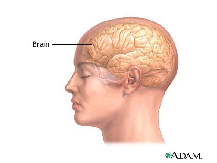
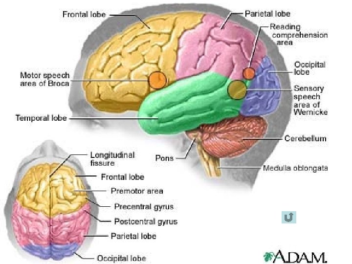
Cerebral Cortex • • • Two hemispheres Separated by longitudinal fissure Right and Left hemispheres Connected by Corpus Callosum Outer gray matter – neurones Inner white matter – nerve fibres, and neuroglia • Sulci and gyri • Frontal parietal temporal and occipital

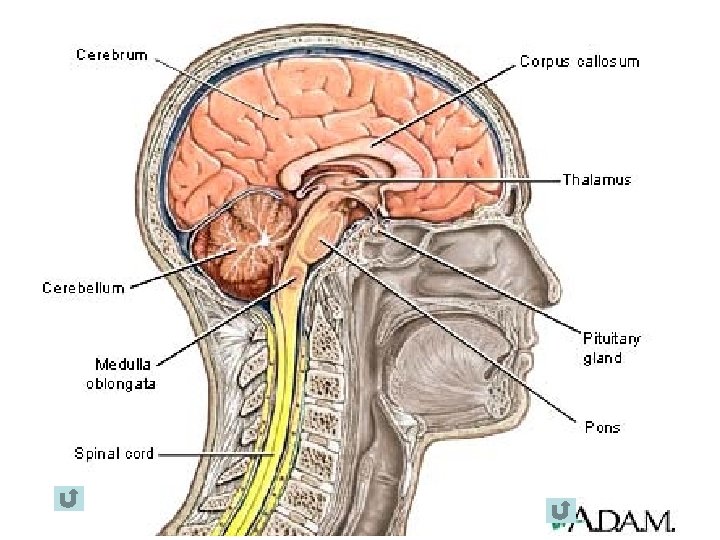
Brain Stem • • Midbrain Pons Medulla oblongata Midbrain connects the pons and the cerebellum with the cerebral hemispheres • Cranial nerves III and IV originate in the midbtrain • Pons in front of cerebellum; between the midbrain and medulla and is a bridge

Cerebellum • Separated from the cerebral hemispheres by tentorium cerebelli • Responsible for coordination of movement – balance position sense (awareness of where each part of the body is)

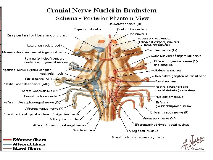
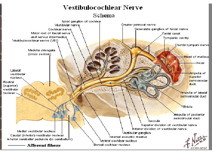
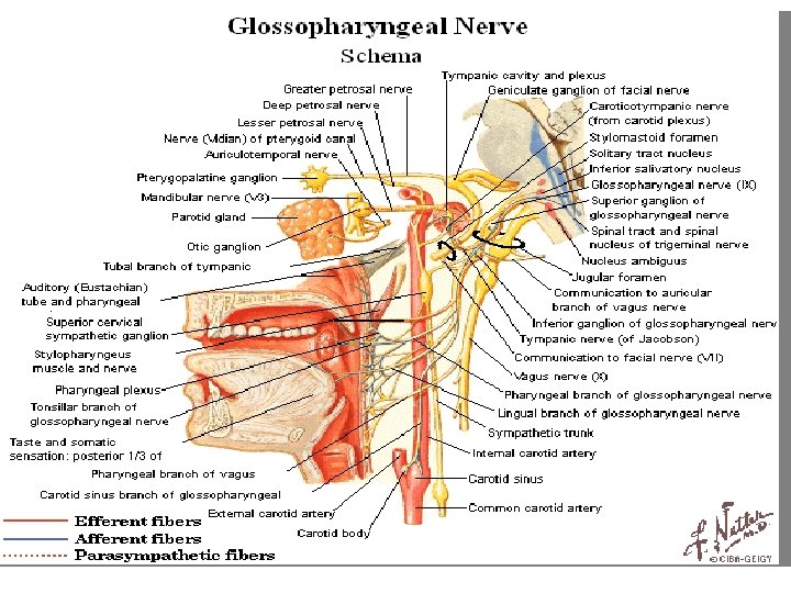
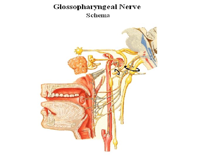
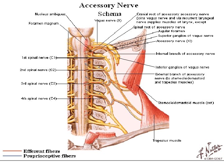
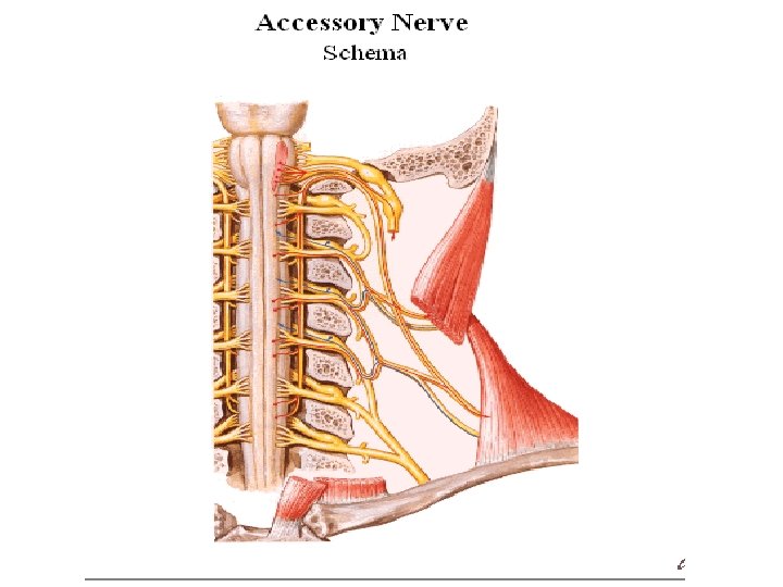
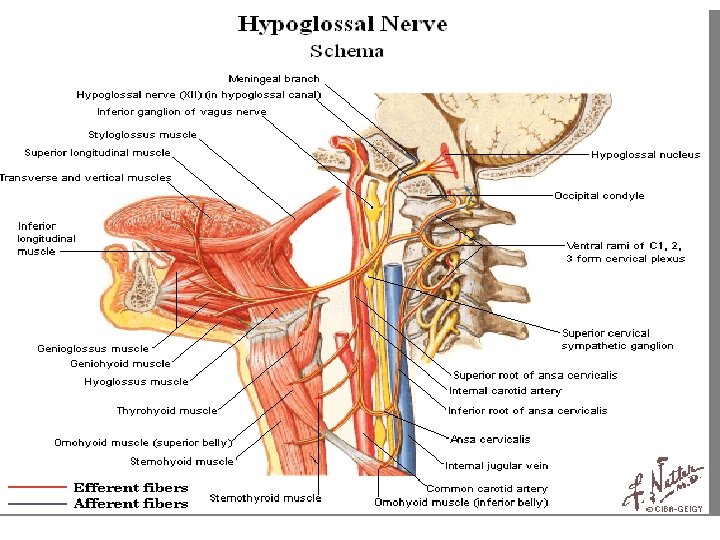
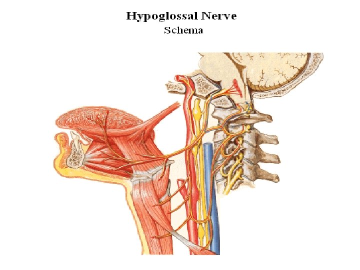
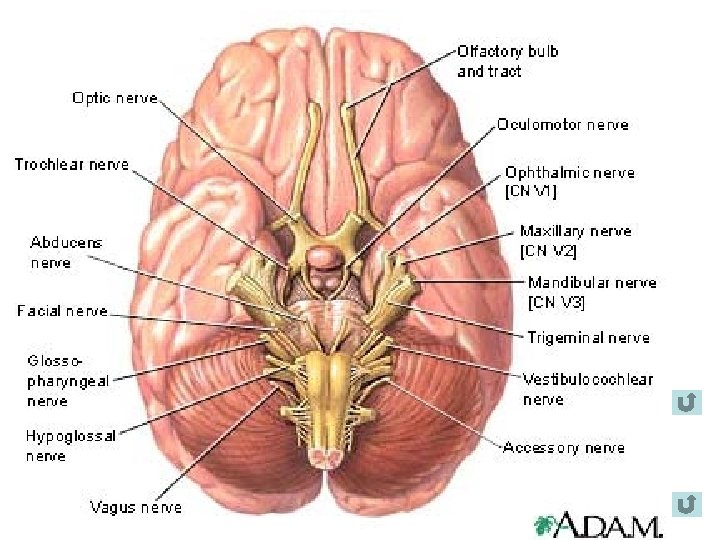
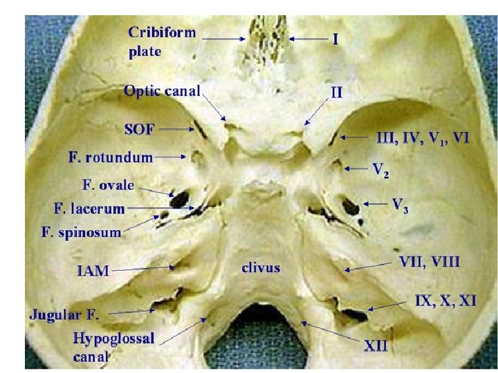
Cranial Nerves • 12 pairs • Emerge from the lower surface of the brain and pass through the foramina in the skull • I, II, VIII – sensory • III, IV , V, XI and XII – motor • V, VII, IX and X are – mixed

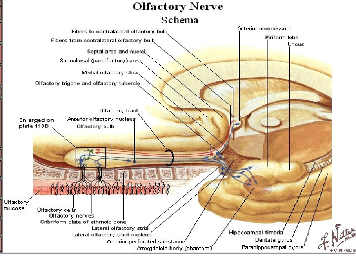
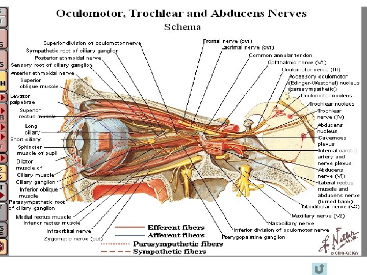
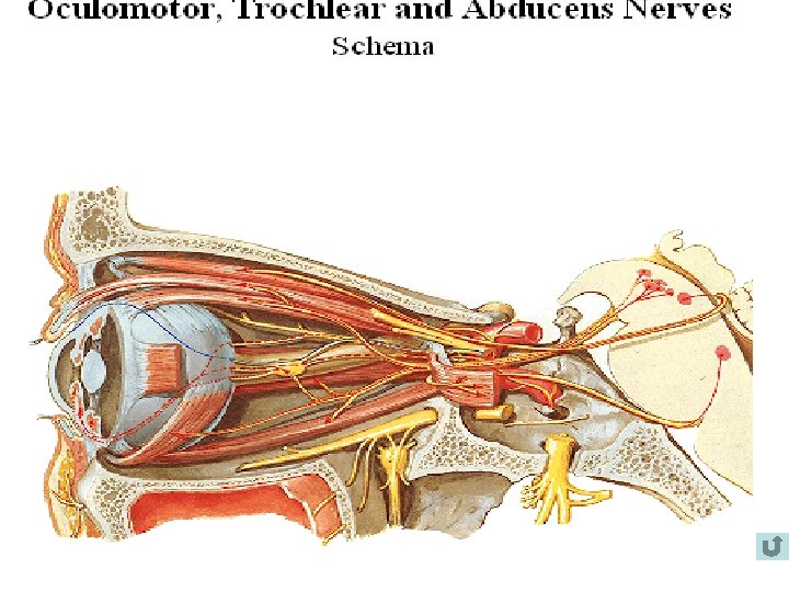
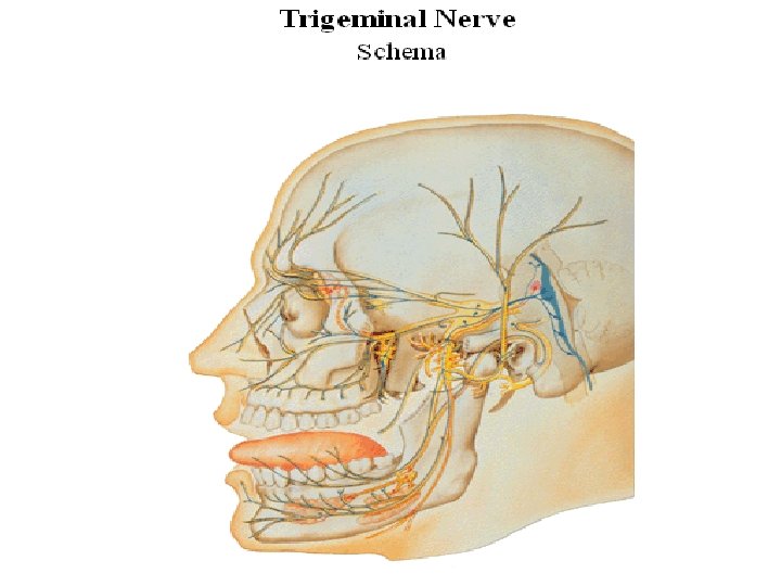
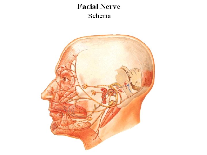
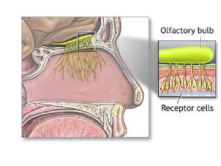
Cranial Nerves 1. Olfactory – sensory - sense of smell 2. Optic nerve – sensory – visual acuity 3. Oculomotor – eye ball movement, lid movement, pupillary constriction lens accommodation 4. Trochlear – motor – muscles that move the eye 5. Trigeminal – mixed – facial sensation, mastication 6. Abducens – motor – muscle that move the eye 7. Facial – mixed – facial expression, salivation, taste 8. Vestibilocochlear – sensory – hearing and equilibrium 9. Glossopharyngeal – mixed – taste, sensation in the pharynx and tongue, pharyngeal muscles 10. Vagus – mixed – muscles of pharynx, larynx, soft palate, thoracic and abdominal viscera – parasympathetic innervation 11. Spinal accessory – motor – sternocleidomastoid trapezius muscles. 12. Hypoglossal – motor – movement of the tongue

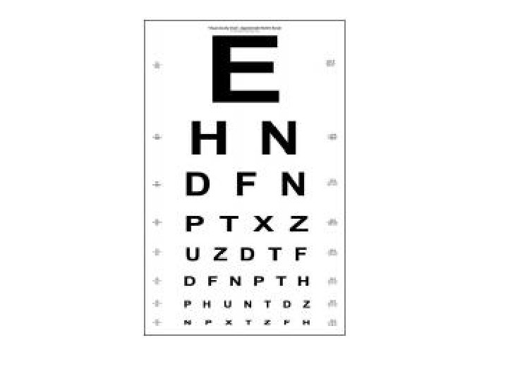
Assessing Cranial Nerve Function • Olfactory Nerve : identify odors with eyes closed – coffee etc • Optic Nerve : snellen eye chart; visual fields : ophthalmoscopic examination • Oculomotor Nerve : III, IV and VI : ocular rotations, conjugate movements, nystagmus. Test for pupillary reflexes and inspect eyelids for ptosis • Trochlear • Agducens


 Frontal bone. b) Frontal sinus. c) Internal frontal spine. d) Foramen caecum. e)")
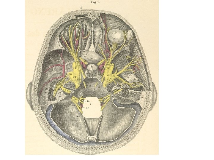
a) Frontal bone. b) Frontal sinus. c) Internal frontal spine. d) Foramen caecum. e) Crista galli. f) Frontal bone, orbital portion. g) Cellulae of the ethmoidal bone. h) Body of the sphenoid bone. i) Greater wing of the sphenoid bone. k) Occipital bone, basilar portion. l) Temporal bone, squamous portion. m) Temporal bone, petrous portion. n) Optic foramen. o) Foramen rotundum. p) Foramen ovale. q) Foramen spinosum. r) Superior orbital fissure. s) Tympanic cavity. t) Internal auditory meatus (broken open from above). u) Malleus, hammer (s. capitulum). v) Incus (s. ambos). w) Cochlea. x) Superior semicircular canal. y) Ocular bulb. z) Lacrimal gland. a) m. Lateral rectus. b) m. Levator palpebrae superioris. g) m. Superior rectus. d) m. Superior oblique. d*) Trochlea for superior oblique. e) m. Medial rectus. z) m. Temporalis (medial surface). h) m. Lateral pterygoid. q) Hypophysis (pituitary gland). i) Foramen magnum. k) Jugular foramen. l) Hypoglossal canal (s. Anterior condyloid foramen). m) Occipital bone (fossae cerebelli). n) Transverse sinus. 1. middle meningeal artery (branch of the maxillary artery). 2. internal carotid artery. 3. lacrymal artery. 4. artery, muscle branch. 5. supraorbital artery. 6. ethmoidal artery. 7. ophthalmic artery. 8. optic nerve (CN II). 9. oculomotor nerve (CN III). 10. trochlear nerve (CN IV). 11. trigeminal nerve (CN V).

23. 24. 25. 26. 27. 28. 29. 30. 31. 32. 33. 34. 35. 36. 37. 38. 23. 24. 25. 26. 27. 28. 29. zygomatic branch, lacrymal nerve. lacrymal branch, lacrymal nerve. ciliary nerves (from the ciliary ganglion [s. ganglion opthalmicum]). nerve to the m. buccinator. deep temporal nerve to the m. masseter. superficial temporal nerve (s. auricularis anterior nerve). maxillary artery. nerves of the external auditory meatus. chorda tympani (of facial nerve). petrosus minor nerve [NA] (s. lesser superficial petrosal nerve). petrosus major nerve [NA] (s. greater superficial petrosal nerve, nervus Vidianus, nervus petrosus superficialis major) [carried in the genu of the facial nerve]. abducens nerve (CN VI). facial nerve (CN VII) inner half of the internal auditory meatus. genu (facial nerve). facial nerve in the Fallopian canal. cochlear nerve (CN VIII). vestibular nerve (CN VIII). glossopharyngeal nerve (CN IX). vagus nerve (CN X). spinal accessory nerve of Willis (CN XI). hypoglossal nerve (CN XII). vertebral artery.


- Slides: 41