
CPC 20180906 VS SzuPei Ho Still Disease Clinical




- Slides: 27
CPC 20180906 VS Szu-Pei Ho
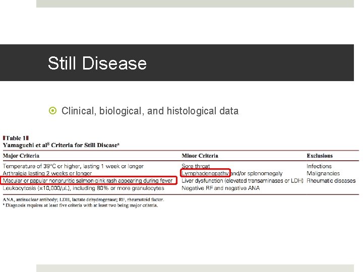
Still Disease Clinical, biological, and histological data
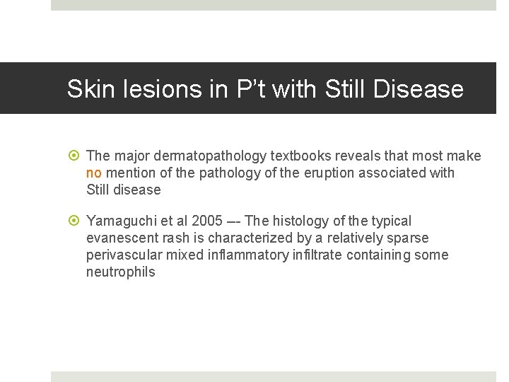
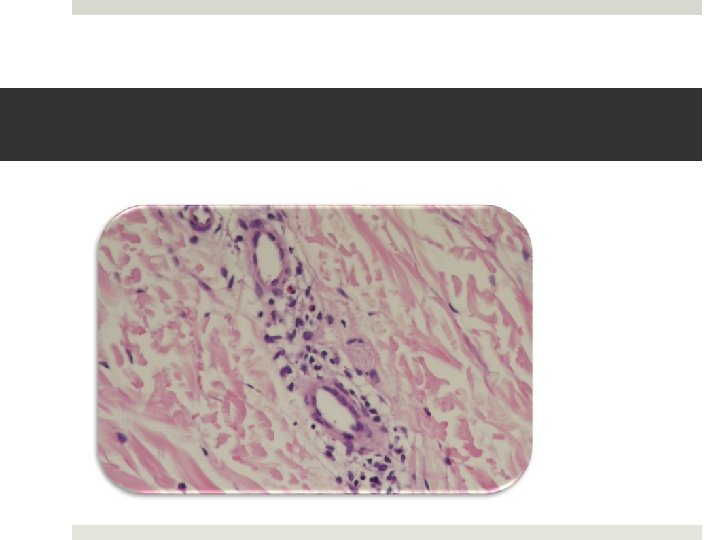
Skin lesions in P’t with Still Disease The major dermatopathology textbooks reveals that most make no mention of the pathology of the eruption associated with Still disease Yamaguchi et al 2005 --- The histology of the typical evanescent rash is characterized by a relatively sparse perivascular mixed inflammatory infiltrate containing some neutrophils
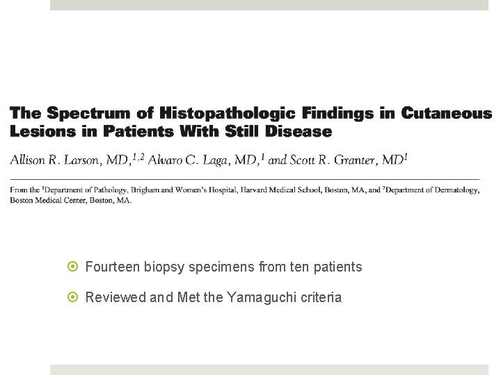
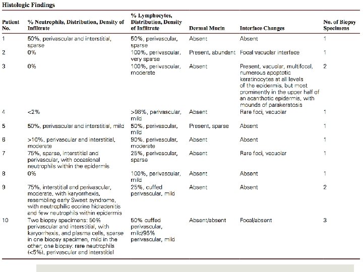
Fourteen biopsy specimens from ten patients Reviewed and Met the Yamaguchi criteria
Clinical Highly characteristic salmon-colored evanescent cutaneous eruption that tends to accompany episodes of fever Pathology A broad histologic spectrum that may be encountered in Still disease and to consider Still disease in the differential diagnosis of neutrophil-rich, lymphocyte-rich, and mixed inflammatory dermatoses
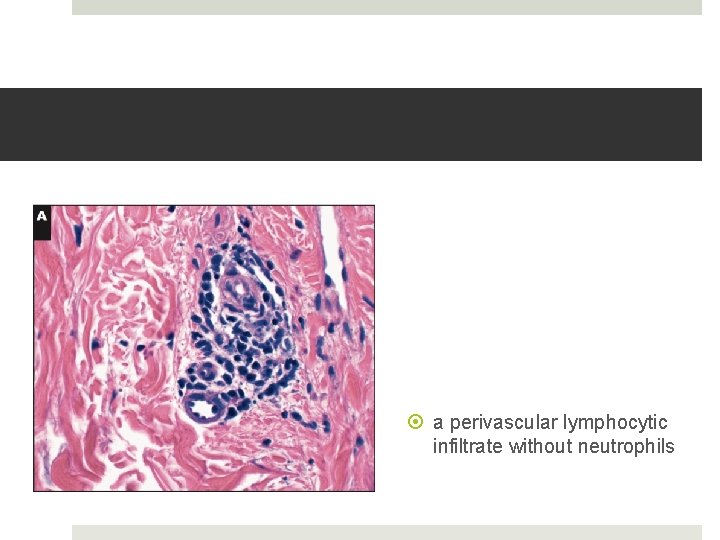
a perivascular lymphocytic infiltrate without neutrophils
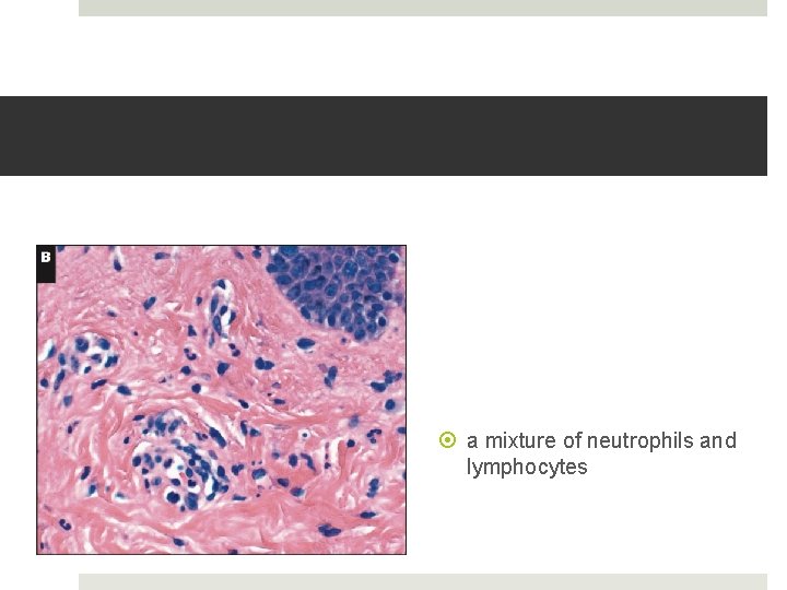
a mixture of neutrophils and lymphocytes
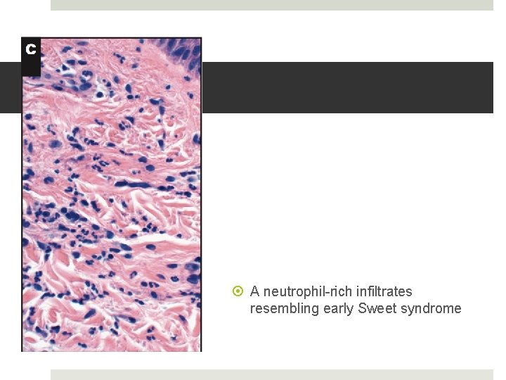
A neutrophil-rich infiltrates resembling early Sweet syndrome
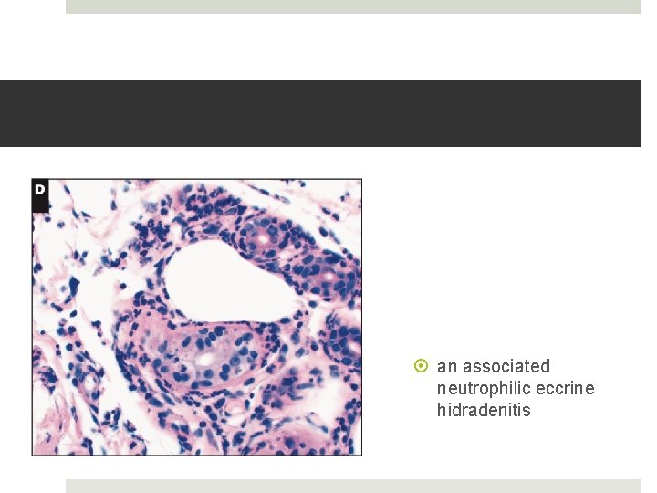
an associated neutrophilic eccrine hidradenitis
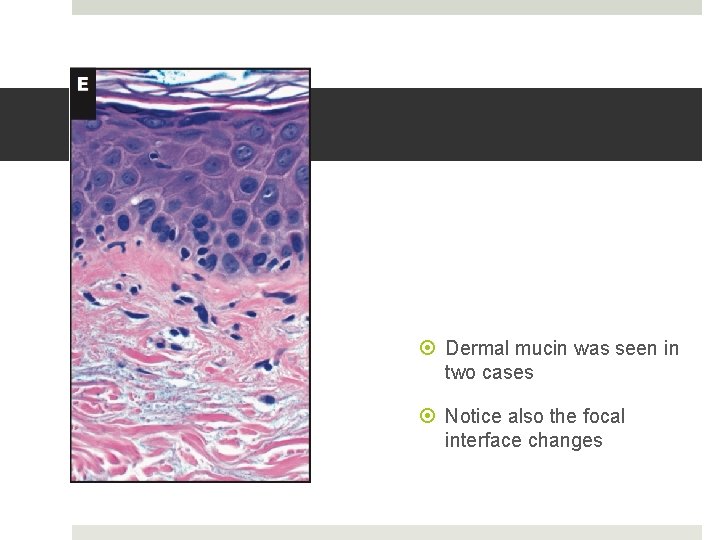
Dermal mucin was seen in two cases Notice also the focal interface changes
D. D. in morphologically aspects Spectrum ranging from lymphocytic-rich to neutrophil-rich infiltrates as well as interface changes, which can be either vacuolar or lichenoid Differential diagnosis almost seems impossibly large and includes broad categories
The role of biopsy The clinical context is important Biopsy of a salmon-colored evanescent rash in a patient that clearly meets criteria for Still disease is unnecessary In other patients, who are more difficult to classify, biopsy may be helpful, if only to exclude other possibilities.
Other clinical D. D. at the time of bx Lupus Erythematosus Parvovirus Dermatomyositis Hypersensitivity Reaction Drug Eruption Serum Sickness Urticaria Sarcoidosis Urticarial Vasculitis Familial Mediterranean Fever Vasculitis Cutaneous Tuberculosis Viral Exanthem Connective Tissue Disease
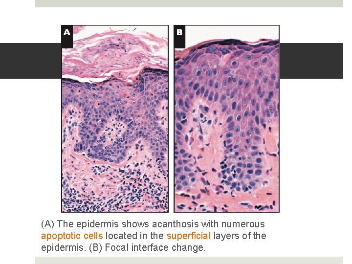
Typical histologic features of the persistent papules and plaques recently described in Still disease More recently, features of the more chronic rash associated with Still disease, so-called persistent papules and plaques, have been better characterized histologically Upper keratinocyte dyskeratosis and scattered superficial dermal neutrophils
(A) The epidermis shows acanthosis with numerous apoptotic cells located in the superficial layers of the epidermis. (B) Focal interface change.
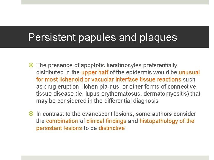
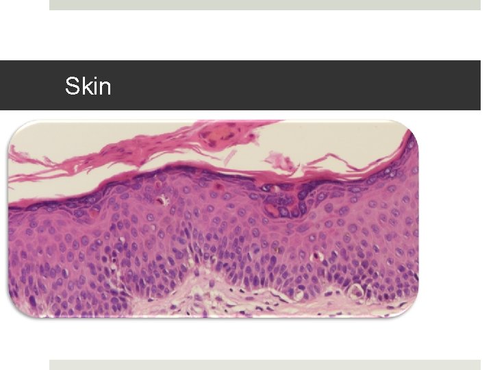
Persistent papules and plaques The presence of apoptotic keratinocytes preferentially distributed in the upper half of the epidermis would be unusual for most lichenoid or vacuolar interface tissue reactions such as drug eruption, lichen pla-nus, or other forms of connective tissue disease (ie, lupus erythematosus, dermatomyositis) that may be considered in the differential diagnosis In contrast to the evanescent lesions, some authors consider the combination of clinical findings and histopathology of the persistent lesions to be distinctive
Biopsy specimens
Skin
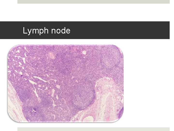
Lymph node
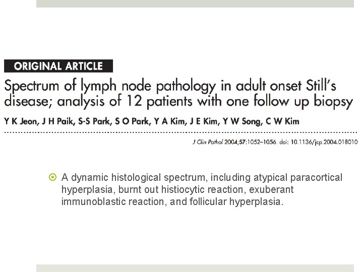
A dynamic histological spectrum, including atypical paracortical hyperplasia, burnt out histiocytic reaction, exuberant immunoblastic reaction, and follicular hyperplasia.
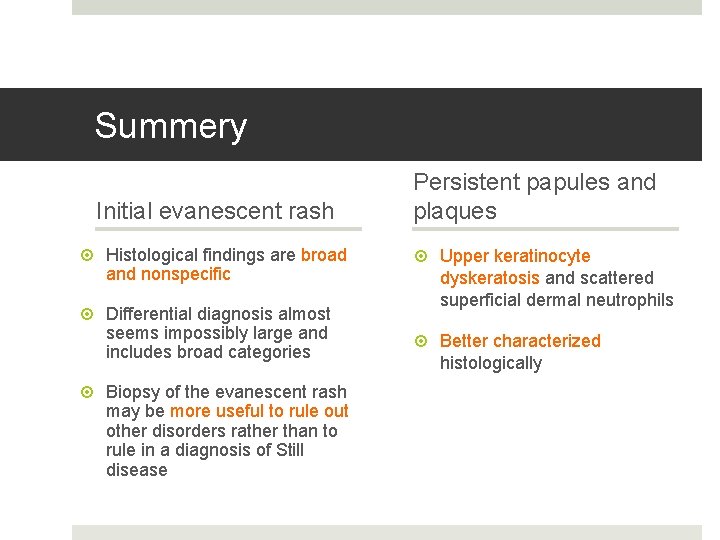
Summery Initial evanescent rash Histological findings are broad and nonspecific Differential diagnosis almost seems impossibly large and includes broad categories Biopsy of the evanescent rash may be more useful to rule out other disorders rather than to rule in a diagnosis of Still disease Persistent papules and plaques Upper keratinocyte dyskeratosis and scattered superficial dermal neutrophils Better characterized histologically
Summery Describe the varied histologic findings in the evanescent rash of Still disease. Correlate pathologic findings with either the evanescent rash or persistent papules and plaques of Still disease. List additional differential diagnostic considerations for the pathologic findings seen in Still disease.
Thanks for your attention
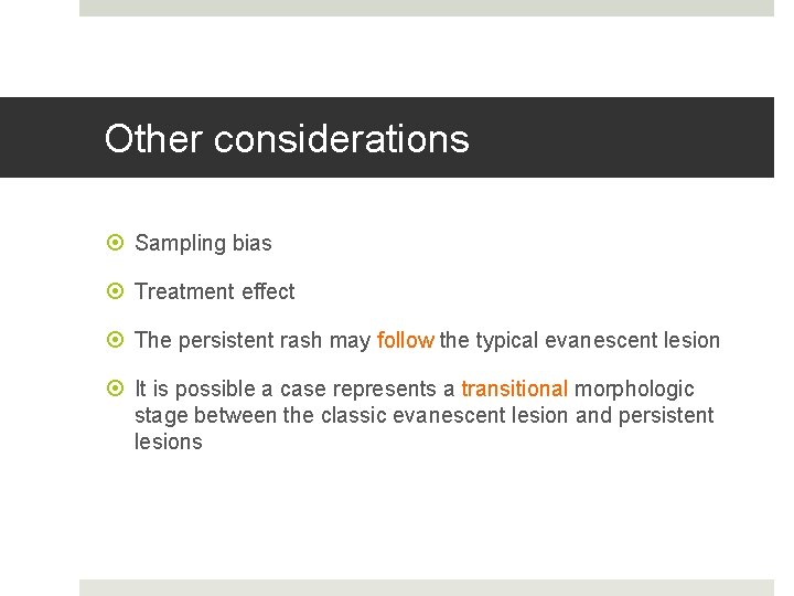
Other considerations Sampling bias Treatment effect The persistent rash may follow the typical evanescent lesion It is possible a case represents a transitional morphologic stage between the classic evanescent lesion and persistent lesions