coronary artery lumen Plaques LV perfusion LV function

 -- 1 - 2 cm Left")
 -- 1 - 2")
 1. Crux 2. Posterior descending artery (PDA) 3. Posterior")


 100 mg 1 h prior")


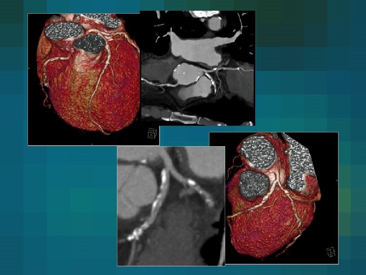
 Calcium Stents")
 Effective energy is shifted to higher")
 motion artifacts (temporal resolution) Calcium/stent obstruction estimation Radiation exposure")

- Slides: 36
coronary artery lumen Plaques LV perfusion LV function
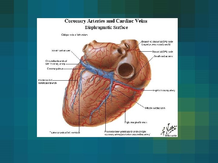
Left posterior aortic sinus Left main artery (LM) -- 1 - 2 cm Left circumflex artery (LCx) 1. Marginal branches 2. Intermedial artery Left anterior descending (LAD) 1. Septal branches 2. Diagonal branches
coronary anatomy Left posterior aortic sinus Left main artery (LM) -- 1 - 2 cm Left anterior descending (LAD) 1. Septal branches 2. Diagonal branches Left circumflex artery (LCx) Anterior aortic sinsus Right coronary artery (RCA) 1. Marginal branches 2. Intermedial artery
coronary anatomy Right dominant (80%) 1. Crux 2. Posterior descending artery (PDA) 3. Posterior lateral branch (PL) Left dominant() Balanced type
coronary anatomy 5 6 7 8 1 9 10 2 3 15 PL 4 PDA 11 13 14 12 Seg. Vessel 01 02 03 04 05 06 07 08 09 10 11 12 13 14 15 RCA RCA PDA LM LAD LAD D 1 D 2 Cx Mo CX MO PL
coronary artery atheroma Ca-X
coronary artery atheroma Modified AHA Calcified nodules Density MDCT >150 High Pictrure MSCT freatures of plaques (Modified AHA calcification) Fibrocalcified plaque Fibreous cap atheroma Thin fibrous cap atheroma Trombus High/Low 50 -100 intermediate 20 -50 low <20 very low
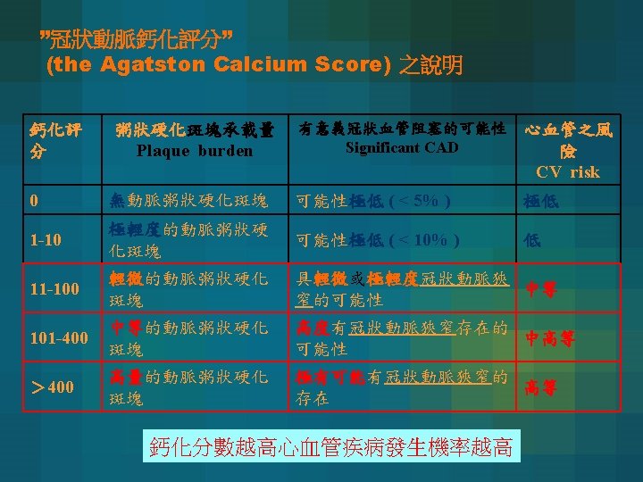
Coronary calcification • The need to detect coronary atherosclerosis early in its course has been well recognized by cardiologists for decades. • The presence and amount of coronary artery calcium have been suggested as a means to assess patients at risk of adverse coronary events. • The presence of coronary calcium is always indicative of the presence of coronary atherosclerosis. • Agatston et al. developed a calcium scoring algorithm that is now widely used in research and clinical practice. • The calcium score is derived from the product of the area of calcification(mm 2) and a factor determined by the maximal X-ray density within this area. (calcification should be at least 1 mm 2 and the X-ray density should exceed the threshold of 130 HU)
Area of plaque × weighting factor = Lesion score ∑ Lesion scores = Vessels score ∑ Vessels scores = Total calcium score (Agatston Score)
How to use the calcium score? • CT calcium quantification can be used for the assessment of long-term risk and primary prevention of future adverse coronary events. • Calcium scoring should not be performed as a ‘standalone’ test but should be integrated into risk assessment with well recognized risk factors. Risk factors: 1, Sex. 2, Age. 3, Family history. 4, Blood lipids. 5, Smoking. 6, Diabetes. 7, Blood pressure. 8, Weight.
冠狀動脈鈣化 性 別 百 分 比 <40 40 -44 45 -49 50 -54 55 -59 60 -64 65 -69 70 -74 >74 男 25 0 0 0 1 4 13 32 64 166 50 1 1 3 15 48 113 180 310 473 75 3 9 36 103 215 410 566 892 1071 90 14 59 154 332 554 994 1299 1774 1982 n 3504 4238 4940 4825 3472 2288 1209 540 235 25 0 0 0 1 3 0 50 0 0 1 3 24 52 75 75 1 1 2 5 23 57 145 210 241 90 3 4 22 55 121 193 410 631 709 n 641 1024 1634 2184 1835 1334 731 438 174 女 年紀 According to Hoff JA et. al JACC 2003; 41; 1008 -12
Level description Low risk 0 -1 risk factor Intermediate 2 risk factor High risk >3 risk factor Risk of event within 10 yrs <10% 10 -20% >20% Estimated 35% prevalence adults 40% 25% Ca. test useful Yes No No Intermediate (1)Calcium score < 80 Lower risk (2)Calcium score > 80 Higher risk
Electrical Activation of the Heart ECG-Triggering/Gating 60% RR 600 ms 400 ms % RR Interval Absolute Reverse Time
Retrospective ECG Gating “ 3 D” Image Data R R Recon Delay Recon R Recon z - Position ous eed u n i t Con can & F l. S a r i Sp Time 加圖
small vessel Challenges in MSCT coronary imaging • size of coronary arteries are small • LCA = 4. 4 mm • LAD = 3. 6 mm distally = 0. 8 mm • CX = 3. 4 mm distally = 1. 5 mm • RCA = 3. 0 mm distally = 1. 0 mm • movement of mid RCA up to 45 mm/sec
small Flat Panel vessel 64 dectector array 4 s 16 s 64 s Rotation 0. 5 0. 42/0. 37 0. 33 Temp resolution 0. 25 0. 21 0. 165 Spatial resolution 1. 0 0. 75 0. 35 Data per rotation 4 16 64 Scan time >40 s 20 s 14 s 16 dectector array
Vascular Enhancement 4 -slice 16 -slice 64 -slice Volume 120 ml 100 ml 80 ml Rate 3 ml/sec 4 ml/sec 5 ml/sec Concentration > 350 mg/ml Antecubital access
Patient Selection Small coronary arteries Fast heart rate >75 bpm Persistent irregular heart rhythm Respiratory impairment and related motion artifacts Diffuse and calcified coronary atherosclerosis Coronary stents Metal objects Renal dysfunction and contrast medium allergy
motion artifacts β- blocking agent - Metoprolol (Beloc ®) 100 mg 1 h prior to exam. p. o. - Propranolol(Dociton ®) 80 mg 1 h prior to exam. p. o. - i. v. Metoprolol 5 mg @1 mg/min i. v. check heart rate @ 5 minutes repeat @ 5 mg (15 mg max. ) NTG 60 -80 Beta-blocker 80 Diastolic Systolic
Patient education 1. Breath holding 2. CM burning sensation 64 dectector array : >9 sec (12) 16 dectector array : >15 sec (20)
coronary anomaly 1%
coronary anomaly bridge Absent right coronary
coronary artery lumen Left Anterior Descending stenosis
coronary artery lumen Proximal LAD stenosis
CABG
CABG L R
CABG
Volume averaging Contrast material (Blooming ) Calcium Stents
Bean Prosthetic valve Contrast material hardening (Streak artifacts) Effective energy is shifted to higher value as the X-rays pass through an object stents Pacemaker wires Calcium Sternal wires Surgical clips
Resolution small vessel (spatial resolution) motion artifacts (temporal resolution) Calcium/stent obstruction estimation Radiation exposure Furture
Thanks for you attention!