
Coronary Artery Disease and Acute Coronary Syndrome Chapter
- Slides: 80
Coronary Artery Disease and Acute Coronary Syndrome Chapter 38 Copyright © 2014 by Mosby, an imprint of Elsevier Inc.
Coronary Artery Disease (CAD) Atherosclerosis Begins as soft deposits of fat that harden with age Referred to as “hardening of arteries” Atheromas (fatty deposits) prefer coronary arteries Also known as ASHD, CVHD, IHD, CHD Copyright © 2014 by Mosby, an imprint of Elsevier Inc.
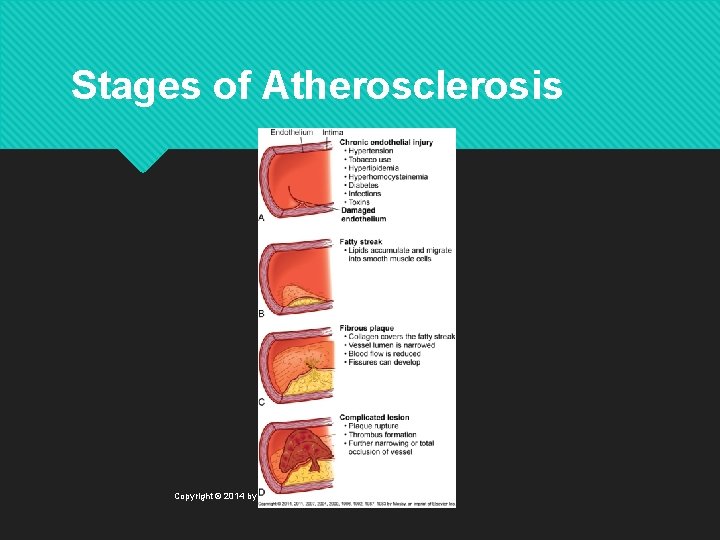
Coronary Artery Disease Etiology and Pathophysiology Atherosclerosis is the major cause of CAD Characterized by lipid deposits within intima of artery Endothelial injury and inflammation play a major role in development Copyright © 2014 by Mosby, an imprint of Elsevier Inc.
Stages of Atherosclerosis Copyright © 2014 by Mosby, an imprint of Elsevier Inc.
Coronary Artery Disease Etiology and Pathophysiology C-reactive protein (CRP) Nonspecific marker of inflammation Increased in many patients with CAD Chronic exposure to CRP associated with unstable plaques and oxidation of LDL cholesterol Copyright © 2014 by Mosby, an imprint of Elsevier Inc.
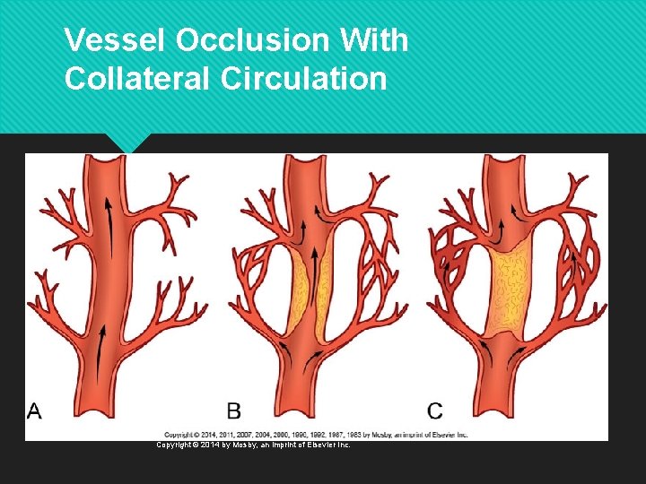
Coronary Artery Disease Etiology and Pathophysiology Collateral circulation Arterial anastomoses (or connections) within the coronary circulation Increased with chronic ischemia May be inadequate with rapid-onset CAD Copyright © 2014 by Mosby, an imprint of Elsevier Inc.
Vessel Occlusion With Collateral Circulation Copyright © 2014 by Mosby, an imprint of Elsevier Inc.
Risk Factors for CAD Nonmodifiable risk factors Age Gender Ethnicity Family history Genetic predisposition Copyright © 2014 by Mosby, an imprint of Elsevier Inc.
Risk Factors for CAD Modifiable risk factors Elevated serum lipids Cholesterol >200 mg/d. L (5. 2 mmol/L) Triglycerides >150 mg/d. L (3. 7 mmol/L) High-density lipoproteins (HDL) Low-density lipoproteins (LDL) Treatment according to guidelines based on 10 -year risk score Copyright © 2014 by Mosby, an imprint of Elsevier Inc.
Risk Factors for CAD Modifiable risk factors Hypertension >140/90 mm Hg or >130/80 mm Hg if diabetes or CKD Begin lifestyle changes for prehypertension Treat stage 1 or 2 hypertension with drugs Copyright © 2014 by Mosby, an imprint of Elsevier Inc.
Risk Factors for CAD Modifiable risk factors Tobacco use Increased catecholamine release ↑ LDL, ↓ HDL, ↑oxygen radicals ↑ Carbon monoxide Second-hand smoke Copyright © 2014 by Mosby, an imprint of Elsevier Inc.
Risk Factors for CAD Modifiable risk factors Physical inactivity Obesity Diabetes Metabolic syndrome Copyright © 2014 by Mosby, an imprint of Elsevier Inc.
Risk Factors for CAD Modifiable risk factors Psychologic states Homocysteine level Substance abuse Copyright © 2014 by Mosby, an imprint of Elsevier Inc.
Collaborative and Nursing Management: CAD Prevention and early treatment Identification of people at high risk Health history, including family history Presence of cardiovascular symptoms Environmental patterns: diet, activity Psychosocial history Values and beliefs about health and illness Copyright © 2014 by Mosby, an imprint of Elsevier Inc.
Collaborative and Nursing Management: CAD Manage high-risk persons by controlling modifiable risk factors Encourage lifestyle changes Education Clarify personal values Set realistic goals Copyright © 2014 by Mosby, an imprint of Elsevier Inc.
Collaborative and Nursing Management: CAD Physical fitness FITT formula: 30 minutes most days plus weight training 2 days a week Regular physical activity contributes to Weight reduction Reduction of >10% in systolic BP In some men more than women, increase in HDL cholesterol Copyright © 2014 by Mosby, an imprint of Elsevier Inc.
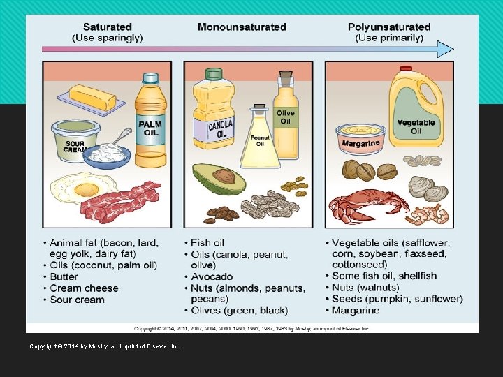
Collaborative and Nursing Management: CAD Nutritional therapy ↓ Saturated fats and cholesterol ↑ Complex carbohydrates and fiber ↓ Red meat, egg yolks, whole milk ↑Omega-3 fatty acids Copyright © 2014 by Mosby, an imprint of Elsevier Inc.
Copyright © 2014 by Mosby, an imprint of Elsevier Inc.
Collaborative and Nursing Management: CAD Lipid-lowering drug therapy If diet and exercise ineffective Statins Inhibit cholesterol synthesis, decrease LDL, increase HDL Monitor for liver damage and myopathy Niacin Lowers LDL and triglyceride by inhibiting synthesis Increases HDL Flushing, pruritus, GI side effects, orthostatic hypotension Copyright © 2014 by Mosby, an imprint of Elsevier Inc.
Collaborative and Nursing Management: CAD Lipid-lowering drug therapy Fibric acid derivatives (Lopid) Decrease triglycerides and increase HDL GI side effects Bile acid sequestrants Increase conversion of cholesterol to bile acids GI side effects; bind with other drugs Ezetimibe (Zetia) Decrease absorption of dietary and biliary cholesterol Copyright © 2014 by Mosby, an imprint of Elsevier Inc.
Collaborative and Nursing Management: CAD Antiplatelet therapy ASA Clopidogrel (Plavix) Copyright © 2014 by Mosby, an imprint of Elsevier Inc.
Gerontologic Considerations CAD Increased incidence and mortality associated with CAD in older adults Strategies to reduce risk and treat CAD are effective Treat hypertension, ↑lipids Smoking cessation Copyright © 2014 by Mosby, an imprint of Elsevier Inc.
Gerontologic Considerations CAD Necessary to modify guidelines for physical activity Longer warm-up Longer periods of low-level activity Longer rest periods Avoid extremes of temperature 30 minutes most days minimum Most likely to change when hospitalized or symptomatic Copyright © 2014 by Mosby, an imprint of Elsevier Inc.
Clinical Manifestations of CAD Angina Progressive disease O 2 demand > O 2 supply → myocardial ischemia Angina = reversible ischemia • Occurs when arteries are blocked 75% or more • Hypoxic within 10 seconds of occlusion • Viable for 20 minutes Copyright © 2014 by Mosby, an imprint of Elsevier Inc.
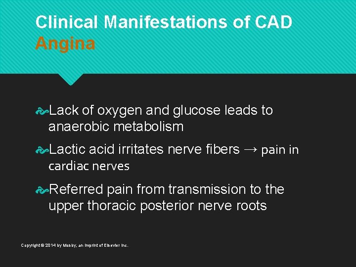
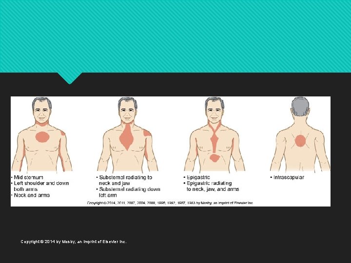
Clinical Manifestations of CAD Angina Lack of oxygen and glucose leads to anaerobic metabolism Lactic acid irritates nerve fibers → pain in cardiac nerves Referred pain from transmission to the upper thoracic posterior nerve roots Copyright © 2014 by Mosby, an imprint of Elsevier Inc.
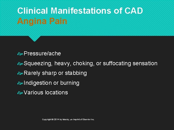
Clinical Manifestations of CAD Angina Pain Pressure/ache Squeezing, heavy, choking, or suffocating sensation Rarely sharp or stabbing Indigestion or burning Various locations Copyright © 2014 by Mosby, an imprint of Elsevier Inc.
Copyright © 2014 by Mosby, an imprint of Elsevier Inc.
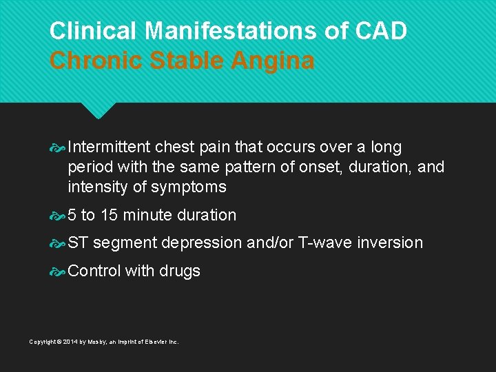
Clinical Manifestations of CAD Chronic Stable Angina Intermittent chest pain that occurs over a long period with the same pattern of onset, duration, and intensity of symptoms 5 to 15 minute duration ST segment depression and/or T-wave inversion Control with drugs Copyright © 2014 by Mosby, an imprint of Elsevier Inc.
Chronic Stable Angina Types of Angina Silent ischemia Ischemia that occurs in the absence of any subjective symptoms Associated with diabetic neuropathy Confirmed by ECG changes Copyright © 2014 by Mosby, an imprint of Elsevier Inc.
Chronic Stable Angina Types of Angina Nocturnal angina Occurs only at night but not necessarily during sleep Angina decubitus Chest pain that occurs only while lying down Relieved by standing or sitting Copyright © 2014 by Mosby, an imprint of Elsevier Inc.
Chronic Stable Angina Types of Angina Prinzmetal’s (variant) angina Occurs at rest usually in response to spasm of major coronary artery Seen in patients with a history of migraine headaches and Raynaud’s phenomenon Spasm may occur in the absence of CAD Copyright © 2014 by Mosby, an imprint of Elsevier Inc.
Chronic Stable Angina Types of Angina Prinzmetal’s (variant) angina Occurs at rest due to spasm of a major coronary artery May occur with or without CAD Not precipitated by increased demand Chest pain with marked, transient ST-segment elevation Copyright © 2014 by Mosby, an imprint of Elsevier Inc.
Chronic Stable Angina Types of Angina Microvascular angina Chest pain occurs in the absence of significant coronary atherosclerosis or coronary spasm Myocardial ischemia associated with abnormalities of the coronary microcirculation Coronary microvascular disease (MVD) affects small, distal coronary arteries Copyright © 2014 by Mosby, an imprint of Elsevier Inc.
Chronic Stable Angina Collaborative Care Goal: ↓ O 2 demand and/or ↑ O 2 supply Short-acting nitrates Dilate peripheral and coronary blood vessels Give sublingually (tablet) or by spray If no relief in 5 minutes, call EMS; if some relief , repeat every 5 minutes for maximum 3 doses Patient teaching Can use prophylactically Copyright © 2014 by Mosby, an imprint of Elsevier Inc.
Chronic Stable Angina Collaborative Care Long-acting nitrates To reduce angina incidence Main side effects: headache, orthostatic hypotension Methods of administration Oral Nitroglycerin (NTG) ointment Transdermal controlled-release NTG Copyright © 2014 by Mosby, an imprint of Elsevier Inc.
Chronic Stable Angina Collaborative Care Angiotensin-converting enzyme inhibitors β-adrenergic blockers Calcium channel blockers Sodium current inhibitor Ranolazine (Ranexa) Copyright © 2014 by Mosby, an imprint of Elsevier Inc.
Chronic Stable Angina Collaborative Care Diagnostic studies Chest x-ray Laboratory studies 12 -lead ECG Calcium-score screening heart scan Echocardiogram Exercise stress test Pharmacologic nuclear imaging Copyright © 2014 by Mosby, an imprint of Elsevier Inc.
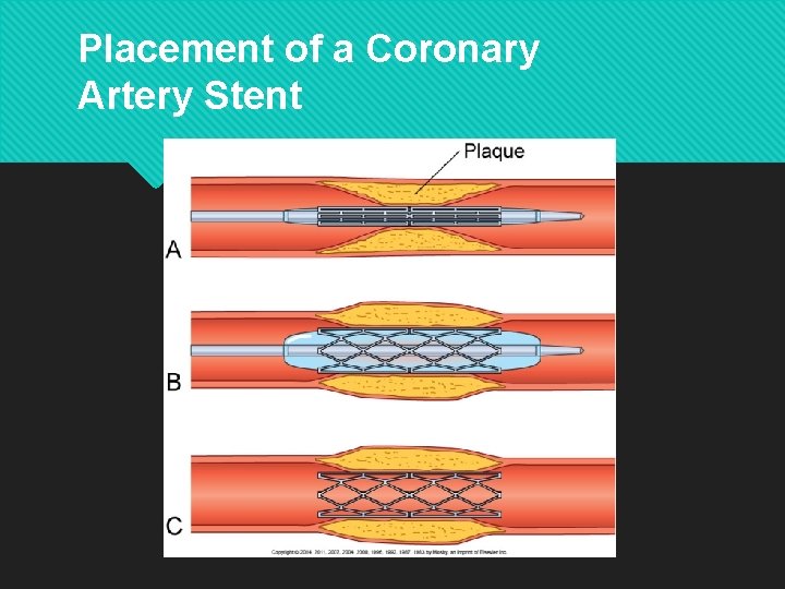
Chronic Stable Angina Nursing/Collaborative Management Cardiac catheterization/coronary angiography Visualize blockages (diagnostic) Open blockages (interventional) Percutaneous coronary intervention (PCI) Balloon angioplasty Stent Copyright © 2014 by Mosby, an imprint of Elsevier Inc.
Placement of a Coronary Artery Stent Copyright © 2014 by Mosby, an imprint of Elsevier Inc.
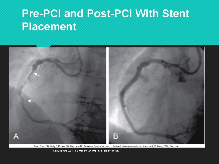
Pre-PCI and Post-PCI With Stent Placement Copyright © 2014 by Mosby, an imprint of Elsevier Inc.
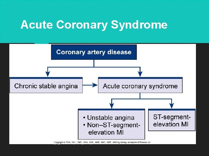
Acute Coronary Syndrome Copyright © 2014 by Mosby, an imprint of Elsevier Inc.
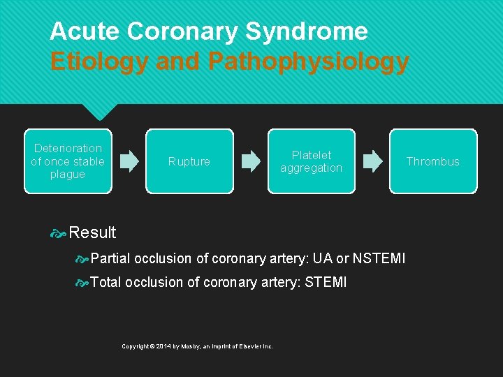
Acute Coronary Syndrome Etiology and Pathophysiology Deterioration of once stable plague Rupture Platelet aggregation Result Partial occlusion of coronary artery: UA or NSTEMI Total occlusion of coronary artery: STEMI Copyright © 2014 by Mosby, an imprint of Elsevier Inc. Thrombus
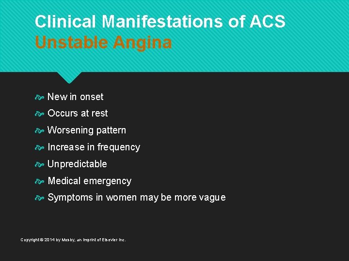
Clinical Manifestations of ACS Unstable Angina New in onset Occurs at rest Worsening pattern Increase in frequency Unpredictable Medical emergency Symptoms in women may be more vague Copyright © 2014 by Mosby, an imprint of Elsevier Inc.
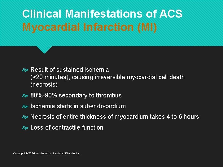
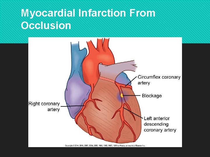
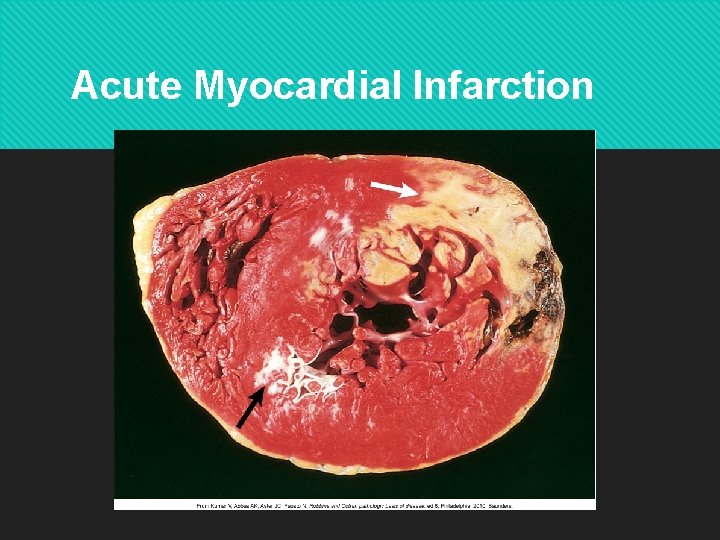
Clinical Manifestations of ACS Myocardial Infarction (MI) Result of sustained ischemia (>20 minutes), causing irreversible myocardial cell death (necrosis) 80%-90% secondary to thrombus Ischemia starts in subendocardium Necrosis of entire thickness of myocardium takes 4 to 6 hours Loss of contractile function Copyright © 2014 by Mosby, an imprint of Elsevier Inc.
Myocardial Infarction From Occlusion Copyright © 2014 by Mosby, an imprint of Elsevier Inc.
Acute Myocardial Infarction Copyright © 2014 by Mosby, an imprint of Elsevier Inc.
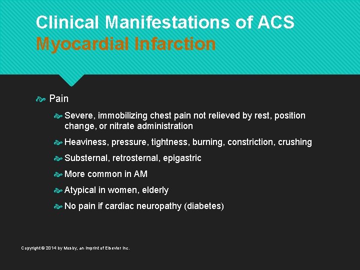
Clinical Manifestations of ACS Myocardial Infarction Pain Severe, immobilizing chest pain not relieved by rest, position change, or nitrate administration Heaviness, pressure, tightness, burning, constriction, crushing Substernal, retrosternal, epigastric More common in AM Atypical in women, elderly No pain if cardiac neuropathy (diabetes) Copyright © 2014 by Mosby, an imprint of Elsevier Inc.
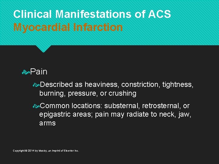
Clinical Manifestations of ACS Myocardial Infarction Pain Described as heaviness, constriction, tightness, burning, pressure, or crushing Common locations: substernal, retrosternal, or epigastric areas; pain may radiate to neck, jaw, arms Copyright © 2014 by Mosby, an imprint of Elsevier Inc.
Clinical Manifestations of ACS Myocardial Infarction Catecholamine release – stimulation of SNS Release of glycogen Diaphoresis Vasoconstriction of peripheral blood vessels Skin: ashen, clammy, and/or cool to touch Copyright © 2014 by Mosby, an imprint of Elsevier Inc.
Clinical Manifestations of ACS Myocardial Infarction Cardiovascular Initially, ↑ HR and BP, then ↓ BP (secondary to ↓ in CO) Crackles Jugular venous distention Abnormal heart sounds S 3 or S 4 New murmur Copyright © 2014 by Mosby, an imprint of Elsevier Inc.
Clinical Manifestations of ACS Myocardial Infarction Nausea and vomiting Reflex stimulation of the vomiting center by severe pain Vasovagal reflex Fever Up to 100. 4° F (38° C) in first 24 hours Systemic inflammatory process caused by myocardial cell death Copyright © 2014 by Mosby, an imprint of Elsevier Inc.
Myocardial Infarction Healing Process Within 24 hours, leukocytes infiltrate the area of cell death Proteolytic enzymes of neutrophils and macrophages begin to remove necrotic tissue by fourth day → thin wall Necrotic zone identifiable by ECG changes and nuclear scanning Collagen matrix laid down Copyright © 2014 by Mosby, an imprint of Elsevier Inc.
Myocardial Infarction Healing Process 10 to 14 days after MI, scar tissue is still weak Myocardium vulnerable to stress Monitor patient carefully as activity level increases Copyright © 2014 by Mosby, an imprint of Elsevier Inc.
Myocardial Infarction Healing Process By 6 weeks after MI, scar tissue has replaced necrotic tissue Area is said to be healed, but less compliant Ventricular remodeling Normal myocardium will hypertrophy and dilate in an attempt to compensate for the infarcted muscle Copyright © 2014 by Mosby, an imprint of Elsevier Inc.
Complications of Myocardial Infarction Dysrhythmias Most common complication Present in 80% of MI patients Can be caused by ischemia, electrolyte imbalances, or SNS stimulation Life-threatening dysrhythmias seen most often with anterior MI, heart failure, or shock Copyright © 2014 by Mosby, an imprint of Elsevier Inc.
Complications of Myocardial Infarction Heart failure Occurs when the pumping power of the heart has diminished Can be subtle or severe Cardiogenic shock Occurs because of severe LV failure Requires aggressive management Copyright © 2014 by Mosby, an imprint of Elsevier Inc.
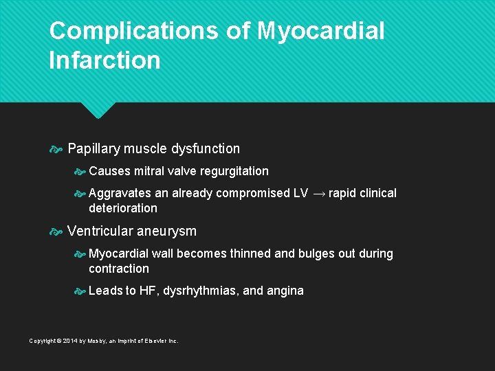
Complications of Myocardial Infarction Papillary muscle dysfunction Causes mitral valve regurgitation Aggravates an already compromised LV → rapid clinical deterioration Ventricular aneurysm Myocardial wall becomes thinned and bulges out during contraction Leads to HF, dysrhythmias, and angina Copyright © 2014 by Mosby, an imprint of Elsevier Inc.
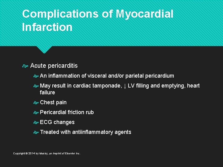
Complications of Myocardial Infarction Acute pericarditis An inflammation of visceral and/or parietal pericardium May result in cardiac tamponade, ↓ LV filling and emptying, heart failure Chest pain Pericardial friction rub ECG changes Treated with antiinflammatory agents Copyright © 2014 by Mosby, an imprint of Elsevier Inc.
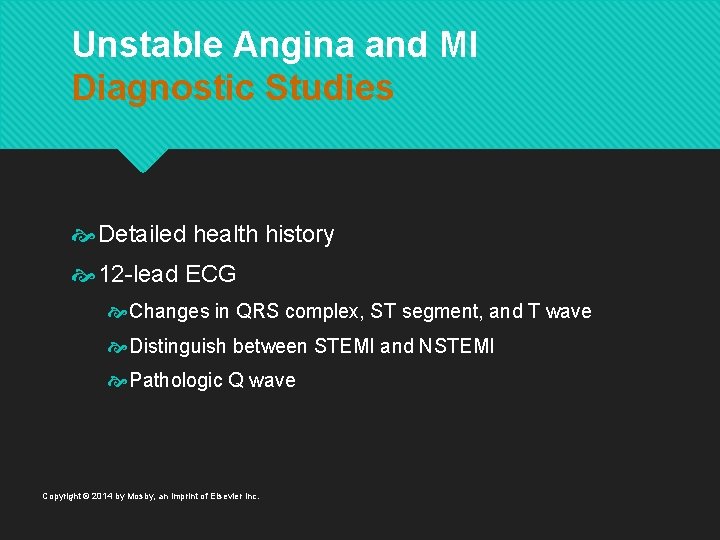
Unstable Angina and MI Diagnostic Studies Detailed health history 12 -lead ECG Changes in QRS complex, ST segment, and T wave Distinguish between STEMI and NSTEMI Pathologic Q wave Copyright © 2014 by Mosby, an imprint of Elsevier Inc.
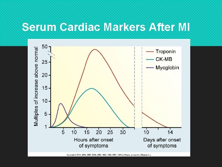
Serum Cardiac Markers After MI Copyright © 2014 by Mosby, an imprint of Elsevier Inc.
Unstable Angina and MI Diagnostic Studies Coronary angiography Exercise or pharmacologic stress testing Echocardiogram Copyright © 2014 by Mosby, an imprint of Elsevier Inc.
Collaborative Care Acute Coronary Syndrome Initial interventions 12 -lead ECG Semi-fowler’s position Oxygen IV access Nitroglycerin (SL) and ASA (chewable) Morphine Copyright © 2014 by Mosby, an imprint of Elsevier Inc.
Collaborative Care Acute Coronary Syndrome Ongoing monitoring Treat dysrhythmias Frequent vital sign monitoring Bed rest/limited activity for 12– 24 hours UA or NSTEMI Aspirin, heparin, and glycoprotein inhibitor Coronary angiography with PCI once stable Copyright © 2014 by Mosby, an imprint of Elsevier Inc.
Collaborative Care Acute Coronary Syndrome STEMI or NSTEMI with + cardiac markers → reperfusion therapy Emergent PCI Treatment of choice for confirmed MI Goal: 90 minutes from door to catheter laboratory Balloon angioplasty + drug-eluting stent(s) Many advantages over CABG Copyright © 2014 by Mosby, an imprint of Elsevier Inc.
Collaborative Care Acute Coronary Syndrome Thrombolytic therapy When PCI not available Stops infarction process by dissolving thrombus Within 6 hours of onset of symptoms Ideally within first hour Given IV Patient selection critical Copyright © 2014 by Mosby, an imprint of Elsevier Inc.
Collaborative Care Acute Coronary Syndrome Drug therapy IV nitroglycerin Morphine sulfate β-adrenergic blockers Angiotensin-converting enzyme inhibitors Antidysrhythmia drugs Cholesterol-lowering drugs Stool softeners Copyright © 2014 by Mosby, an imprint of Elsevier Inc.
Collaborative Care Acute Coronary Syndrome Nutritional therapy Initially NPO Progress to Low salt Low saturated fat Low cholesterol Copyright © 2014 by Mosby, an imprint of Elsevier Inc.
Nursing Management Chronic Stable Angina and ACS Nursing assessment: subjective data Health history CAD/chest pain/angina/ MI Valve disease Heart failure/cardiomyopathy, Hypertension, diabetes, anemia, lung disease, hyperlipidemia Drugs History of present illness Copyright © 2014 by Mosby, an imprint of Elsevier Inc.
Nursing Management Chronic Stable Angina and ACS Nursing assessment: subjective data Family history Indigestion/heartburn; nausea/vomiting Urinary urgency or frequency Straining at stool Palpitations, dyspnea, dizziness, weakness Chest pain Stress, depression, anger, anxiety Copyright © 2014 by Mosby, an imprint of Elsevier Inc.
Nursing Management Chronic Stable Angina and ACS Nursing assessment: objective data Anxious, fearful, restless, distressed Cool, clammy, pale skin Tachycardia or bradycardia Pulsus alternans Pulse deficit Dysrhythmias S 3, S 4, ↑ or ↓ BP, murmur Copyright © 2014 by Mosby, an imprint of Elsevier Inc.
Nursing Management Chronic Stable Angina and ACS Nursing diagnoses Decreased cardiac output Acute pain Anxiety Activity intolerance Ineffective self-health management Copyright © 2014 by Mosby, an imprint of Elsevier Inc.
Nursing Management Chronic Stable Angina and ACS Planning: overall goals Relief of pain Preservation of myocardium Immediate and appropriate treatment Effective coping with illness-associated anxiety Participation in a rehabilitation plan Reduction of risk factors Copyright © 2014 by Mosby, an imprint of Elsevier Inc.
Nursing Management Chronic Stable Angina Acute intervention Semi-Fowler’s position Supplemental oxygen Assess vital signs 12 -lead ECG Administer a nitrate followed by an opioid analgesic, if needed Auscultate heart and breath sounds Copyright © 2014 by Mosby, an imprint of Elsevier Inc.
Nursing Management Chronic Stable Angina Ambulatory and home care Provide reassurance Patient teaching CAD and angina Precipitating factors for angina Risk factor reduction Drugs Copyright © 2014 by Mosby, an imprint of Elsevier Inc.
Nursing Management Acute Coronary Syndrome Acute intervention Pain: nitroglycerin, morphine, oxygen Continuous monitoring • ECG • ST segment • Heart and breath sounds • VS, pulse oximetry, I&O Rest and comfort • Balance rest and activity • Begin cardiac rehabilitation Copyright © 2014 by Mosby, an imprint of Elsevier Inc.
Nursing Management Acute Coronary Syndrome Acute intervention Anxiety reduction Identify source and alleviate Patient teaching important Emotional and behavioral reaction Maximize patient’s social support systems Consider open visitation Copyright © 2014 by Mosby, an imprint of Elsevier Inc.
Nursing Management Acute Coronary Syndrome Coronary revascularization: PCI Monitor for recurrent angina Frequent VS, including cardiac rhythm Monitor catheter insertion site for bleeding Neurovascular assessment Bed rest per institutional policy Copyright © 2014 by Mosby, an imprint of Elsevier Inc.
Nursing Management Acute Coronary Syndrome Ambulatory and home care Cardiac rehabilitation Patient and caregiver teaching Physical activity METs or Borg Scale Monitor heart rate Low-level stress test before discharge Isometric versus isotonic activities Copyright © 2014 by Mosby, an imprint of Elsevier Inc.
Nursing Management Acute Coronary Syndrome Ambulatory and home care Resumption of sexual activity Teach when discuss other physical activity Erectile dysfunction drugs contraindicated with nitrates Prophylactic nitrates before sexual activity When to avoid sex Typically 7– 10 days post MI or when patient can climb two flights of stairs Copyright © 2014 by Mosby, an imprint of Elsevier Inc.
Nursing Management Acute Coronary Syndrome Evaluation Stable vital signs Relief of pain Decreased anxiety Realistic program of activity Effective management of therapeutic regimen Copyright © 2014 by Mosby, an imprint of Elsevier Inc.