CORNEBACTERIUM INFECTIONS FAMILY Corynebacteriaceae Genus Corynebacterium More than

CORNEBACTERIUM INFECTIONS
")
FAMILY: Corynebacteriaceae Genus: Corynebacterium (More than 30 species)

• Pyogenic microorganisms • Opportunistic pathogens • Located in soil and mucous membranes • In general, tissue trauma (sheep skin fissures, cutaneous wounds, tail cuts and during bathing) • Chemotaxonomic similarity to Nocardia, Rhodococcus and Mycobacterium genus • Cell wall; meso diaminopimelic acid (meso-DAP), arabinogalactan and mycolic acid • Nocardia, Mycobacterium and Corynebacterium are called "coryneform" bacteria
 Asporous, nonmotile, facultative")
Gram-positive coccoid rods, pleomorphic, X, V, Y (related to the division) Asporous, nonmotile, facultative anaerobic Babes-Ernst granules (metachromatic, energy-phosphate storage) Most of them catalase positive, oxidase negative Enriched media Blood Agar Hemolysis (narrow full hemolysis) Virulence Factors: Lipid, Exotoxin (Phospholipase D) Intracellular survival in phagocytes, facultative intracellular

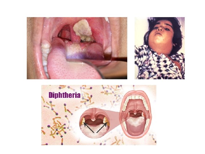
• Corynebacterium equi: Rhodococcus equi • Corynebacterium pyogenes: Trueperella pyogenes – • Old names: Bacillus, Corynebacterium, Actinomyces, Arcanobacterium pyogenes Corynebacterium pseudotuberculosis (1888, Nocard) – Small, non-hemolytic, white-ivory, frozen columns-dry, crumbly – Biotype 1, equine and cattle isolation reducing nitrates to nitrites – Biotype 2, non-reducible, sheep and goat isolation – Corynebacterium renale – There are 3 immunological types, 3 different species are thought to be. – Type I: C. renale, Type II: C. pilosum, Type III: C. cystitidis • Corynebacterium pilosum • Corynebacterium cystitidis – Semitransperent, white colony • Corynebacterium bovis • Corynebacterium kutscheri • Corynebacterium diphteria (1884, Loffler) – Only humans, the agent of diphtheria, fibrinous inflammation, diphtheria toxin creates gray pseudomembrane, has bacteriophage – Kidney fever, throat swelling, coughing, inhaling, swollen lymph nodes, myocarditis in kids

Bacterioscopy


AGENT HOST DİSEASE C. diphteria Human Diphteria C. bovis Cattle Subclinic mastitis C. kutscheri Rats superficial abscess, cazeopurulent foci in liver, lung, and lymph nodules The nitrate non-reducible biotype (biotype 2) Sheep, goat Caseous lymphadenitis The nitrate reducible biotype (biotype 1) Horse and cattle Ulcerative lymphangitis, abscesses C. renale (type 1) Cattle Pyelonephritis Sheep, goat Ulcerative balonopostitis C. pilosum (type 2) Cattle Cystitis, pyelonephritis C. cystitidis (type 3) Cattle Severe cystitis, rarely pyelonephritis C. ulcerans Cattle Mastitis C. pseudotuberculosis

• C. renale group keeps on with the fimbriae • Produces urease • Urea is destroyed, ammonia is formed and therefore p. H is increased • Systis and pyelonephritis develop • The main lesion is abscess in Corynebacterium infections • There is always a lymphatic infection • Abscess: can be transformed into a dry and cheesy form of cream • Abscess content: dead macrophages, fibrous tissue, giant cells, and neutrophils
 • C. pseudotuberculosis non reducing nitrate biotype 2 causes • Incubation")
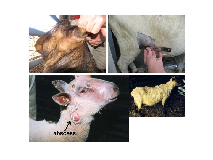
Caseous lymphadenitis (cracks) • C. pseudotuberculosis non reducing nitrate biotype 2 causes • Incubation is about 3 months • Especially seen in sheep and goats • It leads to the disappearance and growth of superficial or internal lymph nodes • In infected animals, poor development is evident and the disease settles in carcasses and causes depreciation of the postings

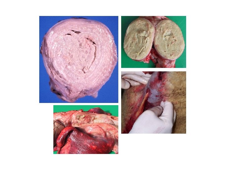
Caseous Lymphadenitis • The disease is transmitted through contaminated bathrooms, forty wound, arthropod bites, • The affected lymph nodes enlarge and when they cut the appearance of the onion ring • The appearance of the onion ring consists of suppuration (pus) formed by inflammatory cells and a new capsule • The encapsulated abscess is caseous. ü Initially it is in green then glazed pastel colour

• Ulcerative Lymphangitis Nitrate + biyotype I C. pseudotuberculosis – In horses, enters through skin wounds – It spreads along the lymph vessels and makes abscess in the lower part of the extremities and the pectoral region – Becomes chronic, abscess hardens, dense, odorless, greenish pus flows by ulceration. • Infectious Acne (canadian horse pox, pigeon fever) – Nitrate + biyotype I C. pseudotuberculosis – Horses are seen in folliculitis, abscesses, pain and limping are seen
 – The urogenital system")
Group of C. renale infections • Bovine Pyelonephritis (Kidney inflammation) – The urogenital system contains commensal – Birth stress and short occurrence of urethra in cattle – The agent reaches the kidney and causes pyelonephritis – Hemorrhage, necrosis, ulceration, all kidneys may be filled with abscess – Fever, anorexia (anorexia), bloody and purulent urine • Ulcerative Balanopostitis – Necrotizing inflammation of surrounding tissues and preputium in coaches – Steered coaches, predispose to intense wool and mohair
 37°C, 24 -48 hours, aerobic incubation")
DIAGNOSIS Blood agar, Mac. Conkey Agar (no reproduction) 37°C, 24 -48 hours, aerobic incubation Hemolysis, aerobic reproduction is evaluated Colony color and type Species other than C. bovis are urease positive C. pseudotuberculosis hemolysis increases when culture with Rhodococcus equi vertically (CAMP positive) To distinguish the biotypes of C. pseudotuberculosis nitrate to nitrite reduction characteristic is evaluated ü Biotype 1: bovine and horse isolates, reduce nitrate to nitrite ü Biotype 2: sheep and goat isolation, can not reduce nitrate to nitrite • •

TREATMENT • Due to the chronic nature of the causative lymphadenitis and the intracellular localization of the organisms, treatment is generally ineffective • Sensitive antibiotics are selected by antibiogram test

RHODOCOCCUS INFECTIONS
 • Genus: Nocardia")
• Family: Nocardiaceae • Genus: Rhodococcus (29 species) • Genus: Nocardia

Rhodococcus equi • Formerly known as Corynebacterium equi • Widespread in soil, soil-based • Only animal pathogens, primary lesions abscess and granulomas • Weakly acid resistant • Gram positive, cocoid bacillus, catalase +, urease + • Asporous, immotile, aerobic, encapsulated, Phospholipase C Colony morphology are S, R, M with salmon color (pink pigment), large, non-hemolytic • Non-fermentative

• Resistant to β-lactamase • In treatment rifampin + erythromycin is effective • Facultative intracellular and the specific surface antigens encoded by the virulence plasmid (Vap) provides virulence • Based on cellular immune response • Mycolic acid in cell wall and capsular polysaccharide are other virulence factors

ü Less than six months in foal, pulmonary abscesses and suppurative bronchopneumonia – Abscess in lung and lung lymph nodes – Airless crowded stables – Abscess and ulcerative intestinal lesions in mesenteric lymph nodes and scattering by excretion – Older than 6 months foals are resistant to pulmonary infection (spread by inhalation) – Mortality exceeds 50% (especially younger than 2 months) – Sudden fever, loss of appetite, bronchopneumonia, dyspnea, wet rallies, diarrhea – Infection may occur in people with immunosuppression – Ruam may be miscible with C. pseudotuberculosis – The vaccine (inactivated? - live? ) ü Horses, superficial abscesses • Pig and Beef; mild cervical lymphadenopathy
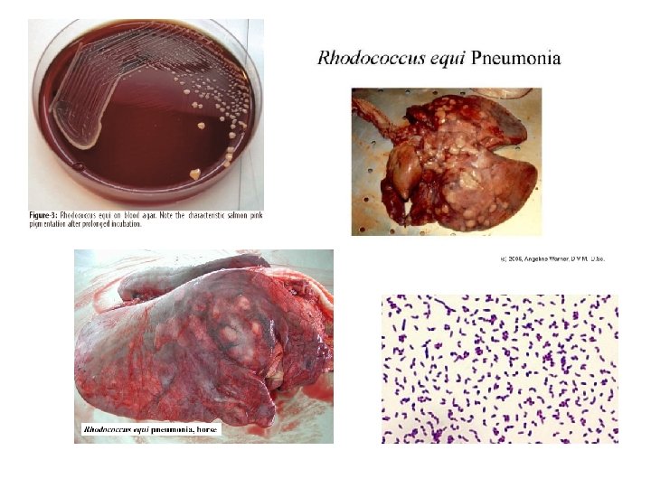

DIAGNOSIS • • • Clinical Findings Necropsy Findings Laboratory Findings Bacterioscopy: Gram-positive coccoid rods Culture: non-hemolytic, colonies in salmon color; No reproduction on Mac. Conkey agar, CAMP test is positive

NOCARDIA INFECTIONS

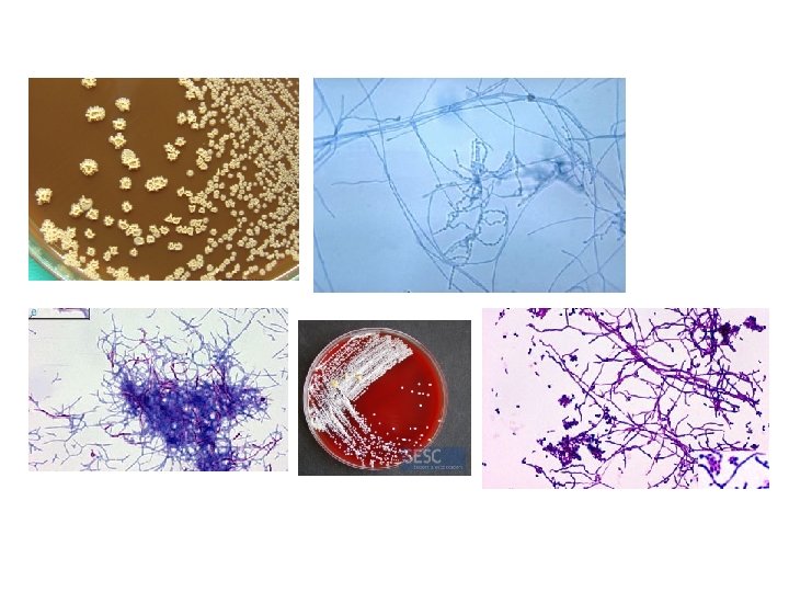
Nocardia • They are Saprophytes found in plants that are in soil and plants. • Cell wall has micolic acid, partially acid-resistant • Gram positive, nonmotile, asporous, mandatory aerobic • It creates branchy and aerial hypha (mushroom-like) • The resting phase is cocobacillus, the active propagating phase is filamentous

• Growth on basic medium and in Sabouraud Dextrose Agar Matte and pigmented (white) agar forms adherent colony • Pellicule on the surface of broth • Urea is positive • Resistant to penicillin, tetracyclines are effective • It survives in phagocytic cells
")
• All animals are sensitive • Very common in humans and animals (dogs) that have immunosuppression • Lymph nodules are always affected, spreading hematogenously and forming a common abscess Nocardia asteroides is the most important species in animals Dog; systemic and cutaneous infections Cattle; mastitis Pig; abortion

DIAGNOSIS • Gram positive rod can be detected by Ziehl. Neelsen staining • It is incubated in aerobic conditions at 37°C for 10 days on blood agar or special medium • Catalase is positive

ACTINOMYCET INFECTIONS

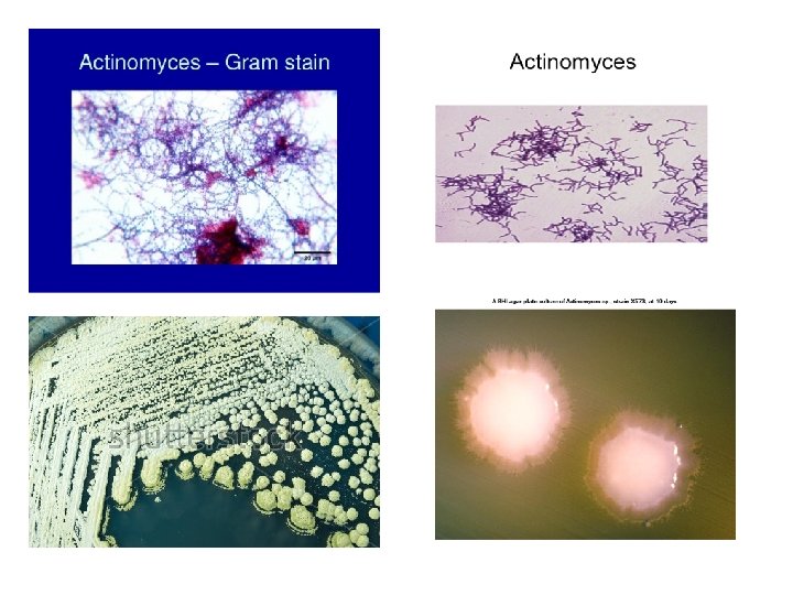
Family: Actinomycetaceae Genus: Actinomyces, Actinobaculum, Trueperella

• They were supposed to be fungi • Opportunistic pathogens • Suppurative lesions • All of these species are found in the mouth, mucous membranes, tooth surfaces and gastrointestinal tract • Infections are caused by wounds in the upper digestive system • Gram positive rod, asporous, immotile • It can culture at 37 degrees on rich medium containing serum and blood • Capnophilites [high CO₂ (10%) lovers] In atmosphere 78% nitrogen, 21% oxygen, 1% other gases present

Important Species and Diseases Species Affected Animals Diseases Caused Actinomyces hordeovulnaris Dogs Cutaneous, visceral abscess Pleurit, peritonite Actinomyces bovis Cattle Bovine Actinomycosis (Lumpy Jaw) Actinomyces viscosus Dog Horse Cattle Canine Aktinomycosis, pyogranuloma Cutaneous pustules, Abortions Actinobaculum suis Pig Cystitis, pyelonephritis Trueperella pyogenes Cattle, sheep Abscess, mastitis, pneumonia, pyometra, arthritis

• Actinomyces species are in the filamentous form • Colonies are adherent on agar, non-hemolytic, similar to fungus colony • Trueperella pyogenes indeterminate hemolysis at the end of 24 hours incubation, forming pin -point B-hemolytic colony after 48 hours
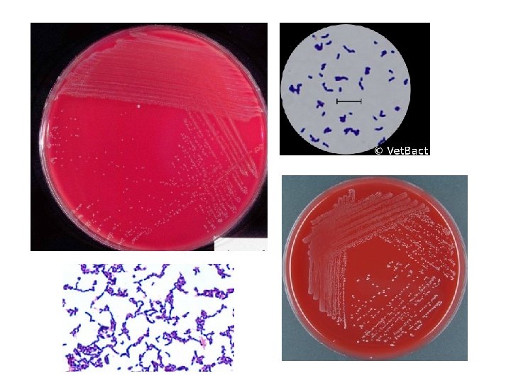

Trueperella pyogenes • Commensal in cattle, sheep and pig nasopharynx • After the injury, the tissues hematogenously • Suppurative lesions, abscess • Chronic abcsess mastitis • Suppurative pneumonia with Mycoplasma and Pasteurella • Septic arthritis, endocarditis, endometritis, pyometra, lymphadenitis, osteomyelitis, peritonitis, omphalitis • Causes mixed infections with F. necrophorum in bovine liver abscess
 • Following trauma due to injury to the")
Actinomyces bovis • Actinomycosis (lumpy jaw) • Following trauma due to injury to the mucous membranes with rough foods or during tooth extraction • Mandibula and other bone tissues of the head • Based on osteomyelitis, normal bone changes to spongiformis • Teething spasm, chewing difficulty and mandibular fractures • Infected bone swells painlessly at 1 -2 weeks, then pain and fistul purulent discharge occurs • It should be distinguished from Actinobasillosis which cause a soft tissue infection of the head ü Appearance of yellowish particles (sulfur granules) several millimeters in diameter in the suspected exudate of the interlabial examination shows actinomycosis

DIAGNOSIS • Incubation on blood agar for 5 days with 10% CO 2 at 37 ° C • Culture on Loeffler medium for Trueperella pyogenes better than other medium • Do not reproduce on Mac. Conkey agar

TREATMENT • Limited lesions in actinomycosis treatment are removed by surgery • Daily 4 -8 gr iodine by oral route • Long-term penicillin treatment

LISTERIA INFECTIONS
 – L. monocytogenes (pathogen) – L. innocua")
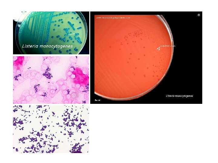
Family: Listeriaceae Genus: Listeria (Joseph Lister, 1940) – L. monocytogenes (pathogen) – L. innocua (pathogen) – L. welshimeri – L. seeligeri – L. ivanovii (pathogen) – L. grayi

• Gram positive, rod, asporous, without capsules • At 22 degrees the peritricular flagella and motile, while at 37 degrees the motion is weak • Catalase +, oxidase • Facultative anaerobic • Zoonotic • Small, S type colonies on blood agar • On Tryptose agar it appears blue-green in oblique-light (Henry's lamp technique) • Most strains have narrow hemolysis • p. H 5. 5 -9. 6 can be tolerated • 16 serotypes according to Somatic O and Flagellar H antigens

Important Species and Diseases Species Host Disease L. monocytogenes Sheep, Goat, Cattle Encephalitis, abortion, septicemia, endophthalmitis Cattle Mastitis Cat, Dog, Horse Abortion, encephalitis Pig Abortion, encephalitis, septicemia Bird Septicemia L. ivanovi Sheep, Cattle Abortion L. innocua Sheep Meningoencephalitis

EPIDEMIOLOGY • Affects people, many animals and poultry • Septicemia, Meningoencephalitis, Abort • Soil contamination and digestion of contaminants are the main route of contamination • Poor quality silage (silage disease) with a p. H higher than 5. 5 • Especially in winter and first spring months, animals of all ages are seen • If the brain stem is affected in the disease, rotation disease (neural listeriosis), paralysis of the stomach, bowing of the head • In cattle and sheep, localized, unilateral (silage-associated) keratoconjunctivitis (ocular listeriosis)

Pathogenesis • Invasive to both phagocytic and non-phagocytic cells • It survives in the cell, multiplies and passes to the other cell • Specific surface proteins «internalins» are responsible for adhesion and invasion to the host cell • listeriolysin O provides Listeria’s phagosome membrane breakdown and leaving the cytolytic toxin – The digestive factor penetrates the intestinal epithelium and M cells and spreads to other tissues via blood and lymph (septicemia). – Transplacental infection in pregnant (stillbirths, anomalies, abortion) – If invasive from oral and nasal cracks, neural listeriosity is caused by cranial nerves (brain stem microaplasia) – Monocytosis is an increase in the number of monocytes in the blood, seen in chronic infections (tuberculosis, brucellosis), in Listeria this is seen only in monogastric animals

Listeriosis in humans • It is rare but 50% mortality can be seen • Especially in immunosuppressed people , in pregnancies and in the elderly; meningitis, encephalitis, low, endocarditis, septicemia • In healthy people; flu-like, mild fever • Infected animals are rarely transmitted • It can grow in the refrigerator temperature and is resistant to salt • Raw milk, cream cheese, uncooked vegetables, meat and fish, frozen foods
,")
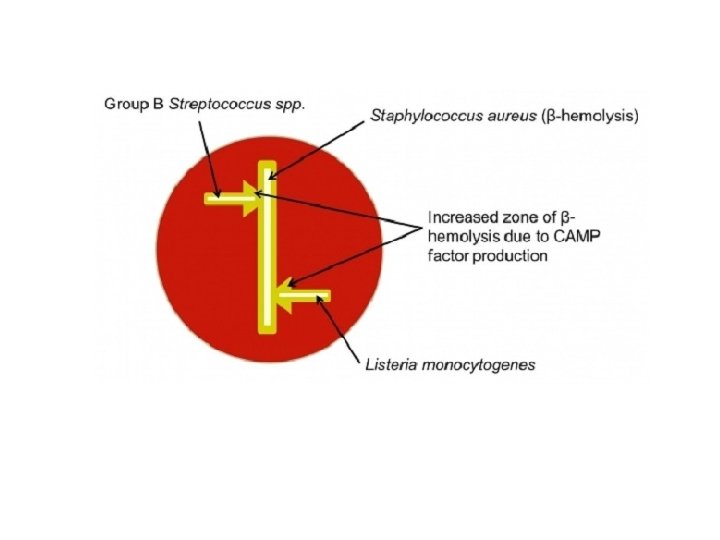
Laboratory Test • Especially, cerebral tissue (medulla, pons, cotyledon, abomasal content, liver and spleen), applying 'cold enrichment' • The sample is homogenized and kept in refrigerator for 12 weeks in nutrient broth at +4 degrees • Small, S-type, blue-green colonies in oblique light, • L. monocytogenes, L. ivanovi, L. seeligeri form narrow hemolysis • When the broth culture is kept at 25°C for 2 -4 hours, the movement of the agent increases (tumbling motility) • CAMP test (with S. aureus)

CAMP TEST TÜRLER With S. aureus With R. equi L. monocytogenes + - L. innocua - - L. welshimeri - - L. seeligeri + - L. ivanovii - + L. grayi - -

Animal Experiment ANTON TEST A drop of liquid culture into the rabbit eye drops in 24 hours keratoconjuntivitis occurs
- Slides: 54